



Avacincaptad pegol sodium

Based on 1 Customer Validation
Avacincaptad pegol (ARC1905) sodium is a 40KDa PEG-conjugated aptamer. Avacincaptad pegol sodium targets complement factor 5 (C5), inhibits the cleavage of C5 into C5a and C5b, limits inflammatory stimulation and complement membrane attack complex (MAC), and is used to study age-related macular degeneration (AMD). Avacincaptad pegol sodium limits irregular cell apoptosis by targeting downstream factors in the complement cascade while preserving the early steps of the complement system.
For research use only. We do not sell to patients.
- Purity : 90.08%
- CAS No.: 1491144-00-3
- Molecular Weight:53500 (Approximately)
-
Storage:
-20°C, sealed storage, away from moisture
* In solvent : -80°C, 6 months; -20°C, 1 month (sealed storage, away from moisture)


To place orders, for customer services and technical support, please contact: MedChemExpress USA
Tel: 609-228-6898 E-mail: [email protected] [email protected]


-
 Biological Activity
-
Chemical Information
-
Solvent & Solubility
- Protocols
- Purity & Documentation
- References
-
 Help & FAQs


-

HY-P3252A
Pegcetacoplan acetateComplement C3/C3b Inhibitor -
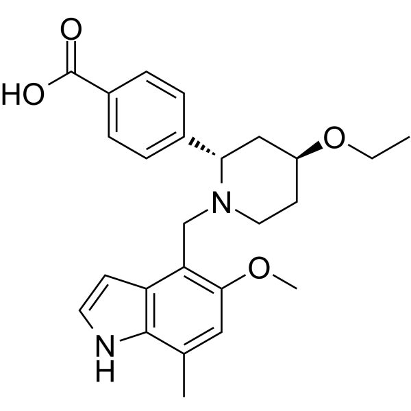
HY-127105
IptacopanFactor B Inhibitor -

HY-P99965
CrovalimabAnti-C5 Recycling Antibody -

HY-P3204
POT-4C3 Receptor Inhibitor -

HY-148457
Avacincaptad pegolC5 Complement Inhibitor -
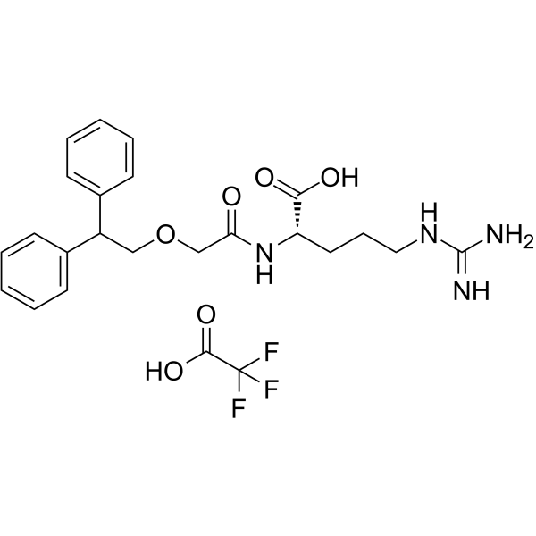
HY-101502A
SB290157 trifluoroacetateC3aR Antagonist -
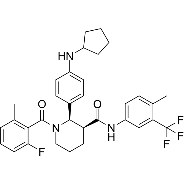
HY-17627
AvacopanC5aR Inhibitor -
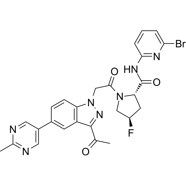
HY-117930
DanicopanFactor D Inhibitor -
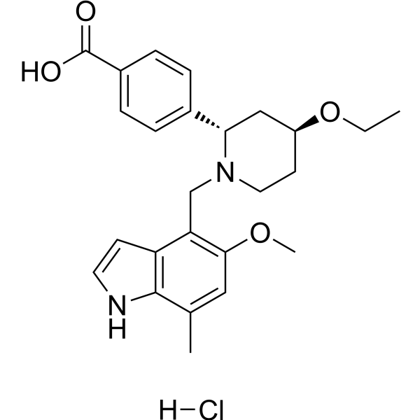
HY-127105A
Iptacopan hydrochlorideFactor B Inhibitor -
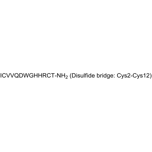
HY-P1036
CompstatinPrimates’ C3 Inhibitor -

HY-P9914
EculizumabComplement C5 Antibody -

HY-P99786
PozelimabAnti-C5 Antibody


Biological Activity
Description
In Vivo
Avacincaptad pegol sodium is delivered by PEGylated RNA to treat geographic atrophy in macular degeneration[5].
MedChemExpress (MCE) has not independently confirmed the accuracy of these methods. They are for reference only.


Chemical Information
-
CAS No. 1491144-00-3
-
Appearance Solid
-
Molecular Weight 53500 (Approximately)
-
Color White to off-white
-
SMILES
[Avacincaptad pegol sodium]
-
Synonyms
ARC1905
-
Sequence
Poly(oxy-1,2-ethanediyl), α-hydro-ω-methoxy-, 5′-ether with RNA ((2′-deoxy-2′-fluoro)C-Gm-(2′-deoxy-2′-fluoro)C-(2′-deoxy-2′-fluoro)C-G-(2′-deoxy-2′-fluoro)C-Gm-Gm-(2′-deoxy-2′-fluoro)U-(2′-deoxy-2′-fluoro)C-(2′-deoxy-2′-fluoro)U-(2′-deoxy-2′-fluoro)C-Am-Gm-Gm-(2′-deoxy-2′-fluoro)C-G-(2′-deoxy-2′-fluoro)C-(2′-deoxy-2′-fluoro)U-Gm-Am-Gm-(2′-deoxy-2′-fluoro)U-(2′-deoxy-2′-fluoro)C-(2′-deoxy-2′-fluoro)U-Gm-Am-Gm-(2′-deoxy-2′-fluoro)U-(2′-deoxy-2′-fluoro)U-(2′-deoxy-2′-fluoro)U-A-(2′-deoxy-2′-fluoro)C-(2′-deoxy-2′-fluoro)C-(2′-deoxy-2′-fluoro)U-Gm-(2′-deoxy-2′-fluoro)C-Gm-(3′→3′)-dT) 5′-[6-[[(2,3-dihydroxypropoxy)carbonyl]amino]hexyl hydrogen phosphate], sodium salt (2:1:39)
-
Shipping
Room temperature in continental US; may vary elsewhere.
-
Storage
-20°C, sealed storage, away from moisture
* In solvent : -80°C, 6 months; -20°C, 1 month (sealed storage, away from moisture)


Solvent & Solubility
In Vitro:
H2O : 100 mg/mL (Need ultrasonic)

Protocols
-
Apoptosis
Apoptosis, also called programmed cell death, is generally characterized by distinct morphological characteristics.
-
TUNEL staining for apoptotic DNA fragmentation
TUNEL staining detects DNA strand breaks by using terminal deoxynucleotidyl transferase to add labeled nucleotides to exposed 3′-OH DNA termini, generating either microscopic staining in fixed cells or tissue sections, or fluorescence/cytometric signal in cell suspensions. TUNEL positivity reflects DNA fragmentation but should not be interpreted alone as definitive apoptosis, because TUNEL can also label necrotic, autolytic, mechanically damaged, or DNA-repair-associated DNA breaks.
-
Annexin V plus membrane-impermeant dye apoptosis staining
Annexin V-based apoptosis assays rely on the detection of phosphatidylserine (PS) externalization from the inner leaflet of the plasma membrane to the outer leaflet, an early biochemical hallmark of apoptosis. Fluorescently labeled Annexin V binds PS in a calcium-dependent manner, enabling identification of early apoptotic cells by flow cytometry or fluorescence microscopy. When combined with a membrane-impermeant DNA-binding dye (e. g. , propidium iodide), this approach allows discrimination between viable (Annexin V−/dye−), early apoptotic (Annexin V+/dye−), and late apoptotic or necrotic (Annexin V+/dye+) cell populations by assessing membrane integrity and PS exposure.
-
Apoptosis Solutions
Apoptosis is a regulated, generally non-lytic cell-death pathway that removes unwanted, damaged, infected, or abnormal cells through coordinated morphological changes, caspase activation, DNA fragmentation, and membrane remodeling. The intrinsic apoptosis pathway is controlled mainly by mitochondrial outer membrane permeabilization, BCL-2 family proteins, cytochrome c release, apoptosome formation, caspase-9 activation, and downstream executioner caspase-3/7 activation. The extrinsic apoptosis pathway is initiated by death receptors such as Fas, TNFR, and TRAIL receptors, which recruit adaptor proteins and activate caspase-8 before engaging executioner caspases or mitochondrial amplification through BID cleavage. Apoptosis is linked to many phenotypes, including cancer cell killing, tissue homeostasis, immune regulation, neurodegeneration, infection response, and treatment-induced cytotoxicity; unresolved questions include how apoptosis interacts with necroptosis, pyroptosis, ferroptos


Purity & Documentation
-
Data Sheet (273 KB)
-
SDS (252 KB)
- English - EN (252 KB)
- Français - FR (252 KB)
- Deutsch - DE (252 KB)
- Norwegian - NO (252 KB)
- Español - ES (252 KB)
- Swedish - SV (252 KB)
- Italian - IT (252 KB)
- Korean - KR (252 KB)
- Portuguese - PT (252 KB)
-
Handling Instructions (1518 KB)
References
[1]. Joshua Ong, et al. The Complement System as a Therapeutic Target in Retinal Disease. Medicina (Kaunas). 2024 Jun 5;60(6):945. [Content Brief]
[3]. Aira Sarkar, et al. Novel and investigational therapies for wet and dry age-related macular degeneration. Drug Discov Today. 2022 Aug;27(8):2322-2332. [Content Brief]
[4]. Padma Sundaram, et al. Therapeutic RNA aptamers in clinical trials. Eur J Pharm Sci. 2013 Jan 23;48(1-2):259-71. [Content Brief]

Calculators
Concentration (start) × Volume (start) = Concentration (final) × Volume (final)


Avacincaptad pegol sodium Related Classifications
HY-147080 Related Classifications
 Powered by Bioz