



Dim16
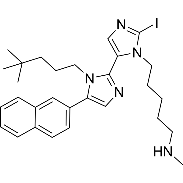
Dim16 is a dual PCSK9 inhibitor and HMG-CoAR inhibitor, with an IC50 of 0.8 nM against human PCSK9 and an IC50 of 146.8 μM against HMG-CoAR. Dim16 disrupts the PCSK9-LDLR protein-protein interaction, inhibits the catalytic activity of HMG-CoAR, enhances cellular uptake of extracellular LDL, and suppresses PCSK9-induced platelet aggregation. Dim16 can be used in research related to hypercholesterolemia.
Nos produits utilisent uniquement pour la recherche. Nous ne vendons pas aux patients.
- CAS No.: 2743448-32-8
- Formule: C29H38IN5
- Masse moléculaire:583.55
-
Stockage:
Please store the product under the recommended conditions in the Certificate of Analysis.

To place orders, for customer services and technical support, please contact: MedChemExpress USA
Tel: 609-228-6898 E-mail: [email protected] [email protected]


-
 Activité biologique
-
Chemical Information
- Protocole
- Pureté et documentation
- Références
-
 Help & FAQs




Activité biologique
Description
IC50 & Target
19 nM (PCSK9)[1]
In Vitro
Dim16 (0.1-1×10⁶ nM; 2 h) potently inhibits the protein-protein interaction between PCSK9 and LDLR in a biochemical assay with an IC50 of 0.8 nM[1].
Dim16 (10-500 μM; 10 min) inhibits HMG-CoAR activity in a biochemical assay with an IC50 of 146.8 μM[1].
Dim16 (0.1 nM-10000 nM; 48 h) is non-toxic to HepG2 cells[1].
Dim16 (1-10 nM; 2 h) restores cell surface LDLR levels in PCSK9-treated HepG2 cells[1].
Dim16 (1-10 nM; 2 h co-incubation with PCSK9, 2 h incubation with fluorescent LDL) restores and enhances LDL uptake in PCSK9-treated HepG2 cells[1].
Dim16 (10 nM; 15 min preincubation with PCSK9) prevents the PCSK9-induced potentiation of epinephrine-stimulated platelet aggregation in human platelet-rich plasma[1].
MedChemExpress (MCE) has not independently confirmed the accuracy of these methods. They are for reference only.
-
Cell Line:HepG2
-
Concentration:0.1 nM, 1 nM, 10 nM, 100 nM, 1000 nM, 10000 nM
-
Incubation Time:48 h
-
Result:Showed no reduction in HepG2 cell vitality at any tested concentration compared to control cells.


Chemical Information
-
CAS No. 2743448-32-8
-
Masse moléculaire 583.55
-
Formule C29H38IN5
-
SMILES
CC(C)(C)CCCN1C(C2=CN=C(N2CCCCCNC)I)=NC=C1C3=CC=C4C=CC=CC4=C3
-
Livraison
Room temperature in continental US; may vary elsewhere.
-
Stockage
Please store the product under the recommended conditions in the Certificate of Analysis.


Pureté et documentation
Références

Calculators
Concentration (start) × Volume (start) = Concentration (final) × Volume (final)


Dim16 Related Classifications
HY-149310 Related Classifications
 Powered by Bioz