



TSI-01

Based on 1 Customer Validation
TSI-01 is a selective LPCAT2 inhibitor, with IC50 values of 0.47 μM and 3.02 μM against LPCAT2 and LPCAT1, respectively. TSI-01 competitively inhibits acetyl-CoA (acetyl-CoA) targeting the lysophosphatidic acid acetyltransferase activity of LPCAT2, and suppresses PAF biosynthesis in calcium ionophore-stimulated mouse peritoneal macrophages. TSI-01 inhibits the proliferation of endometrial cancer cells and promotes endometrial cancer cell apoptosis (apoptosis). TSI-01 can be used for the research of endometrial cancer and PAF-related inflammatory diseases.
Nos produits utilisent uniquement pour la recherche. Nous ne vendons pas aux patients.
- Pureté : 98.87%
- CAS No.: 704878-75-1
- Formule: C14H11Cl2NO4
- Masse moléculaire:328.15
-
Stockage:Powder -20°C, 3 years , 4°C, 2 years ; In solvent -80°C, 6 months , -20°C, 1 month


To place orders, for customer services and technical support, please contact: MedChemExpress USA
Tel: 609-228-6898 E-mail: [email protected] [email protected]


-
 Activité biologique
-
Chemical Information
-
Solvant et solubilité
- Protocole
- Pureté et documentation
- Références
-
 Help & FAQs


-

HY-158790
(R)-HTS-3LPCAT3 Inhibitor -
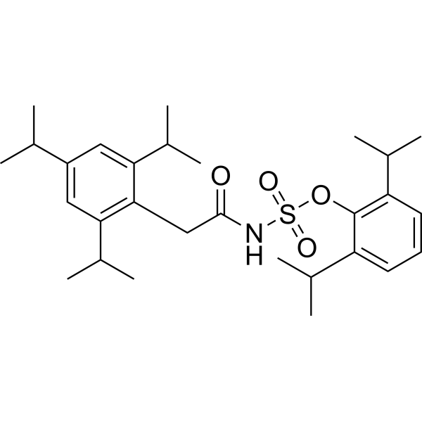
HY-13215
AvasimibeACAT/SOAT Inhibitor -
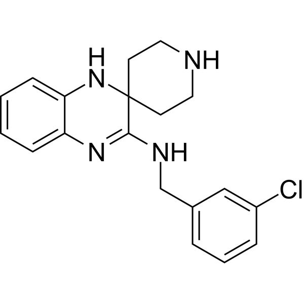
HY-12726
Liproxstatin-1Ferroptosis Inhibitor -

HY-15141
StaurosporinePKC/PKA Inhibitor -
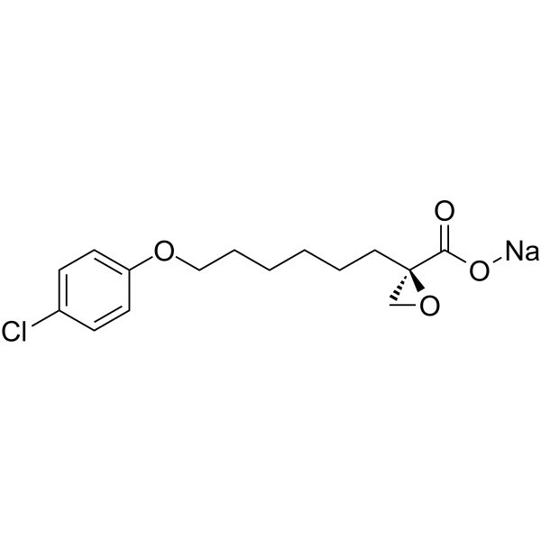
HY-50202A
Etomoxir sodium saltCPT1a Inhibitor -
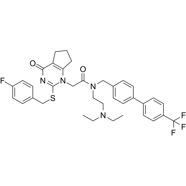
HY-10521
DarapladibLp-PLA2 Inhibitor -
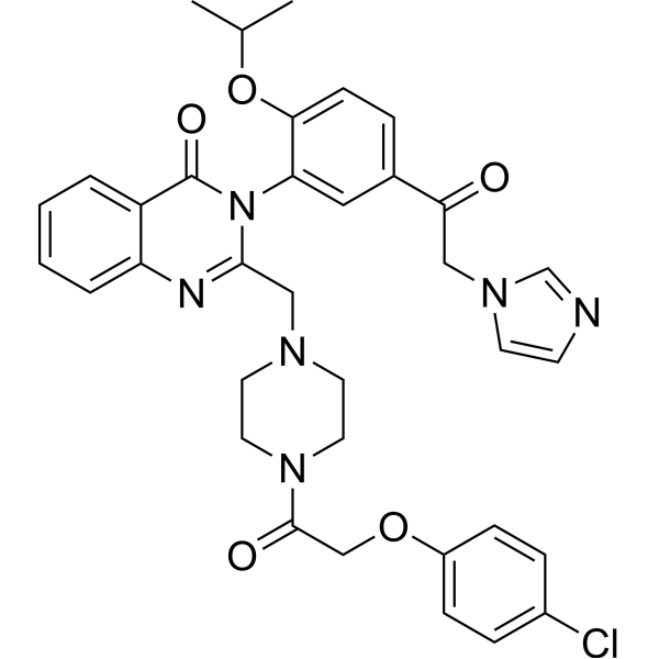
HY-114481
Imidazole ketone erastinFerroptosis Inducer -
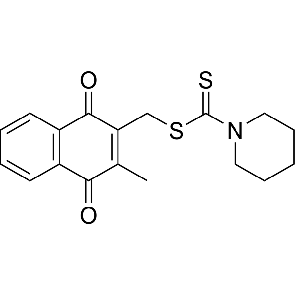
HY-103617
PKM2-IN-1PKM2 Inhibitor -
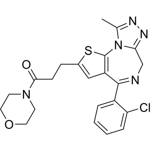
HY-108634
ApafantPAF Receptor Antagonist -
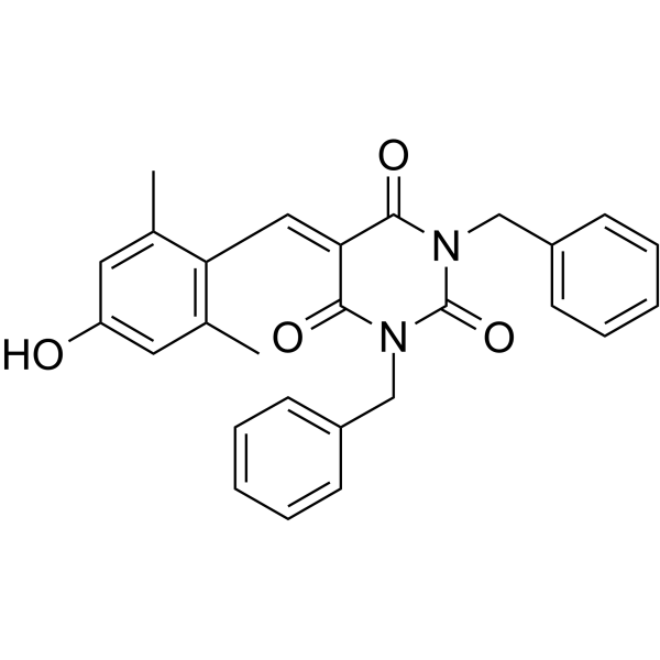
HY-110263
EML 425CBP/p300 Inhibitor -
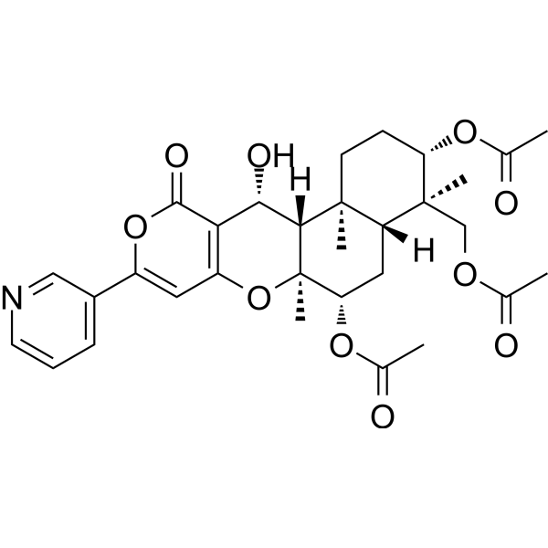
HY-117832
Pyripyropene ASOAT2/ACAT2 Inhibitor -

HY-13215R
Avasimibe (Standard)ACAT/SOAT Inhibitor


Activité biologique
Description
In Vitro
MedChemExpress (MCE) has not independently confirmed the accuracy of these methods. They are for reference only.


Chemical Information
-
CAS No. 704878-75-1
-
Appearance Solid
-
Masse moléculaire 328.15
-
Formule C14H11Cl2NO4
-
Color White to off-white
-
SMILES
O=C(C1=CC=C(N2C(C(Cl)=C(C2=O)Cl)=O)C=C1)OC(C)C
-
Livraison
Room temperature in continental US; may vary elsewhere.
-
Stockage
Powder -20°C 3 years 4°C 2 years In solvent -80°C 6 months -20°C 1 month


Solvant et solubilité
In Vitro:
DMSO : 116.67 mg/mL (355.54 mM; Need ultrasonic; Hygroscopic DMSO has a significant impact on the solubility of product, please use newly opened DMSO)
Please refer to the solubility information to select the appropriate solvent. Once prepared, please aliquot and store the solution to prevent product inactivation from repeated freeze-thaw cycles.
Storage method and period of stock solution: -80°C, 6 months; -20°C, 1 month. When stored at -80°C, please use it within 6 months. When stored at -20°C, please use it within 1 month.
Please refer to the solubility information to select the appropriate solvent. Once prepared, please aliquot and store the solution to prevent product inactivation from repeated freeze-thaw cycles.
Storage method and period of stock solution: -80°C, 6 months; -20°C, 1 month. When stored at -80°C, please use it within 6 months. When stored at -20°C, please use it within 1 month.
Concentration (start) × Volume (start) = Concentration (final) × Volume (final)


Pureté et documentation
-
Fiche technique (273 KB)
-
SDS (396 KB)
- English - EN (396 KB)
- Français - FR (396 KB)
- Deutsch - DE (396 KB)
- Norwegian - NO (396 KB)
- Español - ES (396 KB)
- Swedish - SV (396 KB)
- Italian - IT (396 KB)
- Korean - KR (396 KB)
- Portuguese - PT (396 KB)
-
Instruction de manipulation (2659 KB)
Références
[1]. Zhao T, et al. Overexpression of LPCAT1 enhances endometrial cancer stemness and metastasis by changing lipid components and activating the TGF/β-Smad2/3 signaling pathway. Acta biochimica et biophysica Sinica. 2022 Jun 25;54(7):904-916. [Content Brief]
[2]. Tarui M, et al. Selective inhibitors of a PAF biosynthetic enzyme lysophosphatidylcholine acyltransferase 2. Journal of lipid research. 2014 Jul;55(7):1386-96. [Content Brief]


Complete Stock Solution Preparation Table
Please refer to the solubility information to select the appropriate solvent. Once prepared, please aliquot and store the solution to prevent product inactivation from repeated freeze-thaw cycles.
Storage method and period of stock solution: -80°C, 6 months; -20°C, 1 month. When stored at -80°C, please use it within 6 months. When stored at -20°C, please use it within 1 month.
| Optional Solvent | Concentration Solvent Mass | 1 mg | 5 mg | 10 mg | 25 mg |
|---|---|---|---|---|---|
| DMSO | 1 mM | 3.0474 mL | 15.2369 mL | 30.4739 mL | 76.1847 mL |
| 5 mM | 0.6095 mL | 3.0474 mL | 6.0948 mL | 15.2369 mL | |
| 10 mM | 0.3047 mL | 1.5237 mL | 3.0474 mL | 7.6185 mL | |
| 15 mM | 0.2032 mL | 1.0158 mL | 2.0316 mL | 5.0790 mL | |
| 20 mM | 0.1524 mL | 0.7618 mL | 1.5237 mL | 3.8092 mL | |
| 25 mM | 0.1219 mL | 0.6095 mL | 1.2190 mL | 3.0474 mL | |
| 30 mM | 0.1016 mL | 0.5079 mL | 1.0158 mL | 2.5395 mL | |
| 40 mM | 0.0762 mL | 0.3809 mL | 0.7618 mL | 1.9046 mL | |
| 50 mM | 0.0609 mL | 0.3047 mL | 0.6095 mL | 1.5237 mL | |
| 60 mM | 0.0508 mL | 0.2539 mL | 0.5079 mL | 1.2697 mL | |
| 80 mM | 0.0381 mL | 0.1905 mL | 0.3809 mL | 0.9523 mL | |
| 100 mM | 0.0305 mL | 0.1524 mL | 0.3047 mL | 0.7618 mL |


TSI-01 Related Classifications
HY-112825 Related Classifications
 Powered by Bioz