



Cadonilimab
Based on 1 publication(s) in Google Scholar
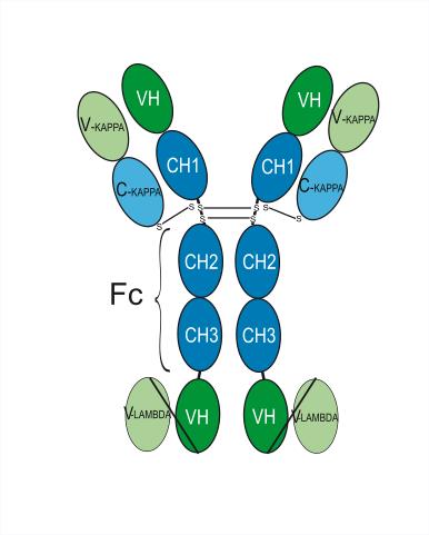
Cadonilimab (AK104) is a humanized tetravalent IgG1 bispecific antibody targeting PD1/CTLA4. Cadonilimab blocks both PD-1 and CTLA-4 pathways, thereby relieving their corresponding immunosuppressive effects and reversing tumor specific T cell exhaustion. Cadonilimab significantly downregulates Fc-mediated effector functions, including antibody-dependent cell-mediated cytotoxicity (ADCC), antibody-dependent cellular phagocytosis (ADCP), complement dependent cytotoxicity (CDC). Cadonilimab can be used for research of metastatic cervical cancer, as well as other malignancies such as gastric cancer, GEJ adenocarcinoma and non-small cell lung cancer (NSCLC).
For research use only. We do not sell to patients.
- Purity : 99.87%
- CAS No.: 2394841-59-7
- Molecular Weight:198.64 kDa
-
Storage:
Please store the product under the recommended conditions in the Certificate of Analysis.


To place orders, for customer services and technical support, please contact: MedChemExpress USA
Tel: 609-228-6898 E-mail: [email protected] [email protected]


-
 Biological Activity
-
Chemical Information
- Protocols
- Purity & Documentation
- References
-
 Help & FAQs


-

HY-P99675
IvonescimabPD-1/VEGF Bispecific Antibody -

HY-P9904
AtezolizumabAnti-PD-L1 Antibody -

HY-P99166
VudalimabPD-1/ CTLA-4 Inhibitor -
HY-P9901
IpilimumabCTLA-4 Inhibitor -

HY-P99380
ZanidatamabAnti- HER2 Antibody -

HY-P99944
SHR-1701PD-L1/TGF β Fusion Protein -
HY-19991
BMS-1PD-1/PD-L1 PPI Inhibitor -

HY-P9906
BevacizumabVEGF-A Inhibitor -

HY-P9902
PembrolizumabPD-1 Inhibitor -
HY-N0242
FraxinelloneSTAT Inhibitor -

HY-P9908
AdalimumabTNF-α Inhibitor -

HY-P9950
OmalizumabHuman IgE Antibody
Publications Citing Use of MedChemExpress (MCE) Cadonilimab
More

Biological Activity
Description
Isotype
IgG1-kappa-[scFv-heavy-lambda]2
Recommend Isotype Controls
Species Reactivity
Human
IC50 & Target
CTLA4 & PDCD1
In Vitro
Cadonilimab (0-100 nM, 10-120 min) maintains a high affinity for Jurkat-CTLA4 and Jurkat-PD1 cells in vitro after iodine modification[4].
MedChemExpress (MCE) has not independently confirmed the accuracy of these methods. They are for reference only. Further protocols information, click here.
In Vivo
MedChemExpress (MCE) has not independently confirmed the accuracy of these methods. They are for reference only.
-
Animal Model:Normal KM mice and B-hPD1/hCTLA4-MC38-hPD-L1 mice[4]
-
Dosage:3.7 MBq (Cadonilimab modified with 124I)
-
Administration:tail i.v., a single dose, 2, 24, 96 and 192 h post-injection acquired static PET scans
-
Result:Resulted in higher tumor to nontumor (T/NT) ratios and increaseed the standard uptake value (SUV) value at the tumor site modified with 124I in mice.
Clinical Trial
| NCT Number | Sponsor | Condition | Start Date |
Phase

|
|---|---|---|---|---|
| NCT01329991 | Plexxikon| | 2011-05 | PHASE1 |
Gene ID
Accession
Conjugated
Unconjugated
Reconsititution
The product can be reconstituted/diluted with sterile PBS or saline.
Format
-
IgG1-kappa-[scFv-heavy-lambda]2
Application
ELISA, FACS, Functional assay
Verified Bioactivity
-
 Immobilized Cadonilimab can bind Human PD-1 Protein (ECD, His Tag). The EC50 for this effect is 0.197 ng/mL.
 -
 Immobilized Cadonilimab can bind Human CTLA-4 Protein (His Tag). The EC50 for this effect is 3.07 ng/mL.
 -
 Immobilized Cadonilimab can bind Human CTLA-4 Protein (His Tag). The EC50 for this effect is 3.07 ng/mL. Flow Cytometry analysis of Raji cells labelling CTLA4 (red) with Cadonilimab (anti-CTLA4) (HY-P99117). Cells were stained with the primary antibody at 1/200 dilution for an hour at 4℃. Goat Anti-Human IgG H&L (AF488) (HY-P83776) was used as the secondary antibody at 1/1,000 dilution for 30 minutes at 4℃. Human IgG1 kappa (HY-P99001, blue) was used as the isotype control, cells without incubation with primary antibody were used as the unlabeled control (black).
 -
 Flow Cytometry analysis of THP-1 cells labelling PDCD1 (red) with Cadonilimab (anti-PDCD1) (HY-P99117). Cells were stained with the primary antibody at 1/200 dilution for an hour at 4℃. Goat Anti-Human IgG H&L (AF488) (HY-P83776) was used as the secondary antibody at 1/1,000 dilution for 30 minutes at 4℃. Human IgG1 kappa (HY-P99001, blue) was used as the isotype control, cells without incubation with primary antibody were used as the unlabeled control (black).


Chemical Information
-
CAS No. 2394841-59-7
-
Appearance Liquid
-
Molecular Weight 198.64 kDa
-
Color Colorless to light yellow
-
SMILES
[Cadonilimab]
-
Synonyms
AK104
-
Shipping
Shipping with dry ice.
-
Formulation
Please refer to the lot-specific COA for specific buffer information.
-
Storage
Please store the product under the recommended conditions in the Certificate of Analysis.


Publications (1)
-
Journal Impact Factor
-
Most Recent

Protocols
-
Phagocytosis Functional Assay
A phagocytosis functional assay measures the ability of phagocytic cells, such as neutrophils, macrophages, monocytes, or microglia/macrophages, to bind and internalize particulate targets including bacteria, yeast particles, beads, or myelin particles. Fluorescent flow-cytometry assays detect target uptake as fluorescence associated with gated phagocytes, while pH-sensitive dyes such as pHrodo increase signal in acidic phagosomal compartments and therefore preferentially report internalized particles rather than particles remaining outside the cell. Microscopy or high-content imaging can be used to confirm intracellular localization and, in some protocols, to follow uptake kinetics.
-
Cell Cytotoxicity Assay
Cytotoxicity assays are usually based on the assessment of cell membrane damage, which can also be indirectly detected by measuring cell viability. Detection methods include MTT assay, CKK-8 assay, LDH assay and ATP assay, etc.
-
Mammalian live/dead viability and cytotoxicity staining
Live/dead viability and cytotoxicity staining assays are based on the simultaneous detection of intracellular esterase activity in metabolically active (viable) cells and membrane integrity loss in non-viable cells. In commonly used dual-staining approaches, membrane-permeant fluorogenic substrates are converted by intracellular esterases into fluorescent products in live cells, while impermeant DNA-binding dyes selectively enter cells with compromised plasma membranes and label nucleic acids in dead or dying cells, enabling discrimination between viable and non-viable populations by fluorescence microscopy or flow cytometry.
-
Research Protocol for Cancer Immunology
Cancer immunology studies how the immune system recognizes, suppresses, edits, or fails to eliminate malignant cells through tumor antigen release, antigen presentation, T-cell priming, immune trafficking, tumor-cell killing, and feedback inhibition in the tumor microenvironment. The cancer-immunity cycle links tumor antigenicity, dendritic-cell priming, CD8+ T-cell infiltration, cytotoxic function, and immune-checkpoint regulation to tumor rejection or immune escape. Immune-checkpoint pathways such as PD-1/PD-L1 and CTLA-4 suppress antitumor T-cell activity and can be therapeutically blocked, but many tumors remain resistant because of poor antigen presentation, weak T-cell infiltration, suppressive myeloid cells, regulatory T cells, and tumor-intrinsic immune-exclusion programs. Unresolved questions include which immune-cell states predict response, how tumor-intrinsic pathways exclude immune cells, how myeloid suppression limits checkpoint blockade, and which combination strategies
-
Cell Viability Determination by MTT Colorimetric Assay
The following protocol uses the MTT colorimetric assay as a classic literature-established method for assessing cell viability/metabolic activity in cultured mammalian cells. MTT[3-(4,5-dimethylthiazol-2-yl)-2,5-diphenyltetrazolium bromide] is reduced by metabolically active cells to a colored formazan product; the amount of formazan is quantified spectrophotometrically and provides an indirect measure of metabolically active viable cells. Importantly, MTT reduction reflects cellular oxidoreductase/metabolic activity rather than an absolute direct count of living cells, so changes in cellular metabolism can alter the signal independently of cell number.


Purity & Documentation
-
Data Sheet (262 KB)
-
SDS (251 KB)
- English - EN (251 KB)
- Français - FR (251 KB)
- Deutsch - DE (251 KB)
- Norwegian - NO (251 KB)
- Español - ES (251 KB)
- Swedish - SV (251 KB)
- Italian - IT (251 KB)
- Korean - KR (251 KB)
- Portuguese - PT (251 KB)
-
Inhibitory Antibodies User Guide (603 KB)
References
[1]. Antonarelli G, et al. Research and Clinical Landscape of Bispecific Antibodies for the Treatment of Solid Malignancies. Pharmaceuticals (Basel). 2021 Aug 31;14(9):884. [Content Brief]
[2]. Kaplon H, et al. Antibodies to watch in 2022. MAbs. 2022 Jan-Dec;14(1):2014296. [Content Brief]
[3]. Chen PY, et al. Case Report: Cadonilimab-related toxic epidermal necrolysis-like reactions successfully treated with supplemental Adalimumab. Front Immunol. 2023 Aug 3;14:1188523. [Content Brief]
[4]. Hou X, et al. Preclinical imaging evaluation of a bispecific antibody targeting hPD1/CTLA4 using humanized mice. Biomed Pharmacother. 2024 Jun;175:116669. [Content Brief]

Calculators
Concentration (start) × Volume (start) = Concentration (final) × Volume (final)


Cadonilimab Related Classifications
HY-P99117 Related Classifications
 Powered by Bioz