



Calcium Orange AM

Based on 1 Customer Validation
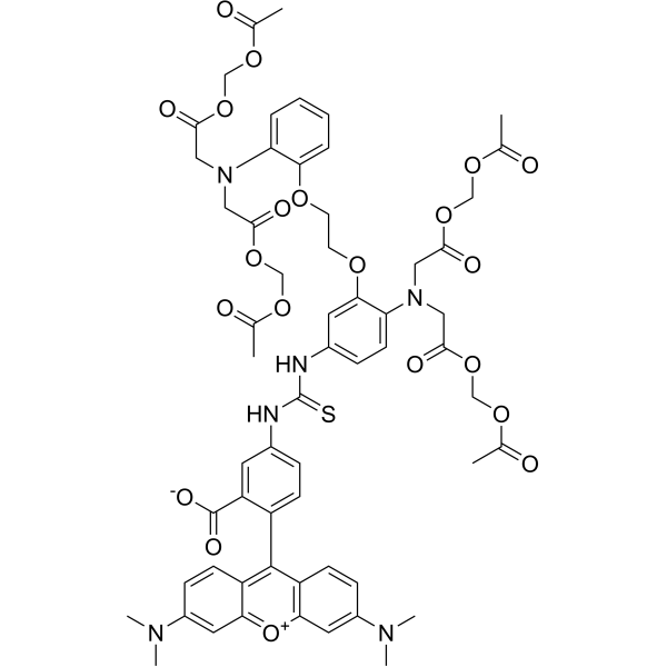
Calcium Orange AM is an intracellular calcium reporter. Specific fluorescence can be detected when free calcium binds to Calcium Orange AM (Ex/Em=549/576 nm). Calcium Orange AM does not enter the vacuoles and does not compartmentalize into acidic vesicles.
For research use only. We do not sell to patients.
- Purity : 98%
- CAS No.: 172646-19-4
- Formula: C59H62N6O21S
- Molecular Weight:1223.22
-
Storage:
-20°C, protect from light
* In solvent : -80°C, 6 months; -20°C, 1 month (protect from light)

To place orders, for customer services and technical support, please contact: MedChemExpress USA
Tel: 609-228-6898 E-mail: [email protected] [email protected]


-
 Biological Activity
-
Chemical Information
- Protocols
- Purity & Documentation
- References
-
 Help & FAQs




Biological Activity
Description
In Vitro
Guide (Following is our recommended protocol. This protocol only provides a guideline, and should be modified according to your specific needs)[2].
1. Calcium Orange AM is freshly prepared in dehydrated DMSO before each experiment.
2. Prepare the recording medium for measuring [Ca2+]i in cells consisted of 20 mM HEPES, 115 mM NaCl, 5.4 mM KCl, 0.8 mM MgCl2, 1.8 mM CaCl2, and 13.8 mM D-glucose adjusted to pH 7.4 by adding 1 M KOH.
3. Exchange the culture medium for 1 mL of a recording medium including 10 μM Calcium Orange AM.
4. Incubate cells under conditions of 5% CO2 and 37°C for 40 min.
5. Remove the recording medium, including calcium orange AM from the dish, and add the culture medium after washing it three times with 1 mL of the recording medium.
6. Incubate the cells for 1 h. After incubation, exchange the culture medium for the recording medium again.
7. Detect fluorescence.
MedChemExpress (MCE) has not independently confirmed the accuracy of these methods. They are for reference only.
Emission (Em)
57
Excitation (Ex)
549


Chemical Information
-
CAS No. 172646-19-4
-
Appearance Solid
-
Molecular Weight 1223.22
-
Formula C59H62N6O21S
-
Color Brown to red
-
SMILES
CN(C1=CC2=[O+]C3=C(C=CC(N(C)C)=C3)C(C4=CC=C(NC(NC5=CC=C(N(CC(OCOC(C)=O)=O)CC(OCOC(C)=O)=O)C(OCCOC6=CC=CC=C6N(CC(OCOC(C)=O)=O)CC(OCOC(C)=O)=O)=C5)=S)C=C4C([O-])=O)=C2C=C1)C
-
Shipping
Room temperature in continental US; may vary elsewhere.
-
Storage
-20°C, protect from light
* In solvent : -80°C, 6 months; -20°C, 1 month (protect from light)


Purity & Documentation
-
Data Sheet (273 KB)
-
SDS (252 KB)
- English - EN (252 KB)
- Français - FR (252 KB)
- Deutsch - DE (252 KB)
- Norwegian - NO (252 KB)
- Español - ES (252 KB)
- Swedish - SV (252 KB)
- Italian - IT (252 KB)
- Korean - KR (252 KB)
- Portuguese - PT (252 KB)
-
Handling Instructions (2659 KB)
References
[1]. Lam CM, et al. Monitoring cytosolic calcium in the dinoflagellate Crypthecodinium cohnii with calcium orange-AM. Plant Cell Physiol. 2005 Jun;46(6):1021-7. [Content Brief]
[2]. Inami W, et al. Intracellular calcium ion concentration measurement using a phase-modulation fluorescence lifetime method with compensation for phase shift due to the presence of proteins. Anal Sci. 2013;29(2):199-203. [Content Brief]

Calculators
Concentration (start) × Volume (start) = Concentration (final) × Volume (final)


Calcium Orange AM Related Classifications
HY-D1629 Related Classifications
 Powered by Bioz