



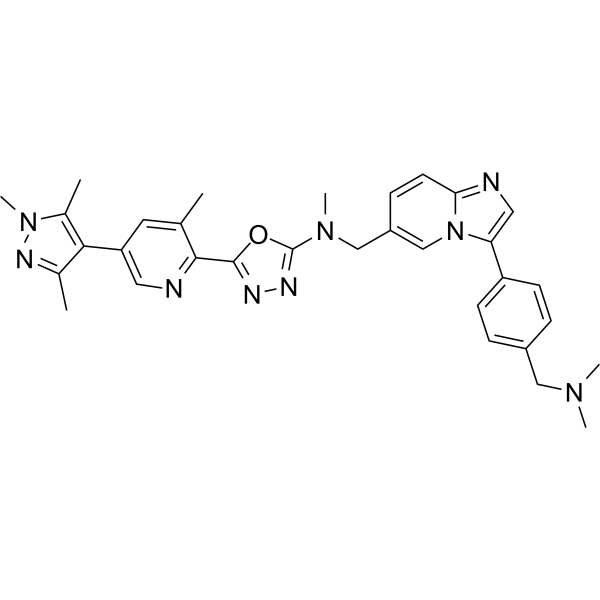
AZ14145845

Based on 1 Customer Validation
AZ14145845 is an orally active dual-target type 1½ kinase inhibitor of Mer/Axl, which inhibits Mer kinase (pIC50 = 9.0) and Axl kinase (pIC50 = 7.9) with high selectivity over Flt3 and Tyro3. AZ14145845 suppresses Mer- and Axl-dependent proliferation in Ba/F3 cells and inhibits efferocytosis in macrophages. AZ14145845 reduces the phagocytosis of photoreceptor outer segments by polarized human retinal epithelial cells. AZ14145845 inhibits tumor growth and induces tumor regression in vivo, and its combination with anti-PD1 antibody and ionizing radiation improves the survival rate of mice with colorectal cancer. AZ14145845 causes retinal degeneration in mice, with pathological changes similar to those in MERTK loss-of-function models. AZ14145845 can be used in studies related to retinal degeneration, lymphoma and colorectal cancer.
For research use only. We do not sell to patients.
- Purity : 98.00%
- CAS No.: 2830555-70-7
- Formula: C32H35N9O
- Molecular Weight:561.68
-
Storage:Powder -20°C, 3 years , 4°C, 2 years ; In solvent -80°C, 6 months , -20°C, 1 month

To place orders, for customer services and technical support, please contact: MedChemExpress USA
Tel: 609-228-6898 E-mail: [email protected] [email protected]


-
 Biological Activity
-
Chemical Information
-
Solvent & Solubility
- Protocols
- Purity & Documentation
- References
-
 Help & FAQs




Biological Activity
Description
|
MERTK 9.0 (pIC50) |
Axl 7.9 (pIC50) |
In Vitro
AZ14145845 potently inhibits the kinase activities of purified MERTK and Axl, with pIC50 values of 9.0 and 7.9, respectively[1].
AZ14145845 inhibits MERTK phosphorylation in transiently transfected Cos-7 cells with a pIC50 of 7.7[1].
AZ14145845 inhibits efferocytosis in macrophages with a pIC50 of 7.6[1].
AZ14145845 (3 μM; 6 h) reduces photoreceptor outer segment phagocytosis by polarized human retinal epithelial ARPE19 cells by 60%[1].
AZ14145845 (compound 32) at 1 μM potently and selectively inhibits Mer and Axl kinases, exhibiting only extremely low activity against 383 tested kinases[2].
AZ14145845 (72 h) potently inhibits the proliferation of Mer-dependent and Axl-dependent Ba/F3 cells (with pGI50 values of 7.6 and 7.0, respectively), while exhibits significantly weaker activity against Flt3-dependent, Tyro3-dependent Ba/F3 cells and parental Ba/F3 cells[2].
AZ14145845 (1 h) potently inhibits intracellular Mer activity (pIC50 = 7.8) and weakly inhibits intracellular Axl activity (pIC50 = 7.0) in transfected COS-7 cells, with no activity against intracellular Flt3 or Tyro3[2].
AZ14145845 potently inhibits Mer-mediated efferocytosis in human primary monocyte-derived macrophages, with a pIC50 of 7.6[2].
AZ14145845 binds to the Mer kinase domain in an I1/2 binding mode, with key interactions occurring at the hinge region, and does not interact with the activation loop of the kinase[2].
AZ14145845 potently inhibits pMerTK (EC50 = 15.8 nM) and pAxl (EC50 = 100 nM), while it shows extremely low activity against Flt3 in biochemical kinase assays (EC50 >31600 nM)[2].
MedChemExpress (MCE) has not independently confirmed the accuracy of these methods. They are for reference only. Further protocols information, click here.
-
Cell Line:Polarized human retinal epithelial ARPE19 cells
-
Concentration:3 μM
-
Incubation Time:6 h
-
Result:Approximately 60% reduction in the number of internalized POS.
Parmacokinetics
In Vivo
AZ14145845 (2 μmol/kg/h; intravenous administration; single 15-minute infusion) binds to melanin, resulting in significantly higher ocular exposure in pigmented rats than in non-pigmented rats[1].
AZ14145845 (compound 32) (3-100 mg/kg; p.o.; twice daily; for 6 consecutive days) exhibits dose-dependent in vivo efficacy in the Ba/F3-Mer xenograft model, with observable tumor regression and tumor growth inhibition; it also binds to the Mer target in vivo in a dose-dependent manner, and significantly reduces phosphorylated Mer levels[2].
AZ14145845 (3-100 mg/kg; p.o.; twice daily; for 5 consecutive days) exhibits dose-dependent in vivo efficacy in the Ba/F3-Axl xenograft model, with significant tumor growth inhibition observed and a significant dose-dependent reduction in phosphorylated Axl levels[2].
AZ14145845 (100 mg/kg; p.o.; twice daily; for 2 weeks) enhances survival when combined with anti-PD1 antibody and ionizing radiation in the MC38 colorectal cancer immunotherapy model[2].
MedChemExpress (MCE) has not independently confirmed the accuracy of these methods. They are for reference only.
-
Animal Model:C57BL/6 J (6-7-week-old female, 20-25 g)[1]
-
Dosage:200 mg/kg
-
Administration:p.o.; twice daily; 28 days
-
Result:Reached quantified concentrations of ~9 µM in the posterior eye region, ~11 µM in the anterior eye region, and ~3 µM in the whole eye.
Induced retinal lesions consistent with atrophy in 4 of 8 treated animals, including marked thinning of the outer nuclear layer, loss of photoreceptors, flattening of RPE cells with loss of apical microvilli, and occasional pyknotic ONL nuclei.
Caused ultrastructural changes including disorganized photoreceptor outer segments at the RPE-POS interface, loss of association between RPE microvilli and POS, and a disorganized debris zone of fragmented discs and microvilli.
Led to diffuse retinal degeneration affecting the entire retina length in one treated animal, and focal lesions affecting 20-25% of the retina length in three animals.
-
Animal Model:Long Evans (male, pigmented); Han Wistar (male, non-pigmented)[1]
-
Dosage:2 μmol/kg/h
-
Administration:i.v.; single 15-minute infusion
-
Result:Achieved an eye partition coefficient (Kp) of 19 in pigmented Long Evans rats and 2.5 in non-pigmented Han Wistar rats, a ~10-fold difference.
-
Animal Model:Female nude mice were implanted on the left flank with 0.1 mL of 2 × 10⁶ Ba/F3-Mer cells in 30% Matrigel[2]
-
Dosage:3 mg/kg; 10 mg/kg; 30 mg/kg; 100 mg/kg
-
Administration:p.o.; twice daily; 6 days; single dose
-
Result:Achieved tumor growth inhibition at 30 mg/kg.
Resulted in tumor regression at 100 mg/kg.
Reduced tumor volume significantly relative to vehicle.
Required a free plasma cover of ~20 hours over the Mer IC50 to achieve 100% tumor growth inhibition (stasis).
Reduced pMer levels in a dose-dependent manner: 30 mg/kg reduced pMer to ~35% of vehicle, 100 mg/kg reduced pMer to ~15% of vehicle.
-
Animal Model:Female nude mice were implanted on the left flank with 0.1 mL of 2 × 10⁶ Ba/F3-Axl cells in 30% Matrigel[2]
-
Dosage:3 mg/kg; 10 mg/kg; 30 mg/kg; 100 mg/kg
-
Administration:p.o.; twice daily; 5 days; single dose
-
Result:Inhibited tumor growth at all doses.
Reduced tumor volume significantly at 30 mg/kg and 100 mg/kg relative to vehicle.
Required a free plasma cover of ~20 hours over the Axl IC50 to achieve 100% tumor growth inhibition (stasis).
Reduced pAxl levels in a dose-dependent manner: 30 mg/kg reduced pAxl to ~30% of vehicle, 100 mg/kg reduced pAxl to ~10% of vehicle relative to vehicle.
-
Animal Model:Female C57BL/6 mice were implanted on the left flank with 1 × 10⁷ MC38 cells, randomized to treatment groups on day 3 post-implantation, and subjected to 2 Gy γ-irradiation for 5 consecutive days[2]
-
Dosage:100 mg/kg
-
Administration:p.o.; twice daily; 2 weeks
-
Result:Improved overall survival when combined with anti-PD1 antibody and ionizing radiation (IR) compared to IR alone.
Showed a clear trend toward improved survival compared to the combination of anti-PD1 antibody and IR, though this comparison was not statistically significant.


Chemical Information
-
CAS No. 2830555-70-7
-
Appearance Solid
-
Molecular Weight 561.68
-
Formula C32H35N9O
-
Color White to off-white
-
SMILES
CC1=C(C2=CN=C(C3=NN=C(N(CC4=CN5C(C=C4)=NC=C5C6=CC=C(CN(C)C)C=C6)C)O3)C(C)=C2)C(C)=NN1C
-
Shipping
Room temperature in continental US; may vary elsewhere.
-
Storage
Powder -20°C 3 years 4°C 2 years In solvent -80°C 6 months -20°C 1 month


Solvent & Solubility
In Vitro:
DMSO : 100 mg/mL (178.04 mM; Need ultrasonic; Hygroscopic DMSO has a significant impact on the solubility of product, please use newly opened DMSO)
Please refer to the solubility information to select the appropriate solvent. Once prepared, please aliquot and store the solution to prevent product inactivation from repeated freeze-thaw cycles.
Storage method and period of stock solution: -80°C, 6 months; -20°C, 1 month. When stored at -80°C, please use it within 6 months. When stored at -20°C, please use it within 1 month.
Please refer to the solubility information to select the appropriate solvent. Once prepared, please aliquot and store the solution to prevent product inactivation from repeated freeze-thaw cycles.
Storage method and period of stock solution: -80°C, 6 months; -20°C, 1 month. When stored at -80°C, please use it within 6 months. When stored at -20°C, please use it within 1 month.
Concentration (start) × Volume (start) = Concentration (final) × Volume (final)
In Vivo:
Select the appropriate dissolution method based on your experimental animal and administration route.
- For the following dissolution methods, please ensure to first prepare a clear stock solution using an In Vitro approach and then sequentially add co-solvents:
- To ensure reliable experimental results, the clarified stock solution can be appropriately stored based on storage conditions. As for the working solution for In Vivo experiments, it is recommended to prepare freshly and use it on the same day.
- The percentages shown for the solvents indicate their volumetric ratio in the final prepared solution. If precipitation or phase separation occurs during preparation, heat and/or sonication can be used to aid dissolution.
Add each solvent one by one: 10% DMSO 40% PEG300 5% Tween-80 45% Saline
Solubility: ≥ 5 mg/mL (8.90 mM); Clear solution
This protocol yields a clear solution of ≥ 5 mg/mL (saturation unknown).
Taking 1 mL working solution as an example, add 100 μL DMSO stock solution (50.0 mg/mL) to 400 μL PEG300, and mix evenly; then add 50 μL Tween-80 and mix evenly; then add 450 μL Saline to adjust the volume to 1 mL.
Preparation of Saline: Dissolve 0.9 g sodium chloride in ddH₂O and dilute to 100 mL to obtain a clear Saline solution.
Add each solvent one by one: 10% DMSO 90% (20% SBE-β-CD in Saline)
Solubility: ≥ 5 mg/mL (8.90 mM); Clear solution
This protocol yields a clear solution of ≥ 5 mg/mL (saturation unknown).
Taking 1 mL working solution as an example, add 100 μL DMSO stock solution (50.0 mg/mL) to 900 μL 20% SBE-β-CD in Saline, and mix evenly.
Preparation of 20% SBE-β-CD in Saline (4°C, storage for one week): 2 g SBE-β-CD powder is dissolved in 10 mL Saline, completely dissolve until clear.
In Vivo Dissolution Calculator
Please enter the basic information of animal experiments:
-
-
-
-
Recommended: Prepare an additional quantity of animals to account for potential losses during experiments.
Please enter your animal formula composition:
-
%DMSO +
Recommended: Keep the proportion of DMSO in working solution below 2% if your animal is weak.
-
%+
-
+%Tween-80 + +
-
%Saline +
The co-solvents required include: DMSO, . All of co-solvents are available by MedChemExpress (MCE). , Tween 80. All of co-solvents are available by MedChemExpress (MCE).
Working solution concentration: 0.22 mg/mL
Method for preparing stock solution: mg drug dissolved in μL DMSO. Stock solution concentration: mg/mL.
1. Take μL DMSO stock solution;
2. Add μL .
μL , mix evenly;
3. Then add μL Tween 80, mix evenly;
4. Then add μL
Please ensure that the stock solution in the first step is dissolved to a clear state, and add co-solvents in sequence. You can use ultrasonic heating (ultrasonic cleaner, recommended frequency 20-40 kHz), vortexing, etc. to assist dissolution.

Protocols
-
Phagocytosis Functional Assay
A phagocytosis functional assay measures the ability of phagocytic cells, such as neutrophils, macrophages, monocytes, or microglia/macrophages, to bind and internalize particulate targets including bacteria, yeast particles, beads, or myelin particles. Fluorescent flow-cytometry assays detect target uptake as fluorescence associated with gated phagocytes, while pH-sensitive dyes such as pHrodo increase signal in acidic phagosomal compartments and therefore preferentially report internalized particles rather than particles remaining outside the cell. Microscopy or high-content imaging can be used to confirm intracellular localization and, in some protocols, to follow uptake kinetics.
-
Patient-Derived Xenograft (PDX)
Patient-derived xenograft (PDX) models are generated by engrafting primary human tumor tissue directly into immunodeficient mice, allowing in vivo propagation of patient tumor biology without initial in vitro adaptation. These models are used to preserve key histopathological and molecular characteristics of the original tumor and enable assessment of tumor growth dynamics and therapeutic response in a living organism. The biological readout is tumor engraftment and subsequent growth in the murine host, which reflects the ability of human tumor cells to survive, vascularize, and expand in an immunocompromised microenvironment.
-
Patient-Derived Orthotopic Xenograft (PDOX)
Patient-derived orthotopic xenograft (PDOX) modeling implants fresh patient tumor tissue or patient-derived tumor cells into the anatomically corresponding organ or tissue site of immunodeficient mice, usually by surgical orthotopic implantation, to preserve patient tumor histology, local microenvironmental context, invasion, metastatic behavior, and treatment-response features better than subcutaneous implantation. PDOX readouts include tumor engraftment, orthotopic tumor growth, local invasion, metastasis, recurrence after resection, histologic similarity to the donor tumor, biomarker retention, molecular concordance, survival, and response or resistance to therapy. PDOX models are used for preclinical drug testing and individualized therapy evaluation, but engraftment success varies by tumor type and specimen quality.
-
Subcutaneous Cell-Line-Derived Xenograft
Subcutaneous cell-line-derived xenograft (CDX) models are established by implanting cultured human cancer cell lines into immunodeficient mice, where the injected cells form localized tumors that can be monitored in vivo as a measure of tumorigenic potential, growth kinetics, and treatment response. These models are widely used in oncology research because they allow reproducible tumor formation and enable comparative assessment of tumor growth between different cell lines or genetic manipulations in a controlled in vivo microenvironment. Subcutaneous implantation of cancer cells in immunodeficient mice is a standard approach for evaluating tumor growth behavior and therapeutic response across multiple cancer types, including prostate, esophageal, pancreatic, and colon cancer models.
-
Orthotopic Cell-Line Xenograft
Orthotopic cell-line xenograft models involve implantation of human cancer cell lines into the anatomically corresponding organ of immunodeficient mice to reproduce tumor growth within a native microenvironment, enabling more clinically relevant tumor behavior compared with subcutaneous models. These models are widely used because orthotopic placement better recapitulates tumor progression, including invasion and metastatic spread, which are often underrepresented in heterotopic implantation systems. Compared with conventional xenografts, orthotopic implantation is described as more technically complex but provides improved simulation of tumor-microenvironment interactions and metastatic behavior, making it particularly valuable for translational oncology research. Surgical orthotopic implantation approaches have been emphasized as enabling faithful reproduction of clinical cancer features, including metastasis and disease progression patterns that align with the tumor’s organ of origi
-
Cell Viability Determination by MTT Colorimetric Assay
The following protocol uses the MTT colorimetric assay as a classic literature-established method for assessing cell viability/metabolic activity in cultured mammalian cells. MTT[3-(4,5-dimethylthiazol-2-yl)-2,5-diphenyltetrazolium bromide] is reduced by metabolically active cells to a colored formazan product; the amount of formazan is quantified spectrophotometrically and provides an indirect measure of metabolically active viable cells. Importantly, MTT reduction reflects cellular oxidoreductase/metabolic activity rather than an absolute direct count of living cells, so changes in cellular metabolism can alter the signal independently of cell number.


Purity & Documentation
-
Data Sheet (295 KB)
-
SDS (251 KB)
- English - EN (251 KB)
- Français - FR (251 KB)
- Deutsch - DE (251 KB)
- Norwegian - NO (251 KB)
- Español - ES (251 KB)
- Swedish - SV (251 KB)
- Italian - IT (251 KB)
- Korean - KR (251 KB)
- Portuguese - PT (251 KB)
-
Handling Instructions (2659 KB)
References


Complete Stock Solution Preparation Table
Please refer to the solubility information to select the appropriate solvent. Once prepared, please aliquot and store the solution to prevent product inactivation from repeated freeze-thaw cycles.
Storage method and period of stock solution: -80°C, 6 months; -20°C, 1 month. When stored at -80°C, please use it within 6 months. When stored at -20°C, please use it within 1 month.
| Optional Solvent | Concentration Solvent Mass | 1 mg | 5 mg | 10 mg | 25 mg |
|---|---|---|---|---|---|
| DMSO | 1 mM | 1.7804 mL | 8.9019 mL | 17.8037 mL | 44.5093 mL |
| 5 mM | 0.3561 mL | 1.7804 mL | 3.5607 mL | 8.9019 mL | |
| 10 mM | 0.1780 mL | 0.8902 mL | 1.7804 mL | 4.4509 mL | |
| 15 mM | 0.1187 mL | 0.5935 mL | 1.1869 mL | 2.9673 mL | |
| 20 mM | 0.0890 mL | 0.4451 mL | 0.8902 mL | 2.2255 mL | |
| 25 mM | 0.0712 mL | 0.3561 mL | 0.7121 mL | 1.7804 mL | |
| 30 mM | 0.0593 mL | 0.2967 mL | 0.5935 mL | 1.4836 mL | |
| 40 mM | 0.0445 mL | 0.2225 mL | 0.4451 mL | 1.1127 mL | |
| 50 mM | 0.0356 mL | 0.1780 mL | 0.3561 mL | 0.8902 mL | |
| 60 mM | 0.0297 mL | 0.1484 mL | 0.2967 mL | 0.7418 mL | |
| 80 mM | 0.0223 mL | 0.1113 mL | 0.2225 mL | 0.5564 mL | |
| 100 mM | 0.0178 mL | 0.0890 mL | 0.1780 mL | 0.4451 mL |


AZ14145845 Related Classifications
HY-132893 Related Classifications
 Powered by Bioz