



APTO-253
Based on 23 publication(s) in Google Scholar
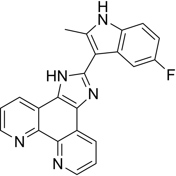
APTO-253 (LOR-253) is a small molecule that inhibits c-Myc expression, stabilizes G-quadruplex DNA, and induces cell cycle arrest and apoptosis in acute myeloid leukemia cells. APTO-253 mediates anticancer activity through induction of the Krüppel-like factor 4 (KLF4) tumor suppressor. APTO-253 has antiarthritic activity.
Nos produits utilisent uniquement pour la recherche. Nous ne vendons pas aux patients.
- Pureté : 99.74%
- CAS No.: 916151-99-0
- Formule: C22H14FN5
- Masse moléculaire:367.38
-
Stockage:Powder -20°C, 3 years , 4°C, 2 years ; In solvent -80°C, 6 months , -20°C, 1 month

To place orders, for customer services and technical support, please contact: MedChemExpress USA
Tel: 609-228-6898 E-mail: [email protected] [email protected]


-
 Activité biologique
-
Chemical Information
-
Solvant et solubilité
- Protocole
- Pureté et documentation
- Références
-
 Help & FAQs


-
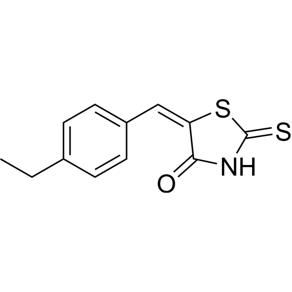
HY-12702
10058-F4c-Myc Inhibitor -
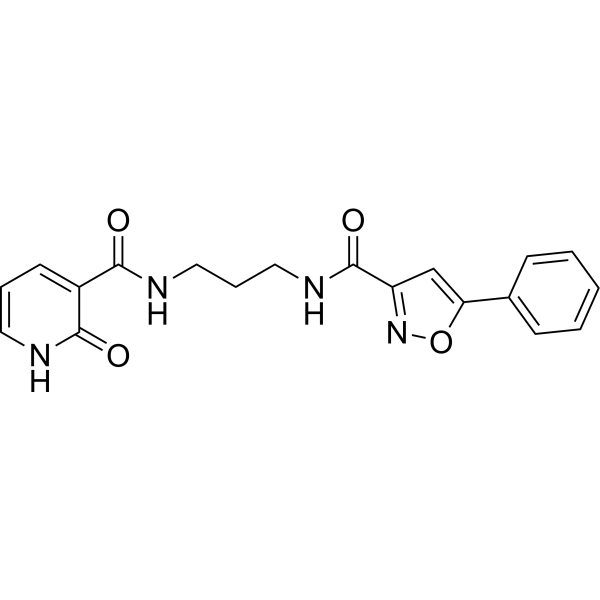
HY-103038
ML327MYC Blocker -
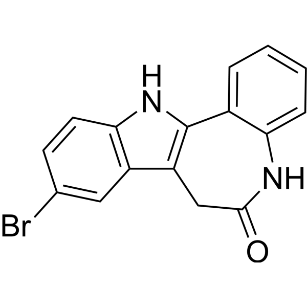
HY-12302
KenpaulloneCDK1/GSK-3β Inhibitor -
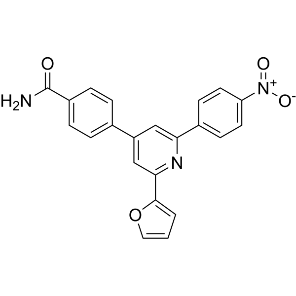
HY-19735
KJ Pyr 9MYC Inhibitor -
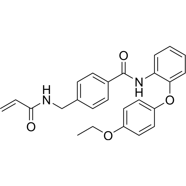
HY-134761
EN4Covalent Ligand of c-MYC -
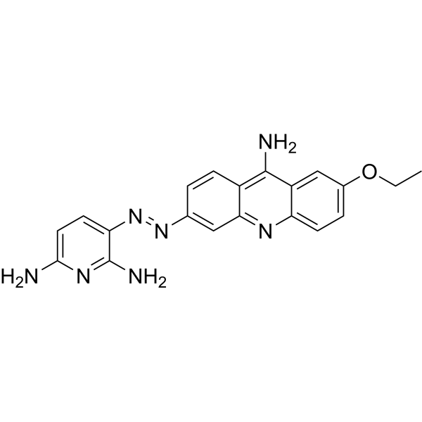
HY-124675
MYCMI-6MYC:MAX Interaction Inhibitor -
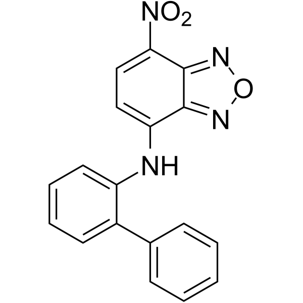
HY-100996
10074-G5c-Myc-Max Inhibitor -
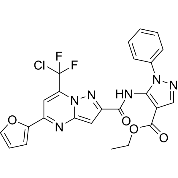
HY-100669
Mycro 3Myc-Max Inhibitor -

HY-100579
Ferrostatin-1Ferroptosis Inhibitor -

HY-10219
RapamycinmTOR Inhibitor -

HY-10227
BortezomibProteasome Inhibitor -
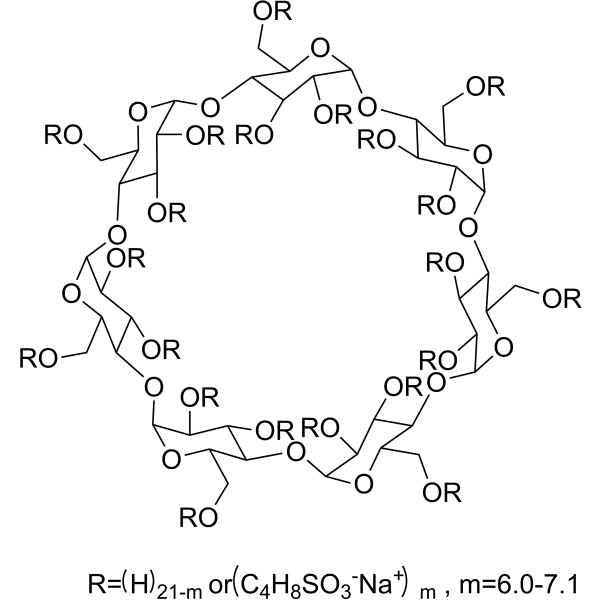
HY-17031
SBE-β-CDExcipient
Publications Citing Use of MedChemExpress (MCE) APTO-253
More- Signal Transduct Target Ther. 2026 Jul 24;11(1):290.
- Ann Rheum Dis. 2021 Apr;80(4):440-450. [Abstract]
- Blood. 2024 Nov 7;144(19):2018-2032. [Abstract]
- Nat Commun. 2023 Sep 2;14(1):5360. [Abstract]
- Nat Commun. 2019 Sep 25;10(1):4369. [Abstract]
- Adv Sci (Weinh). 2025 May;12(19):e2408992. [Abstract]
- J Transl Med. 2024 Oct 10;22(1):922. [Abstract]
- Leukemia. 2025 Sep;39(9):2163-2173. [Abstract]
- Cell Rep. 2026 Mar 24;45(4):117201. [Abstract]
- JCI Insight. 2022 Aug 22;7(16):e160688. [Abstract]
- J Environ Sci. 2025 Nov 28.
- Int Immunopharmacol. 2023 Jul:120:110425. [Abstract]
- Sci Rep. 2026 Jun 29.
- PLoS Pathog. 2025 May 12;21(5):e1013166. [Abstract]
- Mol Cell Biochem. 2021 Apr;476(4):1741-1749. [Abstract]
- FASEB J. 2025 May 31;39(10):e70613. [Abstract]
- J Immunol. 2025 Aug 7:vkaf170. [Abstract]
- Neurotox Res. 2020 Dec;38(4):967-978. [Abstract]
- J Diabetes Res. 2021 Sep 15:2021:7945117. [Abstract]
- Genes (Basel). 2021 Apr 8;12(4):539. [Abstract]
- Research Square Preprint. 2023 Jun 29.
- Research Square Preprint. 2023 Apr 18.
- Patent. US20220332841A1.
 Customer Validation & Images

-
Cell Proliferation/Viability Assay
-
WB
-
WB
-
RT-PCR
-
In Vivo Efficacy Study


Activité biologique
Description
IC50 & Target
In Vitro
APTO-253 (LOR-253) is an inducer of KLF4. APTO-253 (5 μM) induces KLF4 expression, and enhances apoptosis induced by NSC 119875 in both SKOV3 and OVCAR3 cells. APTO-253 (5 μM) also leads to G1 phase arrest and reduces S and G2/M phase cells in SKOV3 and OVCAR3 cells[1].
APTO-253 is cytotoxic to Raji and Raji/253R cell lines, with IC50s of 105 ± 2.4 nM and 1387 ± 94 nM, respectively. APTO-253 (0.5 μM) also causes DNA damage in Raji cells. BRCA1/2 deficient cells are hypersensitive to APTO-253. ABCG2 overexpressed HEK-293 cells are resistant to APTO-253 and inhibition of ABCG2 reverses resistance to APTO-253 in Raji/253R[2].
APTO-253 suppresses the proliferation of acute myeloid leukemia (AML) cell lines and various forms of lymphoma cell lines with IC50s ranging from 57 nM to 1.75 μM. APTO-253 (500 nM) also causes G0/G1 cell cycle arrest, induces apoptosis, and down regulates MYC RNA and protein expression in AML lines. APTO-253 (500 nM) leads to DNA damage response pathways in MV4-11 cells. Futhermore, APTO-253 is a potent stabilizer of Gquadruplex (G4) motifs, and demonstrates the greatest propensity for stabilizing the MYC G4 sequences[3].
MedChemExpress (MCE) has not independently confirmed the accuracy of these methods. They are for reference only.
In Vivo
MedChemExpress (MCE) has not independently confirmed the accuracy of these methods. They are for reference only.
-
Animal Model:DBA/1J male mice (6 weeks) with collagen induced arthritis (CIA)[3]
-
Dosage:15 mg/kg
-
Administration:IV; twice per day for 2 consecutive days per week for 14 days
-
Result:Demonstrated significant preventive and therapeutic activity on arthritis formation.
Essai clinique
| NCT Number | Sponsor | Condition | Start Date |
Phase

|
|---|---|---|---|---|
| NCT01329991 | 2011-05 | PHASE1 |


Chemical Information
-
CAS No. 916151-99-0
-
Appearance Solid
-
Masse moléculaire 367.38
-
Formule C22H14FN5
-
Color Light yellow to yellow
-
SMILES
CC(N1)=C(C(N2)=NC3=C2C4=CC=CN=C4C5=NC=CC=C53)C6=C1C=CC(F)=C6
-
Synonyms
LOR-253; LT-253
-
Livraison
Room temperature in continental US; may vary elsewhere.
-
Stockage
Powder -20°C 3 years 4°C 2 years In solvent -80°C 6 months -20°C 1 month


Publications (23)
-
Journal Impact Factor
-
Most Recent
-
-
Ann Rheum Dis
Parsing multiomics landscape of activated synovial fibroblasts highlights drug targets linked to genetic risk of rheumatoid arthritis. [Abstract]2021 Apr;80(4):440-450. PMID: 33139312
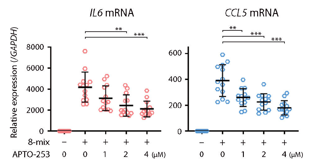
APTO-253 purchased from MedChemExpress. Usage Cited in: Ann Rheum Dis. 2021 Apr;80(4):440-450. [Abstract]
APTO-253 (1-4 μM) reduced mRNA levels of 8-mix SE-contacted genes (IL-6, CCL5) in stimulated RASFs.
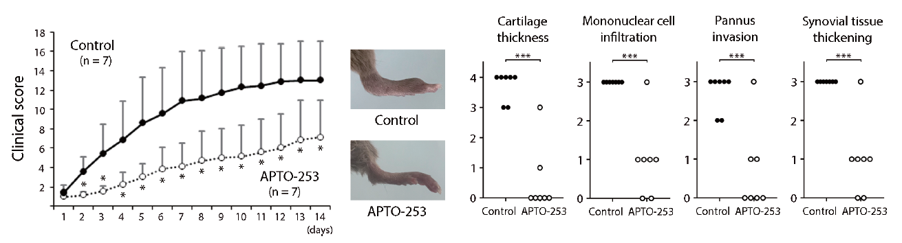
APTO-253 purchased from MedChemExpress. Usage Cited in: Ann Rheum Dis. 2021 Apr;80(4):440-450. [Abstract]
APTO-253 (15 mg/kg; i.v.; twice per day for two consecutive days per week) demonstrated significant therapeutic activity in collagen-induced arthritis (CIA) mice.
-
Blood
Epigenetic Regulation of Non-canonical Menin Targets Modulates Menin Inhibitor Response in Acute Myeloid Leukemia. [Abstract]2024 Nov 7;144(19):2018-2032. PMID: 39158067
APTO-253 purchased from MedChemExpress. Usage Cited in: Blood. 2024 Nov 7;144(19):2018-2032. [Abstract]
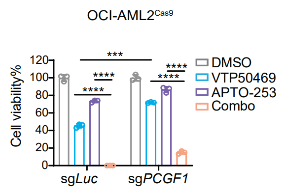
Cell viability of OCI-AML2 cells expressing either sgLuc or sgPCGF1 was assessed under the treatment of APTO-253 (100 nM; 9 d).
-
Nat Commun
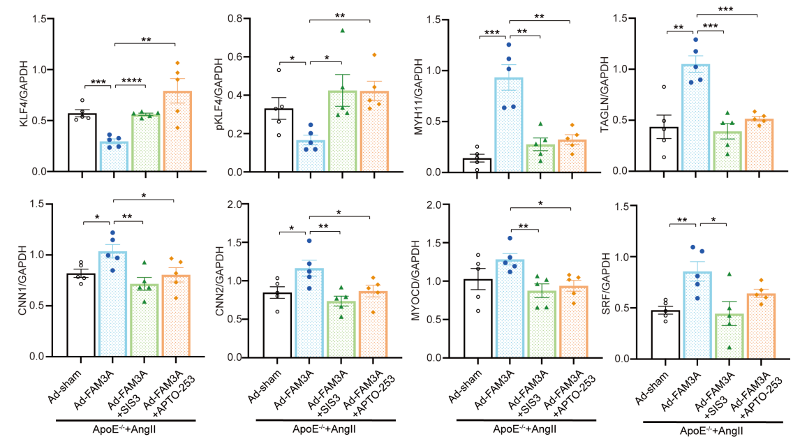
FAM3A reshapes VSMC fate specification in abdominal aortic aneurysm by regulating KLF4 ubiquitination. [Abstract]2023 Sep 2;14(1):5360. PMID: 37660071
APTO-253 purchased from MedChemExpress. Usage Cited in: Nat Commun. 2023 Sep 2;14(1):5360. [Abstract]
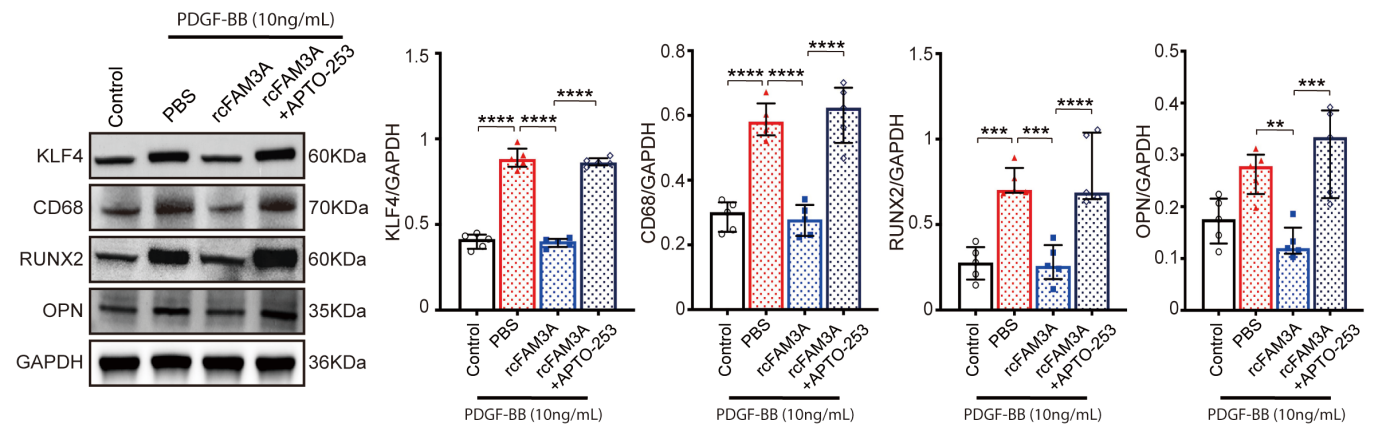
APTO-253 (10 μM) reversed the effects of FAM3A on suppressing the expression of the markers for macrophages (CD68) and osteoblasts (OPN, RUNX2) in VSMCs.
APTO-253 purchased from MedChemExpress. Usage Cited in: Nat Commun. 2023 Sep 2;14(1):5360. [Abstract]
KLF4 inducer APTO-253 (1 μg/g; i.p.; once daily for 4 weeks) significantly abrogated the effects of FAM3A on maintaining the contractile phenotype of VSMCs in aortas from AngII-ApoE−/− murine AAA models.
-
Nat Commun
Chromatin-informed inference of transcriptional programs in gynecologic and basal breast cancers. [Abstract]2019 Sep 25;10(1):4369. PMID: 31554806 -
Adv Sci (Weinh)
Artesunate Inhibits Neointimal Hyperplasia by Promoting IRF4 Associated Macrophage Polarization. [Abstract]2025 May;12(19):e2408992. PMID: 40126336 -
J Transl Med
KLF4 regulates trophoblast function and associates with unexplained recurrent spontaneous abortion. [Abstract]2024 Oct 10;22(1):922. PMID: 39390495 -
Leukemia
Targeting of IRAK4 and GSPT1 enhances therapeutic efficacy in AML via c-Myc destabilization. [Abstract]2025 Sep;39(9):2163-2173. PMID: 40670672 -
Cell Rep
SETDB2-mediated transcriptional repression of IDE in sensory neurons promotes migraine-like pain behaviors in mice. [Abstract]2026 Mar 24;45(4):117201. PMID: 41880325 -
JCI Insight
KLF4 is a therapeutically tractable brake on fibroblast activation that promotes resolution of pulmonary fibrosis. [Abstract]2022 Aug 22;7(16):e160688. PMID: 35852857 -
-
Int Immunopharmacol
Succinate pretreatment attenuates intestinal ischemia-reperfusion injury by inhibiting necroptosis and inflammation via upregulating Klf4. [Abstract]2023 Jul:120:110425. PMID: 37285681 -
-
PLoS Pathog
STAT3, MYC, and EBNA1 cooperate through a ZC3H18 transcriptional network to regulate survival and proliferation of EBV-positive lymphomas. [Abstract]2025 May 12;21(5):e1013166. PMID: 40354417 -
Mol Cell Biochem
2021 Apr;476(4):1741-1749. PMID: 33428060 -
FASEB J
Induction of T-Cell Differentiation by KLF4 in T-Cell Acute Lymphoblastic Leukemia Cells Harboring Activating Mutation in NOTCH3. [Abstract]2025 May 31;39(10):e70613. PMID: 40354086 -
J Immunol
TLR7-mediated immune response of renal myeloid-derived suppressor cells via RUNX1-KLF4 in systemic candidiasis. [Abstract]2025 Aug 7:vkaf170. PMID: 40795213 -
Neurotox Res
2020 Dec;38(4):967-978. PMID: 32870474 -
J Diabetes Res
KLF4 Promotes Diabetic Chronic Wound Healing by Suppressing Th17 Cell Differentiation in an MDSC-Dependent Manner. [Abstract]2021 Sep 15:2021:7945117. PMID: 34568499 -
Genes (Basel)
Krüppel-Like Factor 4 and Its Activator APTO-253 Induce NOXA-Mediated, p53-Independent Apoptosis in Triple-Negative Breast Cancer Cells. [Abstract]2021 Apr 8;12(4):539. PMID: 33918002 -
-
-


Solvant et solubilité
In Vitro:
DMSO : 25 mg/mL (68.05 mM; ultrasonic and warming and heat to 60°C; Hygroscopic DMSO has a significant impact on the solubility of product, please use newly opened DMSO)
Please refer to the solubility information to select the appropriate solvent. Once prepared, please aliquot and store the solution to prevent product inactivation from repeated freeze-thaw cycles.
Storage method and period of stock solution: -80°C, 6 months; -20°C, 1 month. When stored at -80°C, please use it within 6 months. When stored at -20°C, please use it within 1 month.
Please refer to the solubility information to select the appropriate solvent. Once prepared, please aliquot and store the solution to prevent product inactivation from repeated freeze-thaw cycles.
Storage method and period of stock solution: -80°C, 6 months; -20°C, 1 month. When stored at -80°C, please use it within 6 months. When stored at -20°C, please use it within 1 month.
Concentration (start) × Volume (start) = Concentration (final) × Volume (final)
In Vivo:
Select the appropriate dissolution method based on your experimental animal and administration route.
- For the following dissolution methods, please ensure to first prepare a clear stock solution using an In Vitro approach and then sequentially add co-solvents:
- To ensure reliable experimental results, the clarified stock solution can be appropriately stored based on storage conditions. As for the working solution for In Vivo experiments, it is recommended to prepare freshly and use it on the same day.
- The percentages shown for the solvents indicate their volumetric ratio in the final prepared solution. If precipitation or phase separation occurs during preparation, heat and/or sonication can be used to aid dissolution.
Add each solvent one by one: 10% DMSO 40% PEG300 5% Tween-80 45% Saline
Solubility: 2.3 mg/mL (6.26 mM); Suspended solution; Need ultrasonic and warming
This protocol yields a suspended solution of 2.3 mg/mL. Suspended solution can be used for oral and intraperitoneal injection.
Taking 1 mL working solution as an example, add 100 μL DMSO stock solution (23.0 mg/mL) to 400 μL PEG300, and mix evenly; then add 50 μL Tween-80 and mix evenly; then add 450 μL Saline to adjust the volume to 1 mL.
Preparation of Saline: Dissolve 0.9 g sodium chloride in ddH₂O and dilute to 100 mL to obtain a clear Saline solution.
Add each solvent one by one: 10% DMSO 90% (20% SBE-β-CD in Saline)
Solubility: 2.08 mg/mL (5.66 mM); Suspended solution; Need ultrasonic
This protocol yields a suspended solution of 2.08 mg/mL. Suspended solution can be used for oral and intraperitoneal injection.
Taking 1 mL working solution as an example, add 100 μL DMSO stock solution (20.8 mg/mL) to 900 μL 20% SBE-β-CD in Saline, and mix evenly.
Preparation of 20% SBE-β-CD in Saline (4°C, storage for one week): 2 g SBE-β-CD powder is dissolved in 10 mL Saline, completely dissolve until clear.
For the following dissolution methods, please prepare the working solution directly:
It is recommended to prepare fresh solutions and use them promptly within a short period of time.
The percentages shown for the solvents indicate their volumetric ratio in the final prepared solution. If precipitation or phase separation occurs during preparation, heat and/or sonication can be used to aid dissolution.
In Vivo Dissolution Calculator
Please enter the basic information of animal experiments:
-
-
-
-
Recommended: Prepare an additional quantity of animals to account for potential losses during experiments.
Please enter your animal formula composition:
-
%DMSO +
Recommended: Keep the proportion of DMSO in working solution below 2% if your animal is weak.
-
%+
-
+%Tween-80 + +
-
%Saline +
The co-solvents required include: DMSO, . All of co-solvents are available by MedChemExpress (MCE). , Tween 80. All of co-solvents are available by MedChemExpress (MCE).
Working solution concentration: 0.22 mg/mL
Method for preparing stock solution: mg drug dissolved in μL DMSO. Stock solution concentration: mg/mL.
1. Take μL DMSO stock solution;
2. Add μL .
μL , mix evenly;
3. Then add μL Tween 80, mix evenly;
4. Then add μL
Please ensure that the stock solution in the first step is dissolved to a clear state, and add co-solvents in sequence. You can use ultrasonic heating (ultrasonic cleaner, recommended frequency 20-40 kHz), vortexing, etc. to assist dissolution.


Pureté et documentation
-
Fiche technique (285 KB)
-
SDS (396 KB)
- English - EN (396 KB)
- Français - FR (396 KB)
- Deutsch - DE (396 KB)
- Norwegian - NO (396 KB)
- Español - ES (396 KB)
- Swedish - SV (396 KB)
- Italian - IT (396 KB)
- Korean - KR (396 KB)
- Portuguese - PT (396 KB)
-
Instruction de manipulation (2659 KB)
Références
[1]. Local A, et al. APTO-253 Stabilizes G-quadruplex DNA, Inhibits MYC Expression, and Induces DNA Damage in Acute Myeloid Leukemia Cells. Mol Cancer Ther. 2018 Jun;17(6):1177-1186. [Content Brief]
[3]. Haruka Tsuchiya, et al. Parsing multiomics landscape of activated synovial fibroblasts highlights drug targets linked to genetic risk of rheumatoid arthritis. Ann Rheum Dis. 2020 Nov2;annrheumdis-2020-218189. [Content Brief]


Complete Stock Solution Preparation Table
Please refer to the solubility information to select the appropriate solvent. Once prepared, please aliquot and store the solution to prevent product inactivation from repeated freeze-thaw cycles.
Storage method and period of stock solution: -80°C, 6 months; -20°C, 1 month. When stored at -80°C, please use it within 6 months. When stored at -20°C, please use it within 1 month.
| Optional Solvent | Concentration Solvent Mass | 1 mg | 5 mg | 10 mg | 25 mg |
|---|---|---|---|---|---|
| DMSO | 1 mM | 2.7220 mL | 13.6099 mL | 27.2198 mL | 68.0494 mL |
| 5 mM | 0.5444 mL | 2.7220 mL | 5.4440 mL | 13.6099 mL | |
| 10 mM | 0.2722 mL | 1.3610 mL | 2.7220 mL | 6.8049 mL | |
| 15 mM | 0.1815 mL | 0.9073 mL | 1.8147 mL | 4.5366 mL | |
| 20 mM | 0.1361 mL | 0.6805 mL | 1.3610 mL | 3.4025 mL | |
| 25 mM | 0.1089 mL | 0.5444 mL | 1.0888 mL | 2.7220 mL | |
| 30 mM | 0.0907 mL | 0.4537 mL | 0.9073 mL | 2.2683 mL | |
| 40 mM | 0.0680 mL | 0.3402 mL | 0.6805 mL | 1.7012 mL | |
| 50 mM | 0.0544 mL | 0.2722 mL | 0.5444 mL | 1.3610 mL | |
| 60 mM | 0.0454 mL | 0.2268 mL | 0.4537 mL | 1.1342 mL |


APTO-253 Related Classifications
HY-16291 Related Classifications
 Powered by Bioz