



Concanavalin A (agarose)

Based on 1 Customer Validation
Concanavalin A (agarose) is composed of Concanavalin A (HY-P2149) conjugated with agarose. Concanavalin A (ConA) is a selective competitive binder targeting the specific carbohydrate structures of glucose and mannose, inducing mitosis, and exhibiting certain cytotoxicity, hepatotoxicity, and teratogenicity. Concanav in A (agarose) can be used for in vivo blood glucose monitoring in diabetes, and for "fishing out" specific glycoproteins or removing sugar impurities from complex samples.
For research use only. We do not sell to patients.
- CAS No.: 11028-71-0
-
Storage:
Please store the product under the recommended conditions in the Certificate of Analysis.


To place orders, for customer services and technical support, please contact: MedChemExpress USA
Tel: 609-228-6898 E-mail: [email protected] [email protected]


-
 Biological Activity
-
Chemical Information
- Protocols
- Purity & Documentation
- References
-
 Help & FAQs


-

HY-P2149
Concanavalin APlant Lectin -

HY-N7038
PhytohemagglutininT-cell Activator -

HY-W250978
OvalbuminsAllergen -
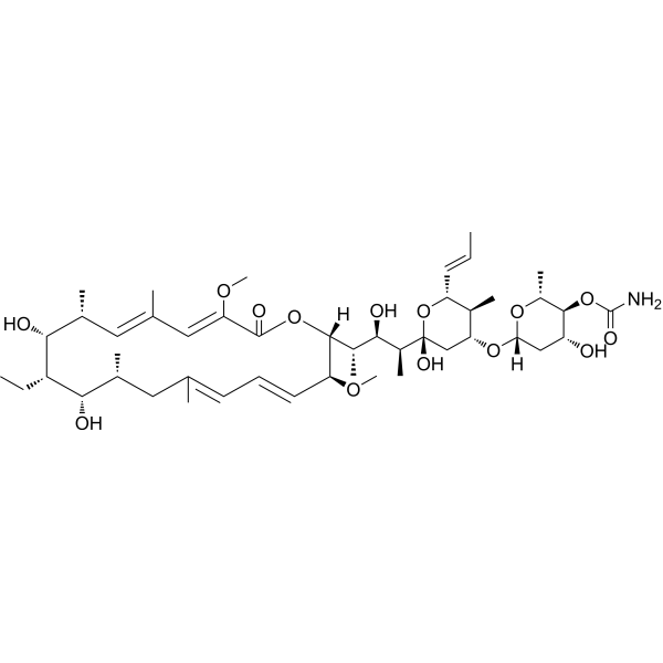
HY-N1724
Concanamycin AAntibiotic -

HY-W250978A
Ovalbumin, low endotoxinAllergen -
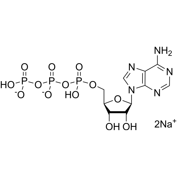
HY-B0345A
ATP disodium saltEndogenous Metabolite -
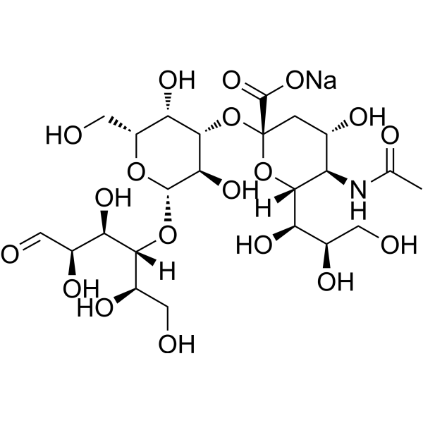
HY-108065A
3'-Sialyllactose sodiumPrebiotic -
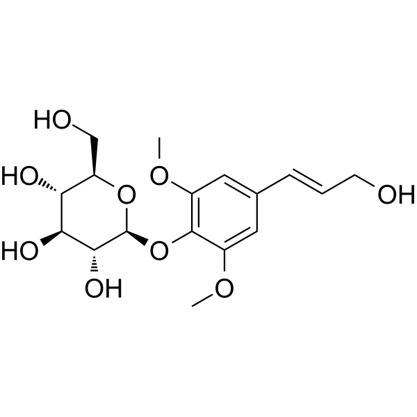
HY-N0824
SyringinPhenolic Glycoside -

HY-W717504
Glucosylceramide, plantAnti-inflammatory Agent -
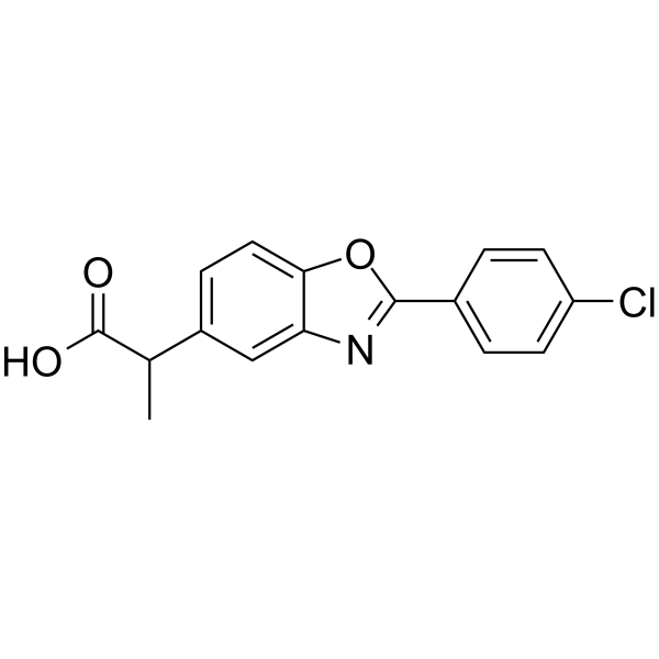
HY-13568
BenoxaprofenNonsteroidal Anti-inflammatory Agent -
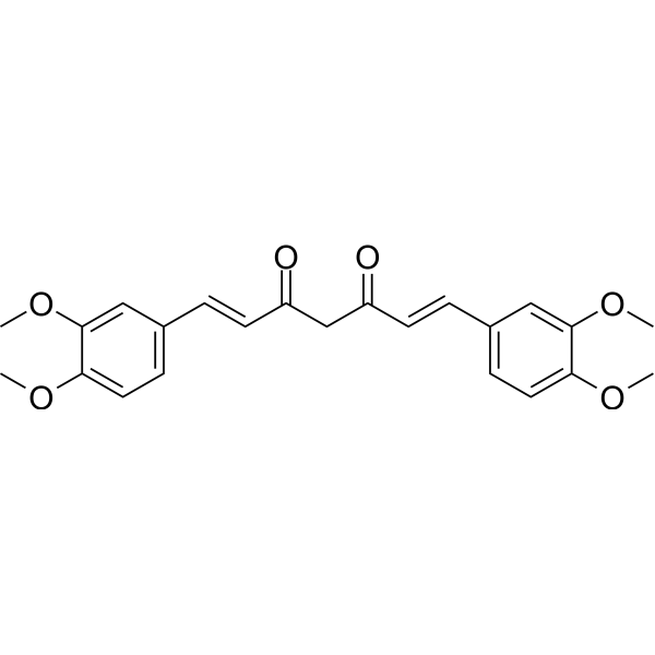
HY-100977
DimethoxycurcuminThioredoxin Reductase Inhibitor -

HY-B2176I
ATP disodium salt, 100 mM Solution, PCR GradeEndogenous Metabolite


Biological Activity
Description
In Vitro
Concanavalin A (agarose) is primarily used for protein purification from complex samples, such as cytokines, membrane proteins, and serum glycoproteins; or for removing unwanted glycoprotein impurities from samples; or for studying the interaction strength between glycoproteins and ConA (e.g., under different elution conditions)[1].
MedChemExpress (MCE) has not independently confirmed the accuracy of these methods. They are for reference only. Further protocols information, click here.
-
Cell Line:Mouse spleen cells, human B lymphocytes, human T lymphocytes, murine T lymphocytes
-
Concentration:3-6 μg/mL (optimal), 10 μg/mL, 25 μg/mL
-
Incubation Time:72 h
-
Result:Concanavalin A (agarose) directly stimulated B cells and T cells to synthesize DNA and proliferate, with the optimal concentration ranging from 3 to 6 μg/mL. The dose-response curve declined sharply when the concentration exceeded 10 μg/mL. It induced the production of interferon in spleen cells from C57BL/6 mice at 25 μg/mL. The mitogenic effect on lymphocytes was age-dependent, with impaired responses observed in lymphocytes from older individuals and murine T lymphocyte subsets.
In Vivo
-
Animal Model:Rabbits (body weight 2 kg, 2.9 kg, 3.1 kg, 1.3 kg)[1]
-
Dosage:Rabbits: 1 mg, 2.4 mg, 4 mg, 7 mg, 15 mg
-
Administration:intravenous injection
-
Result:Caused no symptoms in rabbits.
Administration of 7 mg ConA to a 1.3 kg rabbit resulted in death within 2 days, while 15 mg ConA injected into a 2 kg rabbit induced intravascular erythrocyte agglutination and the rabbit was euthanized.


Chemical Information
-
CAS No. 11028-71-0
-
Appearance Liquid (Density: 1.434±0.06 g/cm3)
-
Color Colorless to light yellow
-
SMILES
[Concanavalin A (agarose)]
-
Shipping
Room temperature in continental US; may vary elsewhere.
-
Storage
Please store the product under the recommended conditions in the Certificate of Analysis.

Protocols
-
Reproductive and Developmental Toxicity Study
Reproductive and developmental toxicity studies detect adverse effects of prenatal or peri/postnatal exposure on maternal condition, pregnancy maintenance, embryo-fetal survival, fetal growth, structural development, and offspring reproductive or developmental endpoints; classic rat protocols generate readouts by comparing treated groups with vehicle, pair-fed, or untreated controls for implantation, resorption, fetal weight, crown-rump length, external morphology, visceral morphology, skeletal ossification, anogenital distance, nipple/areola retention, and postnatal cohort outcomes.
-
Cell Cytotoxicity Assay
Cytotoxicity assays are usually based on the assessment of cell membrane damage, which can also be indirectly detected by measuring cell viability. Detection methods include MTT assay, CKK-8 assay, LDH assay and ATP assay, etc.
-
Mammalian live/dead viability and cytotoxicity staining
Live/dead viability and cytotoxicity staining assays are based on the simultaneous detection of intracellular esterase activity in metabolically active (viable) cells and membrane integrity loss in non-viable cells. In commonly used dual-staining approaches, membrane-permeant fluorogenic substrates are converted by intracellular esterases into fluorescent products in live cells, while impermeant DNA-binding dyes selectively enter cells with compromised plasma membranes and label nucleic acids in dead or dying cells, enabling discrimination between viable and non-viable populations by fluorescence microscopy or flow cytometry.
-
Hepatotoxicity Study
This protocol evaluates hepatotoxicity using complementary in vivo mouse APAP acute liver injury and in vitro hepatocyte-based cytotoxicity readouts. In vivo APAP injury is assessed by serum ALT/AST, liver histology, hepatic glutathione, protein adducts, necrosis, inflammation, and regeneration-related endpoints. In vitro hepatotoxicity is assessed by loss of viability, leakage of ALT/AST/LDH, oxidative-stress markers, mitochondrial function, nuclear morphology, intracellular calcium, and high-content imaging endpoints.
-
Research Protocol for Metabolic Diseases
AMP-activated protein kinase, AMPK, is a conserved cellular energy sensor that responds to reduced cellular energy status and coordinates metabolism by increasing ATP-generating catabolic pathways while suppressing ATP-consuming anabolic processes. In metabolic disease research, the AMPK pathway is experimentally relevant because it regulates hepatic lipid synthesis, fatty acid oxidation, glucose production, skeletal-muscle glucose disposal, mTORC1-linked biosynthesis, autophagy, mitochondrial homeostasis, and whole-body energy balance. The central pathway logic is that energy stress, metformin, exercise-like stimulation, or direct AMPK activators increase AMPKα Thr172 phosphorylation and downstream substrate phosphorylation, including ACC and RAPTOR. Phosphorylation of ACC suppresses lipogenesis and supports fatty acid oxidation, whereas phosphorylation of RAPTOR suppresses mTORC1 signaling and links cellular energy status to growth and protein synthesis control. The pathway is linked
-
Cell Viability Determination by MTT Colorimetric Assay
The following protocol uses the MTT colorimetric assay as a classic literature-established method for assessing cell viability/metabolic activity in cultured mammalian cells. MTT[3-(4,5-dimethylthiazol-2-yl)-2,5-diphenyltetrazolium bromide] is reduced by metabolically active cells to a colored formazan product; the amount of formazan is quantified spectrophotometrically and provides an indirect measure of metabolically active viable cells. Importantly, MTT reduction reflects cellular oxidoreductase/metabolic activity rather than an absolute direct count of living cells, so changes in cellular metabolism can alter the signal independently of cell number.


Purity & Documentation
-
Data Sheet (268 KB)
-
SDS (251 KB)
- English - EN (251 KB)
- Français - FR (251 KB)
- Deutsch - DE (251 KB)
- Norwegian - NO (251 KB)
- Español - ES (251 KB)
- Swedish - SV (251 KB)
- Italian - IT (251 KB)
- Korean - KR (251 KB)
- Portuguese - PT (251 KB)
-
Handling Instructions (2659 KB)
References
[1]. Ballerstadt R, et al. Concanavalin A for in vivo glucose sensing: a biotoxicity review. Biosens Bioelectron. 2006 Aug 15;22(2):275-84. [Content Brief]
[2]. Li W, et, al. Concanavalin A: a potential anti-neoplastic agent targeting apoptosis, autophagy and anti-angiogenesis for cancer therapeutics. Biochem Biophys Res Commun. 2011 Oct 22;414(2):282-6. [Content Brief]
[3]. Cantelli A, et al. Concanavalin A-Rose Bengal bioconjugate for targeted Gram-negative antimicrobial photodynamic therapy. J Photochem Photobiol B. 2020 Mar 13;206:111852. [Content Brief]
[4]. Zhou Y, et al. The Protective Effect of Resveratrol on Concanavalin-A-Induced Acute Hepatic Injury in Mice. Gastroenterol Res Pract. 2015;2015:506390. [Content Brief]

Calculators
Concentration (start) × Volume (start) = Concentration (final) × Volume (final)


Concanavalin A (agarose) Related Classifications
HY-P2149A Related Classifications
 Powered by Bioz