



IL-8/CXCL8 Protein, Human
Based on 7 publication(s) in Google Scholar
Interleukin-8 (IL-8), also known as CXCL8 or NAP-1, is a pro-inflammatory CXC chemokine. IL-8 acts on human neutrophils via two receptors, CXCR1 and CXCR2. IL-8 has a conserved Glu-Leu-Arg (ELR) N-terminal motif, and is an agonist for CXCR1/CXCR2. IL-8 is produced by various cells including leukocytes, endothelial cells, and epithelial cells. IL-8/CXCL8 Protein, Human is produced in E.coil.
- Species: Human
- Source: E. coli
-
Storage:Stored at -20°C for 2 years from date of receipt. After reconstitution, it is stable at 4°C for 1 week or -20°C for longer (with carrier protein). It is recommended to freeze aliquots at -20°C or -80°C for extended storage.


To place orders, for customer services and technical support, please contact: MedChemExpress USA
Tel: 609-228-6898 E-mail: [email protected] [email protected]


-
 Biological Activity
-
Technical Parameters
-
Product Properties
- Documentation
- References
- Help & FAQs


-

HY-P7044
IL-6 Protein, Human -
HY-P7028
IL-1 beta Protein, Human -
HY-P7058
TNF-alpha/TNFSF2 Protein, Human -
HY-P7025
IFN-gamma Protein, Human -
HY-P7237
MCP-1/CCL2 Protein, Human -
HY-P70508
GRO-alpha/CXCL1 Protein, Human (HEK293, His) -
HY-P0224
N-Formyl-Met-Leu-Phe -
HY-P7158
ENA-78/CXCL5 Protein, Human -

HY-18739
Phorbol 12-myristate 13-acetate -
HY-11109
Resatorvid -

HY-10071
Y-27632 -

HY-15763
Erastin


Biological Activity
Description
Interleukin-8 (IL-8), also known as CXCL8 or NAP-1, is a pro-inflammatory CXC chemokine. IL-8 acts on human neutrophils via two receptors, CXCR1 and CXCR2. IL-8 has a conserved Glu-Leu-Arg (ELR) N-terminal motif, and is an agonist for CXCR1/CXCR2. IL-8 is produced by various cells including leukocytes, endothelial cells, and epithelial cells[1][2][3]. IL-8/CXCL8 Protein, Human is produced in E.coil.
Background
IL-8 (CXCL8) belongs to the ELR+ CXC chemokines family. IL-8 is initially produced as a protein of 99 amino acids that undergoes cleavage to form active IL-8 isoforms, a 77 amino acid peptide in non-immune cells or a 72 amino acid peptide in monocytes and macrophages. The gene encoding IL-8 is located on chromosome 4q13-q21. Dimerisation of IL-8 forms the structural basis for receptor binding. IL-8 is expressed by various cells including monocytes, macrophages, leukocytes, endothelial cells, and epithelial cells[1][2][3].
Mature human IL-8/CXCL8 shares 75% amino acid sequence identity with canine IL-8/CXCL8. While, human IL-8 shares 94.95% aa sequence identity with Rhesus Macaque IL-8 protein.
IL-8 is responsible for the recruitment and activation of neutrophils and granulocytes to the site of inflammation. IL-8 is almost undetectable in physiological states, but is rapidly induced by pro-inflammatory cytokines such as TNFα and IL-1β. The function of IL-8 mainly relies on its interaction with specific cell surface GPCR, CXCR1 and CXCR2. In addition, IL-8 is reported to promote integrin β3 upregulation and the invasion of hepatocellular carcinoma cells through activation of the PI3K/Akt pathway. In odontogenic lesions, IL-8 has been proven to be highly expressed in ameloblastoma epithelial cells and irreversible pulpitis. In rheumatoid arthritis and other inflammatory joint diseases IL-8 could bring about the accumulation of neutrophils, which are considered a major source of cartilage-degrading enzymes. IL-8 stimulates the MAPK and tyrosine phosphorylation of cellular proteins. Tumour cells and fibroblasts communicate with each other, including autocrine and paracrine factors, including IL-8, resulting in the upregulation of MMP2 and MMP9 degradable extracellular matrix (ECM) components that trigger tumour invasion[1][2][3][4].
IL-8 is typically known to promote angiogenesis, but it also activates matrix metalloproteinase (MMP) that is involved in metastasisrelated tissue remodelling. IL-8 is induced in lipopolysaccharide (LPS)-stimulated monocytes and shown to induce neutrophil migration. IL-8 exerts multiple effects on biological activities of tumour cells including proliferation, invasion and migration. IL-8 also increases the expression of Akt in androgen-independent prostate cancer (AIPC) cell lines. IL-8 activates MAPK signalling via PI3K in neutrophils, and via transactivation of EGFR resulting in Ras-GTPase activation in ovarian and lung cancer cell lines. There is substantial amount of experimental data suggesting that IL-8 and receptors contribute to elimination of pathogens, but may also contribute significantly to disease-associated processes, including tissue injury, fibrosis, angiogenesis and tumorigenesis[3][5].
In Vitro
Recombinant human IL-8/CXCL8 (100 ng/mL; for 24-96 h) stimulation leads to enhancement of invasion and suppression of late stage apoptosis in MG-63 cells. Moreover, secretions of MMPs by MG-63 cells are also increased upon stimulation. CXCL8 induces the elevations of phosphorylated PI3K and Akt, but not PKC or FAK[6].
In Vivo
Recombinant human IL-8 (10, 30, and 100 μg/kg; single intravenous injection) injected into rhesus monkeys (2-3 years; 2.5-4.5 kg). IL-8 injection results in instant neutropenia that was due to pulmonary sequestration. Within 30 minutes after IL-8 injection, neutrophilia developed with counts up to 10-fold greater than baseline levels. The numbers of hematopoietic progenitor cells (HPCs) increased of blood at 30 minutes after injection of 100 μg/kg IL-8[7].
Verified Bioactivity
1.The ED50 is <150 ng/mL as measured by CHO-K1/Gα15/hCXCR1 cells (human Gα15 and human CXCR1 stably expressed in CHO-K1 cells).
2.Immobilized Human IL-8 at 2 μg/mL (100 μL/well) can bind Anti-IL-8 Antibody. The ED50 for this effect is 20-50 ng/mL.
3.Loaded Eltrekibart (HY-P990737) on AHC2 biosensor, can bind IL-8/CXCL8 Protein, Human with an affinity constant of <1.000E-11 M as determined in BLI assay.
4.Loaded Adakitug (HY-P990598) on AHC2 biosensor, can bind IL-8/CXCL8 Protein, Human with an affinity constant of <1.0E-11 M as determined in BLI assay.
MCE Validation Data
-
Purity - SDS-PAGE
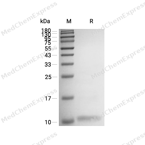
Purity - SDS-PAGE
-
Bioactivity - ELISA
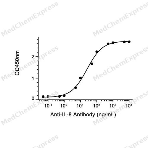
Bioactivity - ELISA
-
Bioactivity - BLI

Bioactivity - BLI
-
Bioactivity - BLI
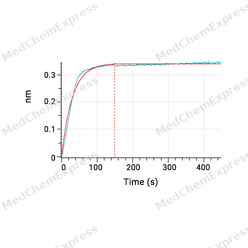
Bioactivity - BLI


Publications (7)
-
Journal Impact Factor
-
Most Recent
-
J Biomed Sci
Fusobacterium nucleatum promotes colorectal cancer liver metastasis via miR-5692a/IL-8 axis by inducing epithelial-mesenchymal transition. [Abstract]2025 Jan 6;32(1):5. PMID: 39757156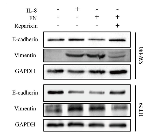
IL-8/CXCL8 Protein, Human purchased from MedChemExpress. Usage Cited in: J Biomed Sci. 2025 Jan 6;32(1):5. [Abstract]
Western blot was performed to evaluate the expression of EMT marker in SW480 and HT29 cells after treated with IL-8, F. nucleatum and Reparixin.
-
Anal Chem
The Dual Nondestructive Amplification Strategy Realizes the Ultrasensitive Detection of Soluble TRAIL in Human Plasma. [Abstract]2025 Nov 18;97(45):25295-25303. PMID: 41195534 -
Cancer Cell Int
SHP2 inhibition enhances the anticancer effect of Osimertinib in EGFR T790M mutant lung adenocarcinoma by blocking CXCL8 loop mediated stemness. [Abstract]2021 Jul 3;21(1):337. PMID: 34217295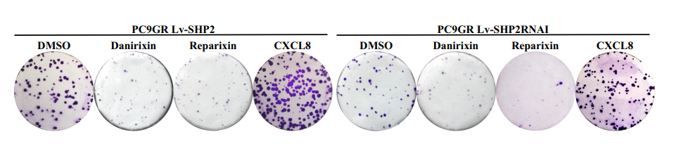
IL-8/CXCL8 Protein, Human purchased from MedChemExpress. Usage Cited in: Cancer Cell Int. 2021 Jul 3;21(1):337. [Abstract]
The clones were dramatically inhibited by Danirixin or Reparixin, while exogenous CXCL8 significantly enhanced the colony formation of PC9GR.
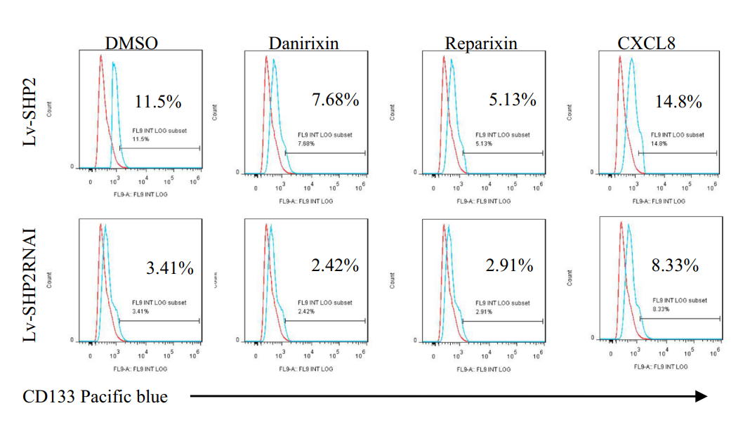
IL-8/CXCL8 Protein, Human purchased from MedChemExpress. Usage Cited in: Cancer Cell Int. 2021 Jul 3;21(1):337. [Abstract]
The percentage of CSCs was reduced in the setting of CXCL8-CXCR1/2 loop blockage, and recombinant CXCL8 could reverse the CSCs proportion.
-
Int J Mol Sci
Mechanistic Study of Matrix Stiffness Promoting Lymph Node Metastasis in Cervical Cancer by Regulating NETs Formation via Piezo1. [Abstract]2026 Jun 16;27(12):5431. PMID: 42353147 -
J Cell Mol Med
IL-8 promotes cell migration through regulating EMT by activating the Wnt/β-catenin pathway in ovarian cancer. [Abstract]2020 Jan;24(2):1588-1598. PMID: 31793192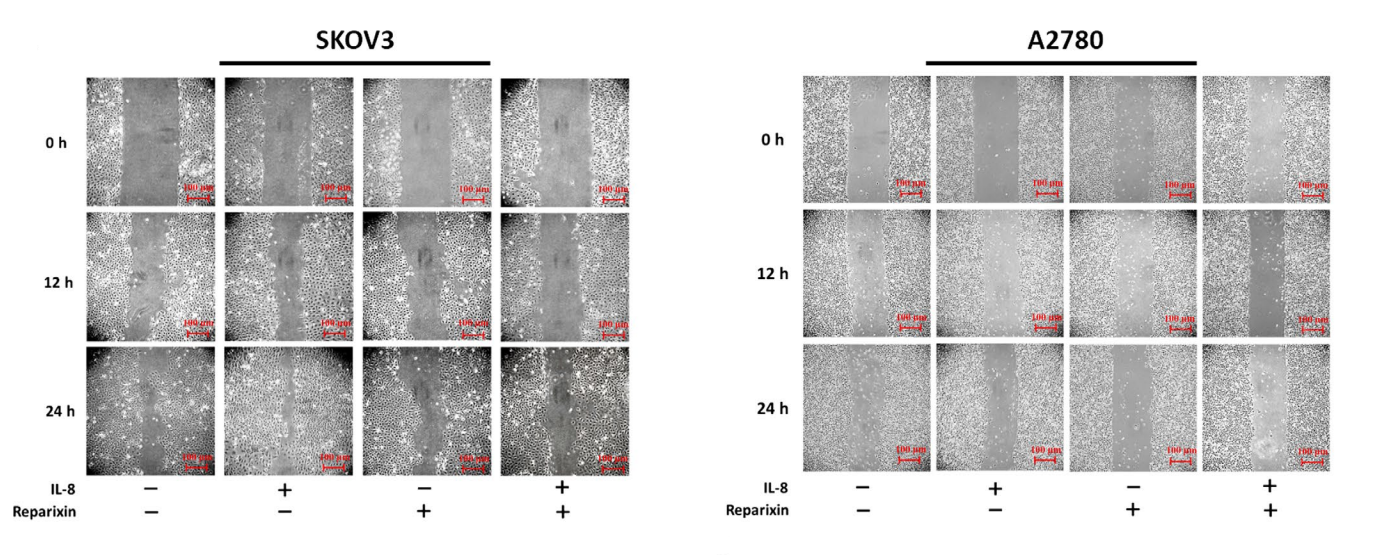
IL-8/CXCL8 Protein, Human purchased from MedChemExpress. Usage Cited in: J Cell Mol Med. 2020 Jan;24(2):1588-1598. [Abstract]
The effects of IL-8 (100 ng/mL; 48 h) on the migration ability of ovarian cancer cells illustrated by the monolayer wound healing assay.
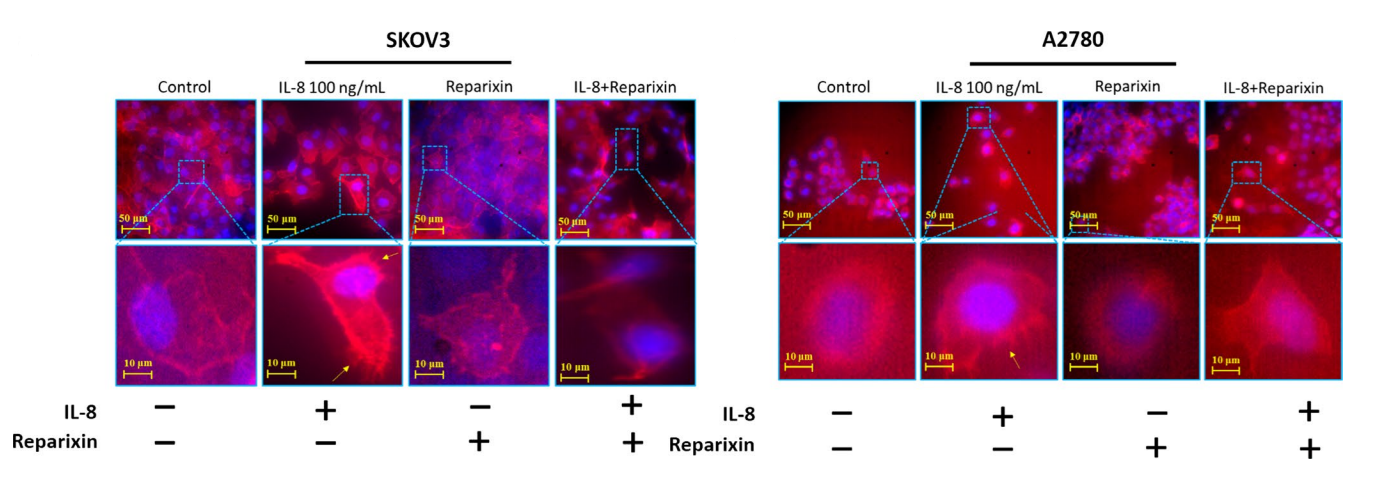
IL-8/CXCL8 Protein, Human purchased from MedChemExpress. Usage Cited in: J Cell Mol Med. 2020 Jan;24(2):1588-1598. [Abstract]
IL-8 promote the cilia formation of SKOV3 cells and A2780 cells. The images showed the cytoskeleton (red) and nucleus (blue).
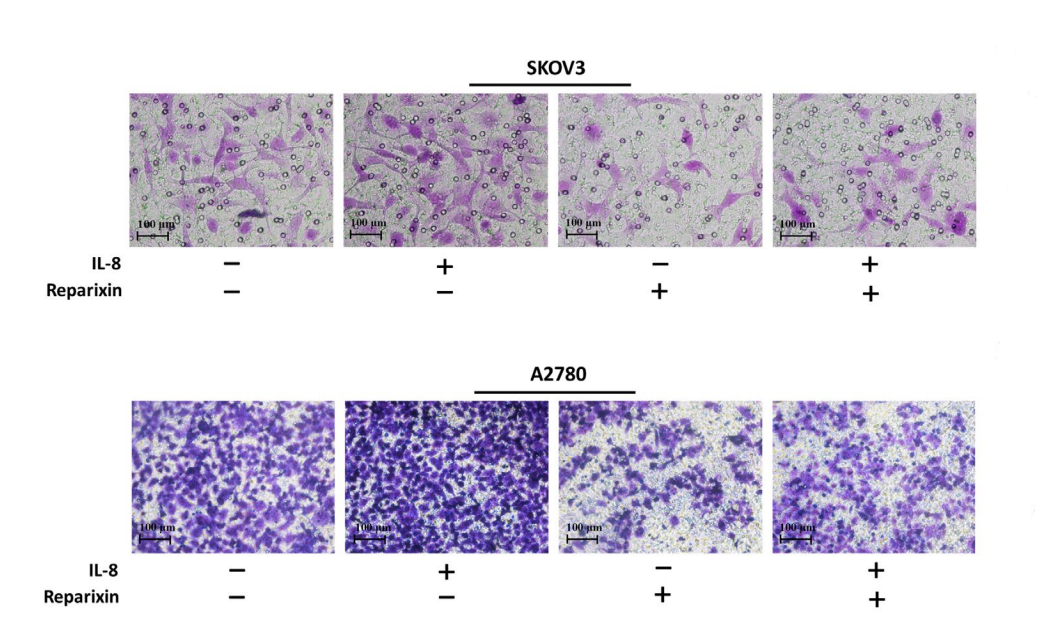
IL-8/CXCL8 Protein, Human purchased from MedChemExpress. Usage Cited in: J Cell Mol Med. 2020 Jan;24(2):1588-1598. [Abstract]
The effects of IL-8 on the migration ability of ovarian cancer cells illustrated by the Transwell assay. Typical optical images of SKOV3 and A2780 illustrated cell migration at 24 h. The cells crossed through the pores of Transwell chamber were stained by crystal violet, Scale bars = 100 μm.
-
-


Technical Parameters
-
Species Human
-
Source E. coli
-
Tag Tag Free
-
Accession
P10145 (A23-S99)
-
Protein Length
Full Length of IL-8 Chain
-
Synonyms
CXCL8; Lung Giant Cell Carcinoma-Derived Chemotactic Protein; Prev. IL8; Alveolar Macrophage Chemotactic Factor I; MDNCF; Tumor Necrosis Factor-Induced Gene 1; NAP-1; Neutrophil-Activating Peptide 1; MONAP; T-Cell Chemotactic Factor; GCP-1; Interleukin 8; Monocyte-Derived Neutrophil Chemotactic Factor; Interleukin-8; Monocyte-Derived Neutrophil-Activating Peptide; Emoctakin; Granulocyte Chemotactic Protein 1; AMCF-I; Chemokine (C-X-C Motif) Ligand 8; B-ENAP; SCYB8; TSG-1; LYNAP; 3-10C; LUCT; K60; LECT; Small Inducible Cytokine Subfamily B, Member 8; IL-8; Beta-Thromboglobulin-Like Protein; GCP1; Neutrophil-Activating Protein 1; NAP1; C-X-C Motif Chemokine 8; NAF; C-X-C Motif Chemokine; Beta Endothelial Cell-Derived Neutrophil Activating Peptide; Protein 3-10C; C-X-C Motif Chemokine Ligand 8; Lymphocyte Derived Neutrophil Activating Peptide
-
AA Sequence
AVLPRSAKELRCQCIKTYSKPFHPKFIKELRVIESGPHCANTEIIVKLSDGRELCLDPKENWVQRVVEKFLKRAENS
-
Predicted Molecular Mass
8.9 kDa
-
Molecular Weight
Approximately 9-11 kDa, based on SDS-PAGE under reducing conditions.
-
Purity
≥ 95%, as determined by reducing SDS-PAGE.


Product Properties
Lyophilized powder
1.Lyophilized from a 0.22 μm filtered solution of PBS.
2.Lyophilized from a 0.22 μm filtered solution of PBS, pH 7.4, 8% trehalose.
3.Lyophilized from a 0.22 μm filtered solution of 50 mM Tris-HCl, 300 mM NaCl, pH 8.0.
Please refer to the lot-specific COA for specific buffer information.
Note: For SPR assay, please replace the buffer. Primary amine components (e.g., Tris, imidazole) can affect protein-coupled chips.
<1 EU/μg, determined by LAL method.
It is not recommended to reconstitute to a concentration less than 100 μg/mL in ddH2O. For long term storage it is recommended to add a carrier protein (0.1% BSA, 5% HSA, 10% FBS or 5% Trehalose).
Stored at -20°C for 2 years from date of receipt. After reconstitution, it is stable at 4°C for 1 week or -20°C for longer (with carrier protein). It is recommended to freeze aliquots at -20°C or -80°C for extended storage.
Room temperature in continental US; may vary elsewhere.


Documentation
-
Data Sheet (268 KB)
-
SDS (254 KB)
- English - EN (254 KB)
- Français - FR (254 KB)
- Deutsch - DE (254 KB)
- Norwegian - NO (254 KB)
- Español - ES (254 KB)
- Swedish - SV (254 KB)
- Italian - IT (254 KB)
- Korean - KR (254 KB)
- Portuguese - PT (254 KB)
-
Handling Instructions (2659 KB)


References
[1]. M Baggiolini, et al. Neutrophil-activating peptide-1/interleukin 8, a novel cytokine that activates neutrophils. J Clin Invest. 1989 Oct;84(4):1045-9. [Content Brief]
[2]. Baggiolini M, et al. Neutrophil-activating peptide-1/interleukin 8, a novel cytokine that activates neutrophils. J Clin Invest. 1989 Oct;84(4):1045-9. [Content Brief]
[3]. M Wolf, et al. Granulocyte chemotactic protein 2 acts via both IL-8 receptors, CXCR1 and CXCR2. Eur J Immunol. 1998 Jan;28(1):164-70. [Content Brief]
[4]. Koch AE, et al. Interleukin-8 as a macrophage-derived mediator of angiogenesis. Science. 1992 Dec 11;258(5089):1798-801. [Content Brief]
[5]. Qian Liu, et al. The CXCL8-CXCR1/2 pathways in cancer. Cytokine Growth Factor Rev. 2016 Oct;31:61-71. [Content Brief]
[6]. Jian-Feng Liu, et al. IL-8 Is Upregulated in the Tissue-Derived EVs of Odontogenic Keratocysts. Biomed Res Int. 2022 Jul 30;2022:9453270. [Content Brief]
[7]. Remo C Russo, et al. The CXCL8/IL-8 chemokine family and its receptors in inflammatory diseases. Expert Rev Clin Immunol. 2014 May;10(5):593-619. [Content Brief]
[8]. Hai Jiang, et al. CXCL8 promotes the invasion of human osteosarcoma cells by regulation of PI3K/Akt signaling pathway. APMIS. 2017 Sep;125(9):773-780. [Content Brief]
[9]. L Laterveer, et al. Rapid mobilization of hematopoietic progenitor cells in rhesus monkeys by a single intravenous injection of interleukin-8. Blood. 1996 Jan 15;87(2):781-8. [Content Brief]

Calculators
Concentration (start) × Volume (start) = Concentration (final) × Volume (final)


 Powered by Bioz

