



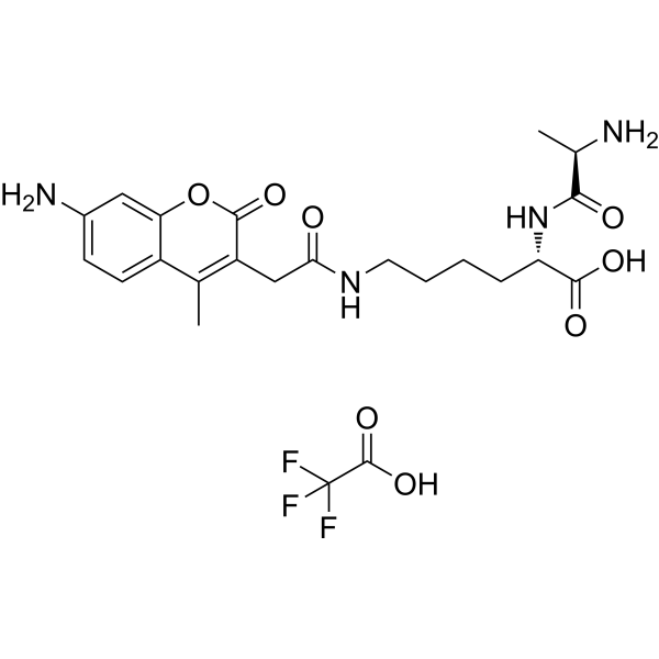
D-Ala-Lys-AMCA TFA
Based on 2 publication(s) in Google Scholar
D-Ala-Lys-AMCA TFA is a known proton-coupled oligopeptide transporter 1 (PEPT1) substrate that emits blue fluorescence. D-Ala-Lys-AMCA TFA may be transported into liver cancer cells and Caco-2 cells based on fluorescence analysis. D-Ala-Lys-AMCA TFA can be used for characterizing PEPT1-specific substrates or inhibitors (Ex/Em = 390/480 nm).
Nur für Forschungszwecke. Wir verkaufen nicht an Patienten.
- Formel: C23H29F3N4O8
- Molecular Weight:546.49
-
Speicherung:
Please store the product under the recommended conditions in the Certificate of Analysis.

To place orders, for customer services and technical support, please contact: MedChemExpress USA
Tel: 609-228-6898 E-mail: [email protected] [email protected]


-
 Biologische Aktivität
-
Chemical Information
- Protokoll
- Reinheit & Dokumentation
- Verweise
-
 Help & FAQs


Publications Citing Use of MedChemExpress (MCE) D-Ala-Lys-AMCA TFA
More

Biologische Aktivität
Beschreibung
In Vitro
D-Ala-Lys-AMCA (25, 50 and 150 μM, 3 h) TFA may be transported into liver cancer and Caco-2 cells[1].
D-Ala-Lys-AMCA (25 μM, 10 min) TFA can be uptaken by absorptive enterocytes of all small intestine segments, whereas there is a complete lack of fluorescence in colonic samples[2].
Guide (Following is our recommended protocol. This protocol only provides a guideline, and should be modified according to your specific needs)[1].
1. Preparation of stock solution:
Use ddH2O or DMSO to prepare a D-Ala-Lys-AMCA stock solution.
2. Preparation of working solution:
Use preheated serum-free cell culture medium or PBS to dilute the stock solution to prepare a 1 mM D-Ala-Lys-AMCA working solution.
Note: Please adjust the concentration of D-Ala-Lys-AMCA working solution according to actual conditions and prepare it before use.
3. Cell staining:
3.1 Add PBS to wash the cells twice, 5 minutes each time.
3.2 Add D-Ala-Lys-AMCA working solution and incubate at 37°C for 1-2 h.
3.3 Aspirate the dye working solution, wash 2-3 times with culture medium, 5 minutes each time, and observe using a fluorescence microscope (Ex/Em = 390/480 nm).
MedChemExpress (MCE) has not independently confirmed the accuracy of these methods. They are for reference only. Further protocols information, click here.
Emission (Em)
480
Excitation (Ex)
390


Chemical Information
-
Molecular Weight 546.49
-
Formel C23H29F3N4O8
-
Versand
Room temperature in continental US; may vary elsewhere.
-
Speicherung
Please store the product under the recommended conditions in the Certificate of Analysis.


Publications (2)
-
Journal Impact Factor
-
Most Recent
-
-
Toxicol Appl Pharmacol
Exploring the potential use of Caenorhabditis elegans as an animal model for evaluating chemical-induced intestinal dysfunction. [Abstract]2024 Dec:493:117140. PMID: 39500396

Protokoll
-
Liver Cancer Modeling
Liver cancer can be classified into primary liver cancer and secondary liver cancer. Secondary liver cancer is the metastatic liver cancer. Primary liver cancer includes hepatocellular carcinoma (HCC), intrahepatic cholangiocarcinoma (ICC) and fibrolamellar HCC, of which HCC is the most common form, accounting for approximately 90% of primary liver cancers[1]. HCC mouse models include chemical agent-induced models, transplanted tumor models, and genetic engineered models.


Reinheit & Dokumentation
Verweise
[1]. Gong Y, et al. Specific expression of proton-coupled oligopeptide transporter 1 in primary hepatocarcinoma-anovel strategy for tumor-targeted therapy. Oncol Lett. 2017 Oct;14(4):4158-4166. [Content Brief]
[2]. Groneberg DA, et al. Intestinal peptide transport: ex vivo uptake studies and localization of peptide carrier PEPT1. Am J Physiol Gastrointest Liver Physiol. 2001 Sep;281(3):G697-704. [Content Brief]

Calculators
Konzentration (Stammlösung) × Volumen (Stammlösung) = Konzentration (Ziellösung) × Volumen (Ziellösung)


D-Ala-Lys-AMCA TFA Related Classifications
HY-111956A Related Classifications
 Powered by Bioz