



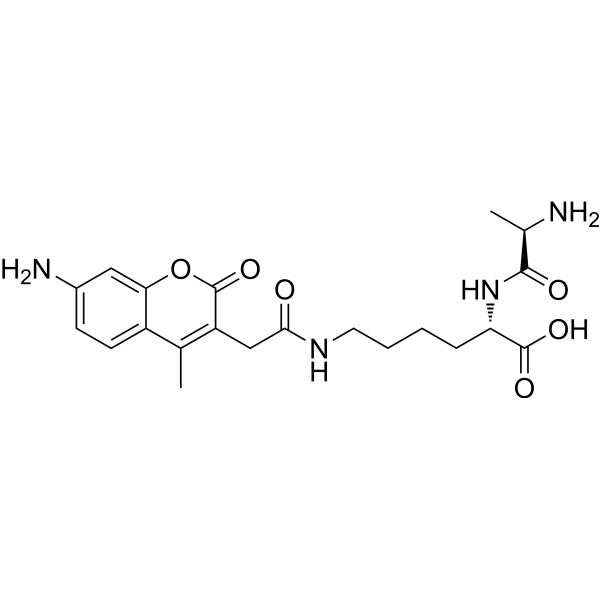
D-Ala-Lys-AMCA
Based on 2 publication(s) in Google Scholar
D-Ala-Lys-AMCA is a known proton-coupled oligopeptide transporter 1 (PEPT1) substrate that emits blue fluorescence. D-Ala-Lys-AMCA may be transported into liver cancer cells and Caco-2 cells based on fluorescence analysis. D-Ala-Lys-AMCA can be used for characterizing PEPT1-specific substrates or inhibitors (Ex/Em = 390/480 nm).
For research use only. We do not sell to patients.
- CAS No.: 375822-19-8
- Formula: C21H28N4O6
- Molecular Weight:432.47
-
Storage:
Please store the product under the recommended conditions in the Certificate of Analysis.

To place orders, for customer services and technical support, please contact: MedChemExpress USA
Tel: 609-228-6898 E-mail: [email protected] [email protected]


-
 Biological Activity
-
Chemical Information
- Protocols
- Purity & Documentation
- References
-
 Help & FAQs


Publications Citing Use of MedChemExpress (MCE) D-Ala-Lys-AMCA
More

Biological Activity
Description
In Vitro
D-Ala-Lys-AMCA (25-150 μM, 3 h) may be transported into liver cancer and Caco-2 cells[1].
D-Ala-Lys-AMCA (25 μM, 10 min) can be uptaken by absorptive enterocytes of all small intestine segments, whereas there is a complete lack of fluorescence in colonic samples[2].
Guide (Following is our recommended protocol. This protocol only provides a guideline, and should be modified according to your specific needs)[1].
1. Preparation of stock solution:
Use ddH2O or DMSO to prepare a D-Ala-Lys-AMCA stock solution.
2. Preparation of working solution:
Use preheated serum-free cell culture medium or PBS to dilute the stock solution to prepare a 1 mM D-Ala-Lys-AMCA working solution.
Note: Please adjust the concentration of D-Ala-Lys-AMCA working solution according to actual conditions and prepare it before use.
3. Cell staining:
3.1 Add PBS to wash the cells twice, 5 minutes each time.
3.2 Add D-Ala-Lys-AMCA working solution and incubate at 37°C for 1-2 h.
3.3 Aspirate the dye working solution, wash 2-3 times with culture medium, 5 minutes each time, and observe using a fluorescence microscope (Ex/Em = 390/480 nm).
MedChemExpress (MCE) has not independently confirmed the accuracy of these methods. They are for reference only.
Emission (Em)
480
Excitation (Ex)
390


Chemical Information
-
CAS No. 375822-19-8
-
Molecular Weight 432.47
-
Formula C21H28N4O6
-
Shipping
Room temperature in continental US; may vary elsewhere.
-
Storage
Please store the product under the recommended conditions in the Certificate of Analysis.


Publications (2)
-
Journal Impact Factor
-
Most Recent
-
-
Toxicol Appl Pharmacol
Exploring the potential use of Caenorhabditis elegans as an animal model for evaluating chemical-induced intestinal dysfunction. [Abstract]2024 Dec:493:117140. PMID: 39500396


Purity & Documentation
References
[1]. Gong Y, et al. Specific expression of proton-coupled oligopeptide transporter 1 in primary hepatocarcinoma-anovel strategy for tumor-targeted therapy. Oncol Lett. 2017 Oct;14(4):4158-4166. [Content Brief]
[2]. Groneberg DA, et al. Intestinal peptide transport: ex vivo uptake studies and localization of peptide carrier PEPT1. Am J Physiol Gastrointest Liver Physiol. 2001 Sep;281(3):G697-704. [Content Brief]

Calculators
Concentration (start) × Volume (start) = Concentration (final) × Volume (final)


D-Ala-Lys-AMCA Related Classifications
HY-111956 Related Classifications
 Powered by Bioz