



Dacarbazine hydrochloride
Based on 16 publication(s) in Google Scholar
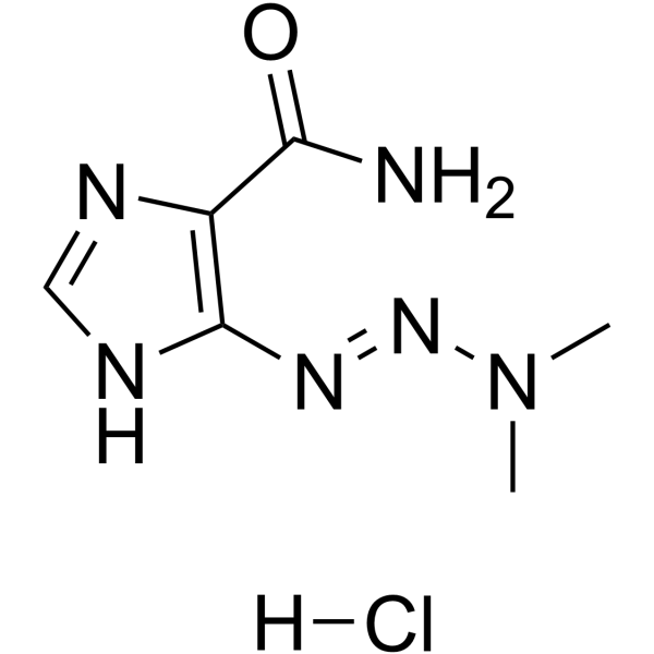
Dacarbazine hydrochloride is a cell cycle nonspecific antineoplastic alkylating agent. Dacarbazine hydrochloride inhibits T and B lymphoblastic response, with IC50 values of 50 and 10 μg/mL, respectively. Dacarbazine hydrochloride can be used for the research of metastatic malignant melanoma.
For research use only. We do not sell to patients.
- CAS No.: 17925-90-5
- Formula: C6H11ClN6O
- Molecular Weight:218.64
-
Storage:
Please store the product under the recommended conditions in the Certificate of Analysis.

To place orders, for customer services and technical support, please contact: MedChemExpress USA
Tel: 609-228-6898 E-mail: [email protected] [email protected]


-
 Biological Activity
-
Chemical Information
- Protocols
- Purity & Documentation
- References
-
 Help & FAQs


Publications Citing Use of MedChemExpress (MCE) Dacarbazine hydrochloride
More- Nature. 2023 Jun;618(7964):374-382. [Abstract]
- Nat Commun. 2024 Sep 10;15(1):7923. [Abstract]
- J Nanobiotechnology. 2023 Oct 19;21(1):383. [Abstract]
- Theranostics. 2020 Jul 25;10(21):9477-9494. [Abstract]
- Adv Sci (Weinh). 2024 Dec;11(48):e2408707. [Abstract]
- Cell Rep Med. 2025 Apr 15;6(4):102053. [Abstract]
- Carbohydr Polym. 2024 Dec 15:346:122645. [Abstract]
- Cell Death Dis. 2025 Apr 9;16(1):268. [Abstract]
- Phytother Res. 2024 Jun;38(6):2800-2817. [Abstract]
- J Ginseng Res. 2024 Nov;48(6):559-569. [Abstract]
- J Ethnopharmacol. 2024 Apr 24:324:117759. [Abstract]
- J Mol Med (Berl). 2019 Aug;97(8):1183-1193. [Abstract]
- Exp Cell Res. 2020 Aug 1;393(1):112054. [Abstract]
- J Cancer Res Clin Oncol. 2025 Dec 17;152(1):12. [Abstract]
- SSRN. 2024 Mar 20.
- Heliyon. 2024 Jan 18;10(3):e24988. [Abstract]
 Customer Validation & Images

-
In Vivo Efficacy Study
-
Cell Proliferation/Viability Assay
-
Cell Proliferation/Viability Assay
-
In Vivo Efficacy Study
-
Histological Imaging/Staining
All Antibiotic Isoforms
More

Biological Activity
Description


Chemical Information
-
CAS No. 17925-90-5
-
Molecular Weight 218.64
-
Formula C6H11ClN6O
-
SMILES
O=C(C1=C(/N=N/N(C)C)NC=N1)N.[H]Cl
-
Synonyms
Imidazole Carboxamide hydrochloride
-
Shipping
Room temperature in continental US; may vary elsewhere.
-
Storage
Please store the product under the recommended conditions in the Certificate of Analysis.


Publications (16)
-
Journal Impact Factor
-
Most Recent
-
Nature
2023 Jun;618(7964):374-382. PMID: 37225988
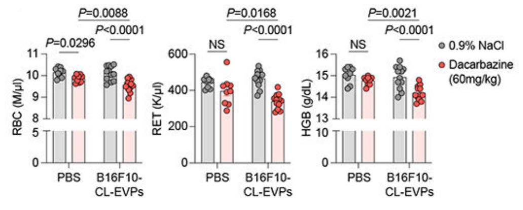
Dacarbazine hydrochloride purchased from MedChemExpress. Usage Cited in: Nature. 2023 Jun;618(7964):374-382. [Abstract]
Dacarbazine (60 mg/kg; i.p.; every other day for 8 d) significantly reduced red blood cell (RBC), reticulocyte (RET), and hemoglobin (HGB) counts in PBS- or B16F10-CL-EVP-educated mice.
-
Nat Commun
2024 Sep 10;15(1):7923. PMID: 39256387 -
J Nanobiotechnology
Combination of ferroptosis and pyroptosis dual induction by triptolide nano-MOFs for immunotherapy of Melanoma. [Abstract]2023 Oct 19;21(1):383. PMID: 37858186
Dacarbazine hydrochloride purchased from MedChemExpress. Usage Cited in: J Nanobiotechnology. 2023 Oct 19;21(1):383. [Abstract]
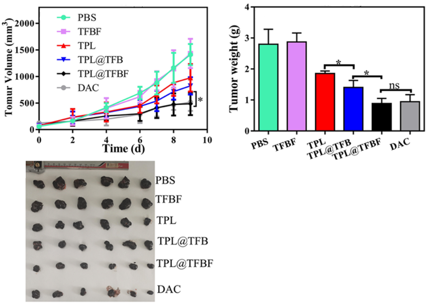
Dacarbazine (DAC) (5 mg/kg; i.v.; once every three days for 3 times) significantly inhibited the growth of subcutaneous B16F10 melanoma xenografts in C57BL/6 mice.
Dacarbazine hydrochloride purchased from MedChemExpress. Usage Cited in: J Nanobiotechnology. 2023 Oct 19;21(1):383. [Abstract]
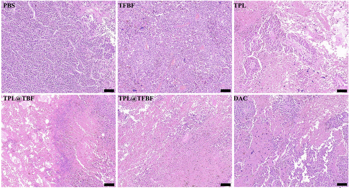
H&E staining of tumors in each group at the end of treatment. The results showed that Dacarbazine (DAC) (5 mg/kg; i.v.; once every three days for 3 times) caused significant cell necrosis and nucleopaenia in tumors of B16F10 tumor-bearing mice. Scale bar, 100 μm.
-
Theranostics
Molecular signatures of BRCAness analysis identifies PARP inhibitor Niraparib as a novel targeted therapeutic strategy for soft tissue Sarcomas. [Abstract]2020 Jul 25;10(21):9477-9494. PMID: 32863940 -
Adv Sci (Weinh)
Patient-Derived Melanoma Immune-Tumoroids as a Platform for Precise High throughput Drug Screening. [Abstract]2024 Dec;11(48):e2408707. PMID: 39475010
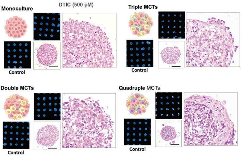
Dacarbazine hydrochloride purchased from MedChemExpress. Usage Cited in: Adv Sci (Weinh). 2024 Dec;11(48):e2408707. [Abstract]
Dacarbazine (DTIC) (500 µM; 72 h) reduced the diameter and metabolic activity of MCTs except monoculture tumoroids, especially the triples and quadruples, resulting in a decrease of approximately 32% and 28% in relative diameter, respectively, and caused a high number of apoptotic cells to detach from the main structure.
-
Cell Rep Med
CAN-Scan: A multi-omic phenotype-driven precision oncology platform identifies prognostic biomarkers of therapy response for colorectal cancer. [Abstract]2025 Apr 15;6(4):102053. PMID: 40187357 -
Carbohydr Polym
Chitosan/dextran-based organohydrogel delivers EZH2 inhibitor to epigenetically reprogram chemo/immuno-resistance in unresectable metastatic melanoma. [Abstract]2024 Dec 15:346:122645. PMID: 39245506 -
Cell Death Dis
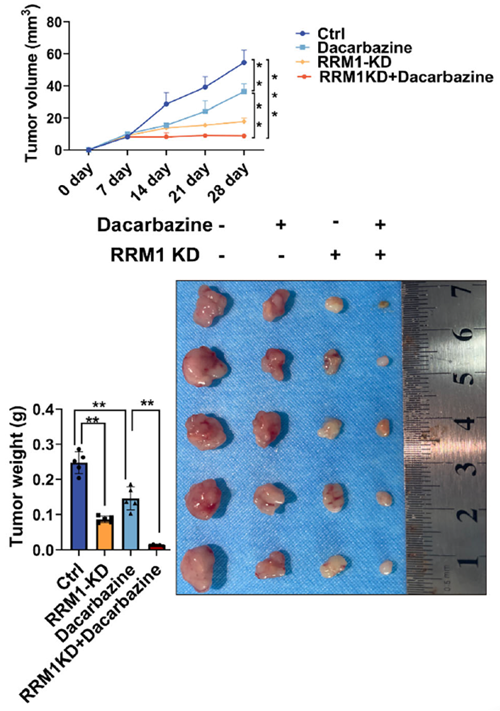
MTCH2 regulates NRF2-mediated RRM1 expression to promote melanoma proliferation and dacarbazine insensitivity. [Abstract]2025 Apr 9;16(1):268. PMID: 40204724
Dacarbazine hydrochloride purchased from MedChemExpress. Usage Cited in: Cell Death Dis. 2025 Apr 9;16(1):268. [Abstract]
Dacarbazine (20 mg/kg; i.p.; 20 d) reduced tumor growth rates and tumor sizes of melanoma xenograft mice.
-
Phytother Res
Isoalantolactone exerts anti-melanoma effects via inhibiting PI3K/AKT/mTOR and STAT3 signaling in cell and mouse models. [Abstract]2024 Jun;38(6):2800-2817. PMID: 38526171 -
J Ginseng Res
20(S)-Ginsenoside Rh2 induces apoptosis and autophagy in melanoma cells via suppressing Src/STAT3 signaling. [Abstract]2024 Nov;48(6):559-569. PMID: 39583170 -
J Ethnopharmacol
Screening of anti-melanoma compounds from Morus alba L.: Sanggenon C promotes melanoma cell apoptosis by disrupting intracellular Ca2+ homeostasis. [Abstract]2024 Apr 24:324:117759. PMID: 38219884 -
J Mol Med (Berl)
2019 Aug;97(8):1183-1193. PMID: 31201471 -
Exp Cell Res
Network-based analysis with primary cells reveals drug response landscape of acute myeloid leukemia. [Abstract]2020 Aug 1;393(1):112054. PMID: 32376287 -
J Cancer Res Clin Oncol
Comprehensive analysis based on spatial domains identifies CD44 as a potential target of puerarin. [Abstract]2025 Dec 17;152(1):12. PMID: 41405724 -
-
Heliyon
2024 Jan 18;10(3):e24988. PMID: 38317912

Protocols
-
Flow cytometric DNA-content cell-cycle staining
Flow cytometric DNA-content cell-cycle staining measures the fluorescence intensity of DNA-bound fluorochromes in single cells or nuclei to estimate DNA content distributions, allowing assignment of populations to G0/G1, S, and G2/M phases by DNA histogram deconvolution. Propidium iodide (PI) intercalates into DNA, and PI fluorescence is proportional to cellular DNA content when staining is performed under conditions that make DNA accessible and minimize non-DNA signal. Cells with G2/M DNA content are expected to show approximately twice the fluorescence intensity of G0/G1 cells, while S-phase cells occupy intermediate fluorescence values. PI-based DNA-content analysis can also detect cells with fractional DNA content, often reported as sub-G1, when DNA fragmentation and extraction during staining reduce retained DNA signal in apoptotic cells. DAPI is an alternative DNA fluorochrome for univariate DNA-content analysis, while bivariate approaches combining DNA content with proliferation
-
Cell Cytotoxicity Assay
Cytotoxicity assays are usually based on the assessment of cell membrane damage, which can also be indirectly detected by measuring cell viability. Detection methods include MTT assay, CKK-8 assay, LDH assay and ATP assay, etc.
-
BrdU Incorporation Assay
Bromodeoxyuridine (BrdU) incorporation assay is based on the principle that BrdU, a thymidine analog, is incorporated into newly synthesized DNA during the S phase of the cell cycle, thereby serving as a marker of DNA replication and cellular proliferation. Incorporated BrdU can be detected using anti-BrdU antibodies following DNA denaturation, enabling visualization or quantification of proliferating cells through immunochemical detection methods such as immunofluorescence or immunohistochemistry.
-
Protocol for Cell Cycle
Cell-cycle analysis by flow cytometry measures DNA content in single cells to estimate the fraction of cells in G0/G1, S, and G2/M phases. Propidium iodide intercalates into DNA, and after RNA removal with RNase, fluorescence intensity reflects cellular DNA content: 2N cells are assigned to G0/G1, cells between 2N and 4N to S phase, and 4N cells to G2/M. DNA-content analysis alone cannot reliably separate G0 from G1 or G2 from M. Ki-67 can distinguish quiescent G0 cells from cycling cells, EdU or BrdU incorporation marks active DNA synthesis in S phase, and phospho-histone H3 staining identifies mitotic cells within the 4N population.


Purity & Documentation
References

Calculators
Concentration (start) × Volume (start) = Concentration (final) × Volume (final)


Dacarbazine hydrochloride Related Classifications
HY-B0078B Related Classifications
 Powered by Bioz