



Dimethyl succinate
Based on 2 publication(s) in Google Scholar
Dimethyl succinate is an orally active cell permeable succinate analogue. Dimethyl succinate can disrupt the TCA cycle by elevating intracellular succinate levels, leading to reduced protein synthesis and impairs myogenic differentiation. Dimethyl succinate can induce apoptosis. Dimethyl succinate can decrease maximal cellular respiration and reserve capacity. Dimethyl succinate can rescue synapse loss in AD. Dimethyl succinate can be used for the researches of metabolic and neurological disease[1][2].
For research use only. We do not sell to patients.
- Purity : 99.95%
- CAS No.: 106-65-0
- Formula: C6H10O4
- Molecular Weight:146.14
-
Storage:
4°C, sealed storage, away from moisture
* In solvent : -80°C, 6 months; -20°C, 1 month (sealed storage, away from moisture)


To place orders, for customer services and technical support, please contact: MedChemExpress USA
Tel: 609-228-6898 E-mail: [email protected] [email protected]


-
 Biological Activity
-
Chemical Information
-
Solvent & Solubility
- Protocols
- Purity & Documentation
- References
-
 Help & FAQs


-
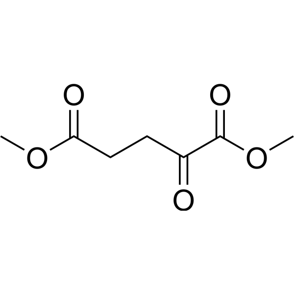
HY-44134
Dimethyl 2-oxoglutarate2-oxoglutarate Derivative -
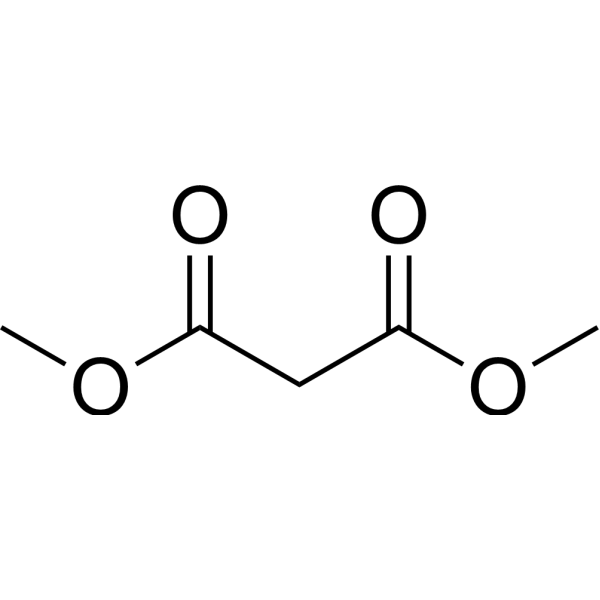
HY-Y1787
Dimethyl malonateSDH Inhibitor -
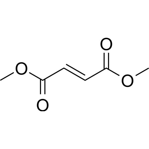
HY-17363
Dimethyl fumarateNrf2 Activator -
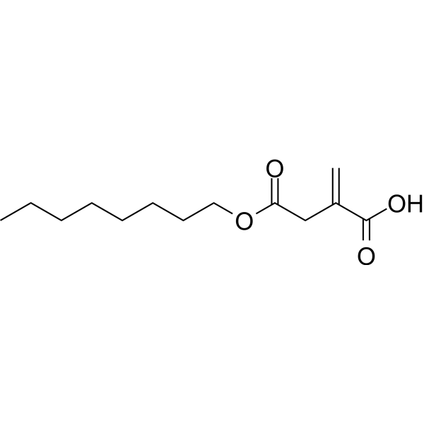
HY-112675
4-Octyl itaconateNrf2 Activator -
HY-B1756
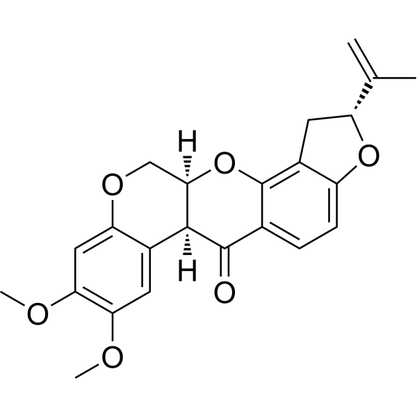
RotenoneMitochondrial Complex I Inhibitor -

HY-10071
Y-27632ROCK Inhibitor -
HY-B0150
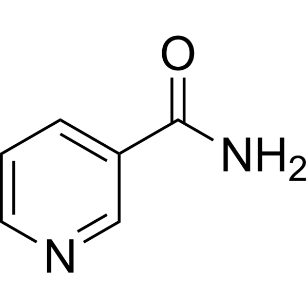
NicotinamideSIRT Inhibitor -
HY-15144
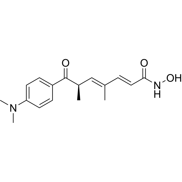
Trichostatin AHDAC Class I/II Inhibitor -
HY-15772
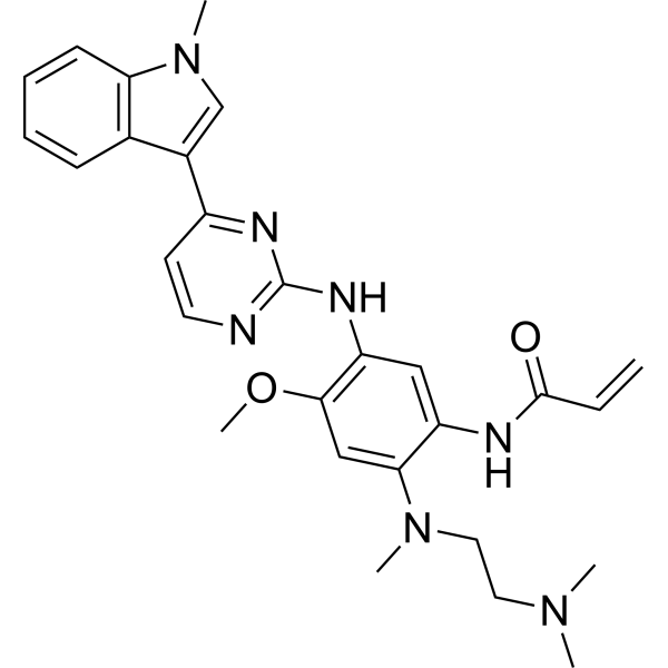
OsimertinibMutant-Selective EGFR Inhibitor -

HY-N6782
OligomycinH+-ATP-Synthase Inhibitor -

HY-100558
Bafilomycin A1V-ATPase Inhibitor -

HY-Y0320
Dimethyl sulfoxide, meets analytical specification of Ch.P.Aprotic Solvent
Publications Citing Use of MedChemExpress (MCE) Dimethyl succinate
More Customer Validation & Images

-
Flow Cytometry


Biological Activity
Description
In Vitro
Dimethyl succinate (16 mM) increases apoptotic cells in C2C12 myoblasts[1].
Dimethyl succinate (4-8 mM, 48 h) increases intracellular succinate, malate and glutamate in C2C12 myoblasts[1].
Dimethyl succinate (8 mM, 48 h) reduces protein anabolism as determined by an O-propargyl-puromycin (OPP) incorporation assay in C2C12 myoblasts[1].
Dimethyl succinate (8 mM) reduces maximal respiration and mitochondria reserve capacity in C2C12 myoblasts[1].
Dimethyl succinate (8 mM) results in an impaired ability to form mature, multinucleated Myosin Heavy Chain-positive structures in C2C12 myoblasts[1].
Dimethyl succinate (5 mM, 48 h) partially rescues synapse loss in AD hiPSC-derived cerebrocortical neurons[2].
MedChemExpress (MCE) has not independently confirmed the accuracy of these methods. They are for reference only. Further protocols information, click here.
-
Cell Line:C2C12 myoblasts
-
Concentration:4 mM, 8 mM, 16 mM
-
Incubation Time:48 hours
-
Result:Showed no significant differences in proliferation over this time period.
-
Cell Line:C2C12 myoblasts
-
Concentration:4 mM, 8 mM, 16 mM
-
Incubation Time:48 hours
-
Result:Only increased apoptosis rates at 16 mM.
In Vivo
MedChemExpress (MCE) has not independently confirmed the accuracy of these methods. They are for reference only.
-
Animal Model:FVB mice; wild type model[1]
-
Dosage:2% in water
-
Administration:Oral; 6 weeks
-
Result:Had no significant difference in total body weight and total lean mass.
Impaired skeletal muscle homeostasis and regeneration.
Exhibited smaller muscle myofiber diameters.


Chemical Information
-
CAS No. 106-65-0
-
Appearance <16°C Solid,>19°C Liquid
-
Molecular Weight 146.14
-
Formula C6H10O4
-
Color Colorless to off-white
-
SMILES
O=C(OC)CCC(OC)=O
-
Shipping
Room temperature in continental US; may vary elsewhere.
-
Storage
4°C, sealed storage, away from moisture
* In solvent : -80°C, 6 months; -20°C, 1 month (sealed storage, away from moisture)


Publications (2)
-
Journal Impact Factor
-
Most Recent
-
Cell Rep
4-octyl itaconate inhibits cytokine-mediated inflammation via alkylation of TYK2 and JAK1. [Abstract]2026 Mar 30;45(4):117179. PMID: 41915469 -
Chin Med
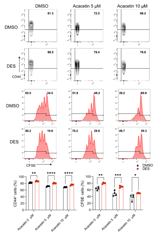
Acacetin alleviates autoimmune myocarditis by regulating CD4+ T cell mitochondrial respiration. [Abstract]2024 May 13;19(1):68. PMID: 38741130
Dimethyl succinate purchased from MedChemExpress. Usage Cited in: Chin Med. 2024 May 13;19(1):68. [Abstract]
Dimethyl succinate (DES) (5 mM; 72 h) reversed the decrease of CD44 expression and proliferation of CD4+ T cells by acacetin.


Solvent & Solubility
In Vitro:
DMSO : 200 mg/mL (1368.55 mM; Need ultrasonic; Hygroscopic DMSO has a significant impact on the solubility of product, please use newly opened DMSO)
H2O : ≥ 100 mg/mL (684.28 mM)
* "≥" means soluble, but saturation unknown.
Please refer to the solubility information to select the appropriate solvent. Once prepared, please aliquot and store the solution to prevent product inactivation from repeated freeze-thaw cycles.
Storage method and period of stock solution: -80°C, 6 months; -20°C, 1 month (sealed storage, away from moisture). When stored at -80°C, please use it within 6 months. When stored at -20°C, please use it within 1 month.
* Note: If you choose water as the stock solution, please dilute it to the working solution, then filter and sterilize it with a 0.22 μm filter before use.
Please refer to the solubility information to select the appropriate solvent. Once prepared, please aliquot and store the solution to prevent product inactivation from repeated freeze-thaw cycles.
Storage method and period of stock solution: -80°C, 6 months; -20°C, 1 month (sealed storage, away from moisture). When stored at -80°C, please use it within 6 months. When stored at -20°C, please use it within 1 month.
* Note: If you choose water as the stock solution, please dilute it to the working solution, then filter and sterilize it with a 0.22 μm filter before use.
Concentration (start) × Volume (start) = Concentration (final) × Volume (final)
In Vivo:
For the following dissolution methods, please prepare the working solution directly:
It is recommended to prepare fresh solutions and use them promptly within a short period of time.
The percentages shown for the solvents indicate their volumetric ratio in the final prepared solution. If precipitation or phase separation occurs during preparation, heat and/or sonication can be used to aid dissolution.
Add each solvent one by one: Saline
Solubility: ≥ 100 mg/mL (684.28 mM); Clear solution

Protocols
-
Apoptosis
Apoptosis, also called programmed cell death, is generally characterized by distinct morphological characteristics.
-
TUNEL staining for apoptotic DNA fragmentation
TUNEL staining detects DNA strand breaks by using terminal deoxynucleotidyl transferase to add labeled nucleotides to exposed 3′-OH DNA termini, generating either microscopic staining in fixed cells or tissue sections, or fluorescence/cytometric signal in cell suspensions. TUNEL positivity reflects DNA fragmentation but should not be interpreted alone as definitive apoptosis, because TUNEL can also label necrotic, autolytic, mechanically damaged, or DNA-repair-associated DNA breaks.
-
Annexin V plus membrane-impermeant dye apoptosis staining
Annexin V-based apoptosis assays rely on the detection of phosphatidylserine (PS) externalization from the inner leaflet of the plasma membrane to the outer leaflet, an early biochemical hallmark of apoptosis. Fluorescently labeled Annexin V binds PS in a calcium-dependent manner, enabling identification of early apoptotic cells by flow cytometry or fluorescence microscopy. When combined with a membrane-impermeant DNA-binding dye (e. g. , propidium iodide), this approach allows discrimination between viable (Annexin V−/dye−), early apoptotic (Annexin V+/dye−), and late apoptotic or necrotic (Annexin V+/dye+) cell populations by assessing membrane integrity and PS exposure.
-
Apoptosis Solutions
Apoptosis is a regulated, generally non-lytic cell-death pathway that removes unwanted, damaged, infected, or abnormal cells through coordinated morphological changes, caspase activation, DNA fragmentation, and membrane remodeling. The intrinsic apoptosis pathway is controlled mainly by mitochondrial outer membrane permeabilization, BCL-2 family proteins, cytochrome c release, apoptosome formation, caspase-9 activation, and downstream executioner caspase-3/7 activation. The extrinsic apoptosis pathway is initiated by death receptors such as Fas, TNFR, and TRAIL receptors, which recruit adaptor proteins and activate caspase-8 before engaging executioner caspases or mitochondrial amplification through BID cleavage. Apoptosis is linked to many phenotypes, including cancer cell killing, tissue homeostasis, immune regulation, neurodegeneration, infection response, and treatment-induced cytotoxicity; unresolved questions include how apoptosis interacts with necroptosis, pyroptosis, ferroptos
-
Research Protocol for Neurological Diseases
PINK1/Parkin-mediated mitophagy pathway is a mitochondrial quality-control signaling axis in which mitochondrial depolarization stabilizes PINK1 on damaged mitochondria, activates Parkin recruitment and E3 ubiquitin ligase activity, promotes ubiquitination of outer mitochondrial membrane proteins, recruits selective autophagy adaptors, and drives lysosomal degradation of damaged mitochondria. In neurological disease research, this pathway is experimentally important because neurons, especially dopaminergic neurons, are highly dependent on mitochondrial integrity, and defective mitochondrial turnover can lead to mitochondrial dysfunction, oxidative stress, impaired neuronal survival, α-synuclein accumulation, and neuroinflammatory damage-associated signals. The genetic disease link is strongest in Parkinson’s disease because mutations in PRKN/parkin cause autosomal recessive juvenile parkinsonism, mutations in PINK1 cause hereditary early-onset Parkinson’s disease, and Drosophila studie


Purity & Documentation
-
Data Sheet (274 KB)
-
SDS (393 KB)
- English - EN (393 KB)
- Français - FR (393 KB)
- Deutsch - DE (393 KB)
- Norwegian - NO (393 KB)
- Español - ES (393 KB)
- Swedish - SV (393 KB)
- Italian - IT (393 KB)
- Korean - KR (393 KB)
- Portuguese - PT (393 KB)
-
Handling Instructions (2659 KB)
References
[1]. Arneson-Wissink PC, et al. The wasting-associated metabolite succinate disrupts myogenesis and impairs skeletal muscle regeneration. JCSM Rapid Commun. 2020;3(2):56-69. [Content Brief]
[2]. Andreyev AY, et al. Metabolic Bypass Rescues Aberrant S-nitrosylation-Induced TCA Cycle Inhibition and Synapse Loss in Alzheimer's Disease Human Neurons. Adv Sci (Weinh). 2024;11(12):e2306469. [Content Brief]


Complete Stock Solution Preparation Table
Please refer to the solubility information to select the appropriate solvent. Once prepared, please aliquot and store the solution to prevent product inactivation from repeated freeze-thaw cycles.
Storage method and period of stock solution: -80°C, 6 months; -20°C, 1 month (sealed storage, away from moisture). When stored at -80°C, please use it within 6 months. When stored at -20°C, please use it within 1 month.
| Optional Solvent | Concentration Solvent Mass | 1 mg | 5 mg | 10 mg | 25 mg |
|---|---|---|---|---|---|
| H2O / DMSO | 1 mM | 6.8428 mL | 34.2138 mL | 68.4275 mL | 171.0688 mL |
| 5 mM | 1.3686 mL | 6.8428 mL | 13.6855 mL | 34.2138 mL | |
| 10 mM | 0.6843 mL | 3.4214 mL | 6.8428 mL | 17.1069 mL | |
| 15 mM | 0.4562 mL | 2.2809 mL | 4.5618 mL | 11.4046 mL | |
| 20 mM | 0.3421 mL | 1.7107 mL | 3.4214 mL | 8.5534 mL | |
| 25 mM | 0.2737 mL | 1.3686 mL | 2.7371 mL | 6.8428 mL | |
| 30 mM | 0.2281 mL | 1.1405 mL | 2.2809 mL | 5.7023 mL | |
| 40 mM | 0.1711 mL | 0.8553 mL | 1.7107 mL | 4.2767 mL | |
| 50 mM | 0.1369 mL | 0.6843 mL | 1.3686 mL | 3.4214 mL | |
| 60 mM | 0.1140 mL | 0.5702 mL | 1.1405 mL | 2.8511 mL | |
| 80 mM | 0.0855 mL | 0.4277 mL | 0.8553 mL | 2.1384 mL | |
| 100 mM | 0.0684 mL | 0.3421 mL | 0.6843 mL | 1.7107 mL |
* Note: If you choose water as the stock solution, please dilute it to the working solution, then filter and sterilize it with a 0.22 μm filter before use.


Dimethyl succinate Related Classifications
HY-Y0808 Related Classifications
 Powered by Bioz