



Trimyristin

Based on 1 Customer Validation
Trimyristin is an orally active compound. Trimyristin can be isolated from the seeds of nutmeg (Myristica fragrans). Trimyristin inhibits the activities of acetylcholinesterase (AChE), ACP and ALP, with IC50 values of 0.11, 0.16 and 0.18 mM, respectively. Trimyristin exerts competitive-noncompetitive inhibition on acetylcholinesterase, uncompetitive inhibition on ACP, and competitive/noncompetitive inhibition on ALP. Trimyristin restores the downregulated acetylcholinesterase concentration in the cerebral cortex of rats exposed to sodium arsenite. Trimyristin can be used in studies related to fascioliasis and neurotoxicity.
For research use only. We do not sell to patients.
- Purity : 99.72%
- CAS No.: 555-45-3
- Formula: C45H86O6
- Molecular Weight:723.16
-
Storage:
4°C, sealed storage, away from moisture
* In solvent : -80°C, 6 months; -20°C, 1 month (sealed storage, away from moisture)

To place orders, for customer services and technical support, please contact: MedChemExpress USA
Tel: 609-228-6898 E-mail: [email protected] [email protected]


-
 Biological Activity
-
Chemical Information
-
Solvent & Solubility
- Protocols
- Purity & Documentation
- References
-
 Help & FAQs


-
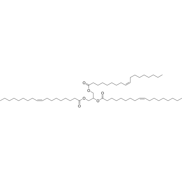
HY-N1981
TrioleinMonoacid Triglyceride -
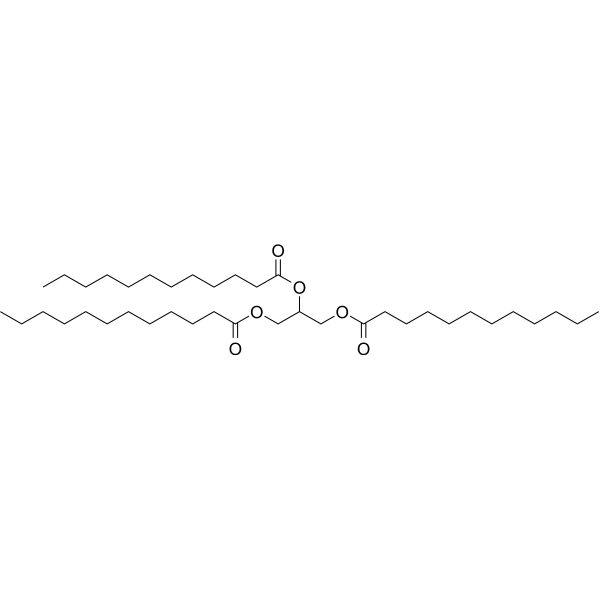
HY-126573
TrilaurinNeoplasm Inhibitor -
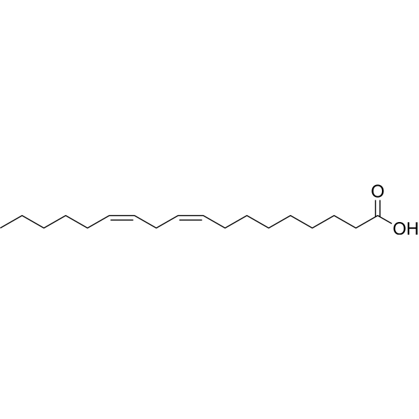
HY-N0729
Linoleic acidHuman Endogenous Metabolite -

HY-N2511R
Trimyristin (Standard)Reference Standard -
HY-W013061
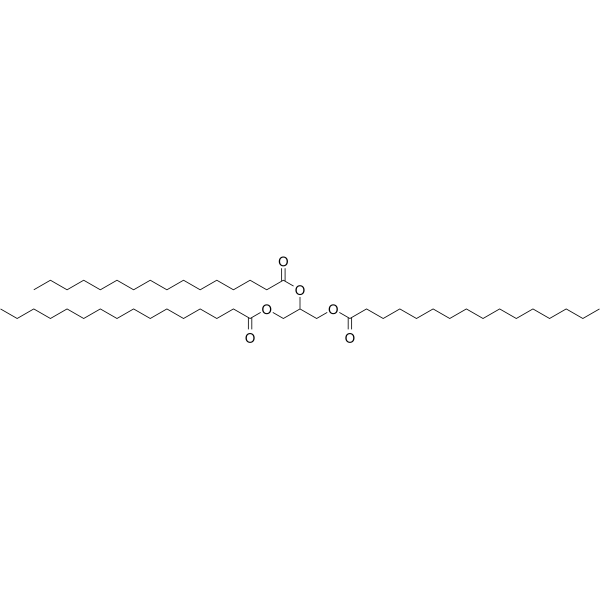
Propane-1,2,3-triyl tripalmitateGlycerol Tristearate Lymphatic Transport Promoter -

HY-N0729D
Linoleic acid,suitable for cell cultureNo Target-Effect Identified -
HY-N0729R
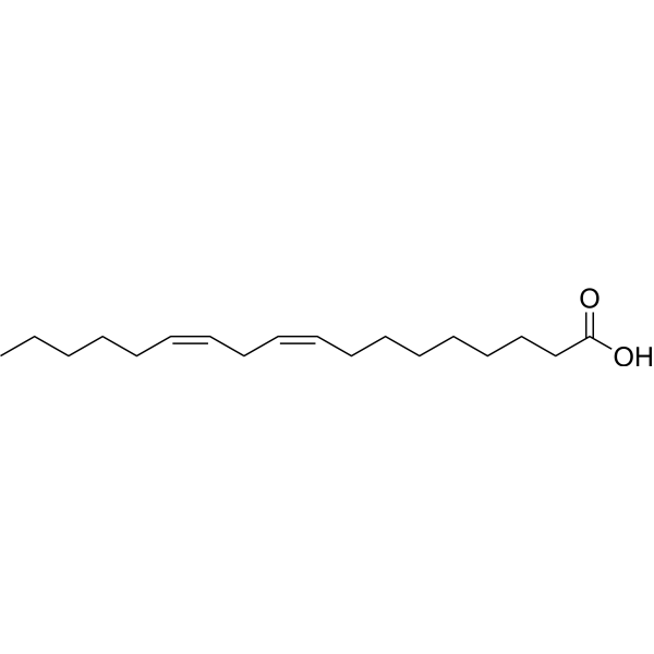
Linoleic acid (Standard)Standard of Linoleic acid -
HY-B1804
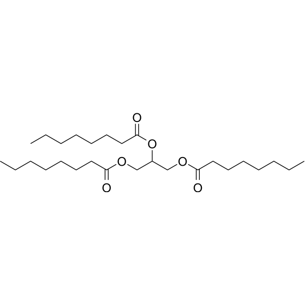
TricaprilinKetogenic Agent -

HY-N1981R
Triolein (Standard)Monoacid Triglyceride -
HY-B2167
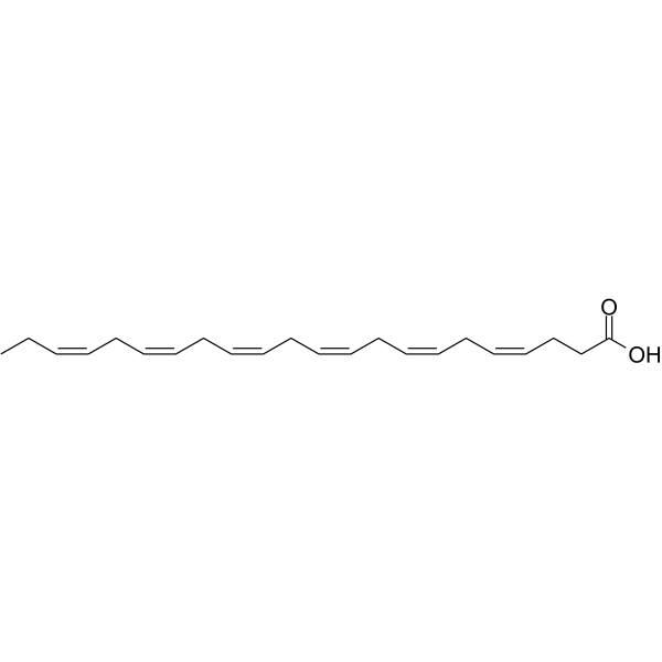
Docosahexaenoic acidomega-3 Fatty Acid -
HY-128393
TrilinoleinAnticancer Agent -
HY-N1446
Oleic acidMonounsaturated Fatty Acid


Biological Activity
Description
IC50 & Target
[1]|
AChE 0.11 mM (IC50) |
ACP 0.16 mM (IC50) |
ALP 0.18 mM (IC50) |
In Vitro
Trimyristin (0.04-0.15 mM; 15 min preincubation) potently inhibits AChE (IC50 = 0.11 mM), ACP (IC50 = 0.16 mM), and ALP (IC50 = 0.18 mM) in nervous tissue homogenates of Lymnaea acuminata snails via competitive noncompetitive inhibition of AChE and ALP, and uncompetitive inhibition of ACP[1].
MedChemExpress (MCE) has not independently confirmed the accuracy of these methods. They are for reference only. Further protocols information, click here.
In Vivo
Trimyristin (15-30 mg/kg; p.o.; daily; 6 weeks) restores sodium arsenite-induced downregulated cerebral cortex acetylcholinesterase concentrations to baseline control levels in adult male Wistar rats[2].
Trimyristin (15-30 mg/kg; p.o.; daily; 9 weeks) shows that chronic treatment with 30 mg/kg significantly reduces cerebral cortex acetylcholinesterase concentrations in adult male Wistar rats, while 15 mg/kg maintains control-level concentrations[2].
MedChemExpress (MCE) has not independently confirmed the accuracy of these methods. They are for reference only.
-
Animal Model:Lymnaea acuminata Lamarck (adults, length 2.25 cm)[1]
-
Dosage:6.21 mg/L (40% of 24 h LC50); 12.43 mg/L (80% of 24 h LC50); 2.80 mg/L (40% of 96 h LC50); 5.60 mg/L (80% of 96 h LC50)
-
Administration:aquatic exposure; 24 or 96 hours
-
Result:Reduced acetylcholinesterase (AChE) activity to 90%, 82%, 73%, and 63% of control levels after exposure to 40% 24 h LC50, 80% 24 h LC50, 40% 96 h LC50, and 80% 96 h LC50 of trimyristin, respectively.
Reduced acid phosphatase (ACP) activity to 87%, 80%, 73%, and 64% of control levels after exposure to 40% 24 h LC50, 80% 24 h LC50, 40% 96 h LC50, and 80% 96 h LC50 of trimyristin, respectively.
Reduced alkaline phosphatase (ALP) activity to 92%, 84%, 71%, and 61% of control levels after exposure to 40% 24 h LC50, 80% 24 h LC50, 40% 96 h LC50, and 80% 96 h LC50 of trimyristin, respectively.
Caused statistically significant (P < .05) concentration- and time-dependent inhibition of AChE, ACP, and ALP, with maximum inhibition of 37% for AChE, 36% for ACP, and 39% for ALP observed at the 80% 96 h LC50 dose.
-
Animal Model:Wistar (adult male, 150-250 g, average 200 g, sodium arsenite-induced neurotoxicity)[2]
-
Dosage:15 mg/kg; 30 mg/kg
-
Administration:p.o.; daily; 6 weeks
-
Result:Significantly upregulated and restored cerebral cortex AChE concentrations to baseline control levels.
Restored normal cytoarchitecture of cerebral cortices, with no significant inflammation or degenerative changes.
-
Animal Model:Wistar (adult male, 150-250 g, average 200 g)[2]
-
Dosage:15 mg/kg; 30 mg/kg
-
Administration:p.o.; daily; 9 weeks
-
Result:Maintained AChE concentrations at levels comparable to control groups (15 mg/kg).
Caused a statistically significant reduction in cerebral cortex AChE concentrations relative to control groups (30 mg/kg).


Chemical Information
-
CAS No. 555-45-3
-
Appearance Solid
-
Molecular Weight 723.16
-
Formula C45H86O6
-
Color White to off-white
-
SMILES
CCCCCCCCCCCCCC(OC(COC(CCCCCCCCCCCCC)=O)COC(CCCCCCCCCCCCC)=O)=O
-
Structure Classification
-
Initial Source
-
Shipping
Room temperature in continental US; may vary elsewhere.
-
Storage
4°C, sealed storage, away from moisture
* In solvent : -80°C, 6 months; -20°C, 1 month (sealed storage, away from moisture)


Solvent & Solubility
In Vitro:
Acetone : 5 mg/mL (6.91 mM; Need ultrasonic)
DMF : 2.5 mg/mL (3.46 mM; Need ultrasonic)
DMSO : < 1 mg/mL (insoluble or slightly soluble)
Please refer to the solubility information to select the appropriate solvent. Once prepared, please aliquot and store the solution to prevent product inactivation from repeated freeze-thaw cycles.
Storage method and period of stock solution: -80°C, 6 months; -20°C, 1 month (sealed storage, away from moisture). When stored at -80°C, please use it within 6 months. When stored at -20°C, please use it within 1 month.
Please refer to the solubility information to select the appropriate solvent. Once prepared, please aliquot and store the solution to prevent product inactivation from repeated freeze-thaw cycles.
Storage method and period of stock solution: -80°C, 6 months; -20°C, 1 month (sealed storage, away from moisture). When stored at -80°C, please use it within 6 months. When stored at -20°C, please use it within 1 month.
Concentration (start) × Volume (start) = Concentration (final) × Volume (final)

Protocols
-
Neurotoxicity Study
This protocol assesses in vitro neurotoxicity by combining neuronal viability, mitochondrial/metabolic activity, neurite outgrowth, and optional neuronal network function readouts. Calcein-AM or resazurin/PrestoBlue readouts estimate viable or metabolically active cells; βIII-tubulin immunofluorescence detects neuronal morphology and neurite networks; TMRE detects mitochondrial membrane potential; and MEA recordings detect functional changes in neuronal network activity.
-
Cell Cytotoxicity Assay
Cytotoxicity assays are usually based on the assessment of cell membrane damage, which can also be indirectly detected by measuring cell viability. Detection methods include MTT assay, CKK-8 assay, LDH assay and ATP assay, etc.
-
Mesenchymal stromal/stem cell osteogenic differentiation
Mesenchymal stromal/stem cells can be induced toward an osteoblast-like lineage in vitro by culture in osteogenic medium containing dexamethasone, ascorbic acid or ascorbate-2-phosphate, and β-glycerophosphate; the differentiation process is commonly evaluated by alkaline phosphatase activity, osteogenic marker expression, collagenous matrix formation, and calcium-rich matrix mineralization. The main readouts are alkaline phosphatase activity as an early osteogenic marker and Alizarin Red S staining as a calcium-deposit readout for mineralized extracellular matrix; Alizarin Red S can be inspected microscopically or extracted and measured colorimetrically at 405 nm.


Purity & Documentation
-
Data Sheet (272 KB)
-
SDS (393 KB)
- English - EN (393 KB)
- Français - FR (393 KB)
- Deutsch - DE (393 KB)
- Norwegian - NO (393 KB)
- Español - ES (393 KB)
- Swedish - SV (393 KB)
- Italian - IT (393 KB)
- Korean - KR (393 KB)
- Portuguese - PT (393 KB)
-
Handling Instructions (2659 KB)
References


Complete Stock Solution Preparation Table
Please refer to the solubility information to select the appropriate solvent. Once prepared, please aliquot and store the solution to prevent product inactivation from repeated freeze-thaw cycles.
Storage method and period of stock solution: -80°C, 6 months; -20°C, 1 month (sealed storage, away from moisture). When stored at -80°C, please use it within 6 months. When stored at -20°C, please use it within 1 month.
| Optional Solvent | Concentration Solvent Mass | 1 mg | 5 mg | 10 mg | 25 mg |
|---|---|---|---|---|---|
| DMF / Acetone | 1 mM | 1.3828 mL | 6.9141 mL | 13.8282 mL | 34.5705 mL |
| Acetone | 5 mM | 0.2766 mL | 1.3828 mL | 2.7656 mL | 6.9141 mL |


Trimyristin Related Classifications
HY-N2511 Related Classifications
 Powered by Bioz