



AZD2858
Based on 9 publication(s) in Google Scholar
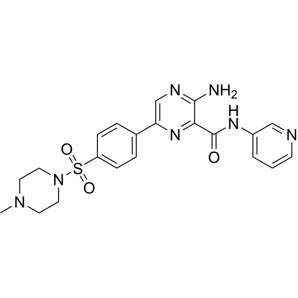
AZD2858 is a potent, orally active GSK-3 inhibitor, with IC50s of 0.9 and 5 nM for GSK-3α and GSK-3β, respectively, used in the research of fracture healing.
For research use only. We do not sell to patients.
- Purity: 99.44%
- CAS No.: 486424-20-8
- Formula: C21H23N7O3S
- Molecular Weight:453.52
-
Storage:Powder -20°C, 3 years , 4°C, 2 years ; In solvent -80°C, 1 year , -20°C, 6 months

To place orders, for customer services and technical support, please contact: MedChemExpress USA
Tel: 609-228-6898 E-mail: [email protected] [email protected]


-
 Biological Activity
-
Chemical Information
-
Solvent & Solubility
- Protocol
- Purity & Documentation
- References
-
 Help & FAQs
-


Kinase Inhibitor Library
HY-L009
-
PI3K/Akt/mTOR Compound Library
HY-L015
-
Protein Tyrosine Kinase Compound Library
HY-L016
-
Stem Cell Signaling Compound Library
HY-L017
-
Wnt/Hedgehog/Notch Compound Library
HY-L020
-
Anti-Aging Compound Library
HY-L034
-
Reprogramming Compound Library
HY-L039
-
Oxygen Sensing Compound Library
HY-L045
-
Glycolysis Compound Library
HY-L058
-
Cytoskeleton Compound Library
HY-L060
-
Orally Active Compound Library
HY-L061
-
Anti-Alzheimer's Disease Compound Library
HY-L069
-
Anti-Pancreatic Cancer Compound Library
HY-L077
-
Anti-Cancer Metabolism Compound Library
HY-L083
-
Neurodegenerative Disease-related Compound Library
HY-L086
-
Anti-Obesity Compound Library
HY-L087
-
Glucose Metabolism Compound Library
HY-L092
-
Targeted Diversity Library
HY-L099
-
Antidepressant Compound Library
HY-L108
-
Human Metabolite Library
HY-L123
-
Anti-Pulmonary Fibrosis Compound Library
HY-L125
-
Cancer Stem Cells Compound Library
HY-L135
-
Metabolic Enzyme Compound Library
HY-L146
-
Serine/Threonine Kinase Inhibitor Library
HY-L164
-
Multi-Target Compound Library
HY-L176
-
Bioactive Compound Library Max
HY-L181
-
MCE Bioactive Compound Library
HY-L001V
-
Bioactive Compound Library
HY-L001
-
Protein Kinase Compound Library
HY-L196
-
Cell Proliferation Compound Library
HY-L201
-
High-Throughput Bioactive Compound Library
HY-L205
-
Mass Spectrometry Human Metabolite Library
HY-L215
-
Posttranslational Modification Library
HY-L226


Publications Citing Use of MedChemExpress (MCE) AZD2858
More- Cell Res. 2022 Jun;32(6):513-529. [Abstract]
- Oncogene. 2023 Nov;42(47):3503-3513. [Abstract]
- ACS Appl Mater Interfaces. 2025 May 28;17(21):30600-30612. [Abstract]
- Nanoscale Horiz. 2026 Jun 8;11(6):1614-1628. [Abstract]
- Oncol Rep. 2019 Jul;42(1):91-102. [Abstract]
- FASEB J. 2025 Nov 30;39(22):e71267. [Abstract]
- J Biol Chem. 2016 Dec 2;291(49):25529-25541. [Abstract]
- J Cell Physiol. 2019 Sep;234(9):15472-15486. [Abstract]
- University of Saskatchewan. 2023 Aug.
 Customer Validation & Images

-
WB
-
WB
-
WB


Biological Activity
|
GSK-3α 0.9 nM (IC50) |
GSK-3β 5 nM (IC50) |
CDK5/p25 356 nM (IC50) |
Haspin 366 nM (IC50) |
CDK5/p35 387 nM (IC50) |
DYRK2 491 nM (IC50) |
CDK2/cyclin A 810 nM (IC50) |
CDK1/cyclin B 1246 nM (IC50) |
PIM3 1269 nM (IC50) |
TLK2 1381 nM (IC50) |
PKD2 2462 nM (IC50) |
CDK2/cyclin E 3310 nM (IC50) |
Aurora-A 4966 nM (IC50) |
|
Cell Line

|
Type | Value | Description | References |
|---|---|---|---|---|
| NIH3T3 | IC50 |
76 nM
Compound: 18
|
Inhibition of GSK3beta-mediated Tau phosphorylation in mouse 3T3 cells after 4 hrs by Western blot analysis
Inhibition of GSK3beta-mediated Tau phosphorylation in mouse 3T3 cells after 4 hrs by Western blot analysis
|
[PMID: 22489897] |
AZD2858 (1 μM) increases β-catenin levels after a short period of time in human osteoblast cells. AZD2858 inhibits GSK-3β dependent phosphorylation with an IC50 of 68 nM. AZD2858 (10 nM) has no effect on β-catenin levels[1]. AZD2858 increases TAZ expression and osterix expression both by 1.4-fold, with EC50 of 440 nM and 1.2 μM, respectively, in hADSC. AZD2858 also induces a marked increase in osteogenic mineralisation in hADSC[3]. AZD2858 (AR28) demonstrates from 70- to greater than 6000-fold selectivity over a panel of other kinases and an IC50 of 5 nM. AR28 inhibits GSK-3 in murine cells and indicates activation of the canonical Wnt/β-catenin signaling cascade. AR28 (50, 10, and 1 nM) enhances the clonogenic ability of mesenchymal progenitors with osteogenic and adipogenic potential. AR28 (50 μM) also enhances the differentiation ability of mesenchymal progenitors to the osteogenic but not adipogenic lineage in vitro[4].
MedChemExpress (MCE) has not independently confirmed the accuracy of these methods. They are for reference only.
MedChemExpress (MCE) has not independently confirmed the accuracy of these methods. They are for reference only.


Chemical Information
-
CAS No. 486424-20-8
-
Appearance Solid
-
Molecular Weight 453.52
-
Formula C21H23N7O3S
-
Color Brown to breen
-
SMILES
O=C(C1=NC(C2=CC=C(S(=O)(N3CCN(C)CC3)=O)C=C2)=CN=C1N)NC4=CC=CN=C4
-
Shipping
Room temperature in continental US; may vary elsewhere.
-
Storage
Powder -20°C 3 years 4°C 2 years In solvent -80°C 1 year -20°C 6 months


Publications (9)
-
Journal Impact Factor
-
Most Recent
-
Cell Res
Derivation of totipotent-like stem cells with blastocyst-like structure forming potential. [Abstract]2022 Jun;32(6):513-529. PMID: 35508506 -
Oncogene
D-mannose induces TFE3-dependent lysosomal degradation of EGFR and inhibits the progression of NSCLC. [Abstract]2023 Nov;42(47):3503-3513. PMID: 37845392 -
ACS Appl Mater Interfaces
TiO2 Nanotube Implants Modified with Silk Fibroin and Mesoporous Silica Nanocomposite Coatings Enable Efficient Drug Release to Promote Osteogenesis. [Abstract]2025 May 28;17(21):30600-30612. PMID: 40289330 -
Nanoscale Horiz
2026 Jun 8;11(6):1614-1628. PMID: 41910746 -
Oncol Rep
Promotion of ovarian cancer cell invasion, migration and colony formation by the miR‑21/Wnt/CD44v6 pathway. [Abstract]2019 Jul;42(1):91-102. PMID: 31115569 -
FASEB J
2025 Nov 30;39(22):e71267. PMID: 41277295 -
J Biol Chem
Concerted Action of PGC-1-related Coactivator (PRC) and c-MYC in the Stress Response to Mitochondrial Dysfunction. [Abstract]2016 Dec 2;291(49):25529-25541. PMID: 27789709
AZD2858 purchased from MedChemExpress. Usage Cited in: J Biol Chem. 2016 Dec 2;291(49):25529-25541. [Abstract]
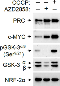
Treatment of cells with AZD2858 leads to the induction of both PRC and c-MYC under conditions where the NRF-2α control is unaffected. Human log phase U2OS cells are plated and treated with either vehicle, the GSK-3 inhibitor, AZD2858, or with CCCP for 24 h. Total cell extracts are subjected to immunoblotting using rabbit anti-PRC(1047-1379), mouse anti-c-MYC, rabbit anti-pGSK-3α/β(Ser9/21), rabbit anti-GSK-3α/β or rabbit anti-NRF-2α as the control.
-
J Cell Physiol
PLCε knockdown overcomes drug resistance to androgen receptor antagonist in castration-resistant prostate cancer by suppressing the wnt3a/β-catenin pathway. [Abstract]2019 Sep;234(9):15472-15486. PMID: 30684266
AZD2858 purchased from MedChemExpress. Usage Cited in: J Cell Physiol. 2019 Sep;234(9):15472-15486. [Abstract]
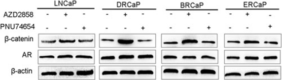
Western blot analysis was used to confirm the total protein expression of β-catenin and AR in the LNCaP, DRCaP, BRCaP, and ERCaP cells treated with the wnt/β-catenin activator (AZD2858, 5 nM, 48 hr) or the wnt/β-catenin inhibitor (PNU74654, 129.8 μM, 48 hr).
AZD2858 purchased from MedChemExpress. Usage Cited in: J Cell Physiol. 2019 Sep;234(9):15472-15486. [Abstract]
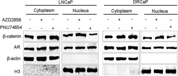
Western blot analysis was used to confirm the cytoplasmic and nuclear protein expression of β-catenin and AR in the LNCaP, DRCaP, BRCaP, and ERCaP cells treated with the wnt/β-catenin activator (AZD2858, 5 nM, 48 hr) or the wnt/β-catenin inhibitor (PNU74654, 129.8 μM, 48 hr).
-


Solvent & Solubility
DMSO : 12.5 mg/mL (27.56 mM; Need ultrasonic; Hygroscopic DMSO has a significant impact on the solubility of product, please use newly opened DMSO)
Please refer to the solubility information to select the appropriate solvent. Once prepared, please aliquot and store the solution to prevent product inactivation from repeated freeze-thaw cycles.
Storage method and period of stock solution: -80°C, 1 year; -20°C, 6 months. When stored at -80°C, please use it within 1 year. When stored at -20°C, please use it within 6 months.
Please refer to the solubility information to select the appropriate solvent. Once prepared, please aliquot and store the solution to prevent product inactivation from repeated freeze-thaw cycles.
Storage method and period of stock solution: -80°C, 1 year; -20°C, 6 months. When stored at -80°C, please use it within 1 year. When stored at -20°C, please use it within 6 months.
Concentration (start) × Volume (start) = Concentration (final) × Volume (final)
Select the appropriate dissolution method based on your experimental animal and administration route.
- For the following dissolution methods, please ensure to first prepare a clear stock solution using an In Vitro approach and then sequentially add co-solvents:
- To ensure reliable experimental results, the clarified stock solution can be appropriately stored based on storage conditions. As for the working solution for In Vivo experiments, it is recommended to prepare freshly and use it on the same day.
- The percentages shown for the solvents indicate their volumetric ratio in the final prepared solution. If precipitation or phase separation occurs during preparation, heat and/or sonication can be used to aid dissolution.
Add each solvent one by one: 10% DMSO 40% PEG300 5% Tween-80 45% Saline
Solubility: 1.25 mg/mL (2.76 mM); Suspended solution; Need ultrasonic
This protocol yields a suspended solution of 1.25 mg/mL. Suspended solution can be used for oral and intraperitoneal injection.
Taking 1 mL working solution as an example, add 100 μL DMSO stock solution (12.5 mg/mL) to 400 μL PEG300, and mix evenly; then add 50 μL Tween-80 and mix evenly; then add 450 μL Saline to adjust the volume to 1 mL.
Preparation of Saline: Dissolve 0.9 g sodium chloride in ddH₂O and dilute to 100 mL to obtain a clear Saline solution.
Add each solvent one by one: 10% DMSO 90% (20% SBE-β-CD in Saline)
Solubility: ≥ 1.25 mg/mL (2.76 mM); Clear solution
This protocol yields a clear solution of ≥ 1.25 mg/mL (saturation unknown).
Taking 1 mL working solution as an example, add 100 μL DMSO stock solution (12.5 mg/mL) to 900 μL 20% SBE-β-CD in Saline, and mix evenly.
Preparation of 20% SBE-β-CD in Saline (4°C, storage for one week): 2 g SBE-β-CD powder is dissolved in 10 mL Saline, completely dissolve until clear.
Please enter the basic information of animal experiments:
-
-
-
-
Recommended: Prepare an additional quantity of animals to account for potential losses during experiments.
Please enter your animal formula composition:
-
%DMSO +
Recommended: Keep the proportion of DMSO in working solution below 2% if your animal is weak.
-
%+
-
+%Tween-80 + +
-
%Saline +
The co-solvents required include: DMSO, . All of co-solvents are available by MedChemExpress (MCE). , Tween 80. All of co-solvents are available by MedChemExpress (MCE).
Working solution concentration: 0.22 mg/mL
Method for preparing stock solution: mg drug dissolved in μL DMSO. Stock solution concentration: mg/mL.
1. Take μL DMSO stock solution;
2. Add μL .
μL , mix evenly;
3. Then add μL Tween 80, mix evenly;
4. Then add μL
Please ensure that the stock solution in the first step is dissolved to a clear state, and add co-solvents in sequence. You can use ultrasonic heating (ultrasonic cleaner, recommended frequency 20-40 kHz), vortexing, etc. to assist dissolution.

Protocol
The potency of compounds at GSK-3β and cyclin-dependent protein kinase 2 (CDK2, kinase with closest homology to GSK-3β) is assessed using Z-LYTE™ Kinase assay kit in the presence of 7 and 80 μM ATP respectively. A ratiometric method is used to calculate the ratio of donor emission (445 nm) to acceptor emission (520 nm) after excitation of the donor fluorophore at 400 nm to quantitate the reaction progress. Kinase selectivity with AR79, AZD2858 and AZ13282107 are determined using the KinaseProfiler Service or University of Dundee Kinase. Over 80 different kinases are assessed at a single concentration of 1 or 10 μM of AR79, AZD2858 and AZ13282107. Concentration-inhibition 10-point curves to compounds that show activity are constructed to determine pIC50 estimations. Also, in some kinase assays these pIC50 estimations are converted to binding affinity values (pKi) using the Cheng-Prussoff equation to correct for the concentration of ATP used[3].
MedChemExpress (MCE) has not independently confirmed the accuracy of these methods. They are for reference only.
Each rat is dosed orally with vehicle or AZD2858 using a plastic gavage tube. The dose volume is 10 mL/kg. The vehicle consists of deionized water adjusted to pH 3.5±0.1. Formulations are adjusted to pH 3.5±0.1. The doses are 0, 0.2, 2 or 20 mg/kg respectively and administered either twice daily (TD), once daily (OD), every other day (O/2D), or every fourth day (O/4D) for 14 days. In total, the protocol results in 13 groups with 8 animals in each group (104 animals). At 7 days after the start of the study, and again 2 days prior to the scheduled terminal necropsy, each animal is injected subcutaneously with a bicarbonate buffered calcein solution (8 mg/kg, 1 mL/kg)[1].
MedChemExpress (MCE) has not independently confirmed the accuracy of these methods. They are for reference only.


Purity & Documentation
-
Data Sheet (281 KB)
-
SDS (644 KB)
- English - EN (644 KB)
- Français - FR (644 KB)
- Deutsch - DE (644 KB)
- Norwegian - NO (644 KB)
- Español - ES (644 KB)
- Swedish - SV (644 KB)
- Italian - IT (644 KB)
- Korean - KR (644 KB)
- Portuguese - PT (644 KB)
-
Handling Instructions (2659 KB)
References
[1]. Marsell R, et al. GSK-3 inhibition by an orally active small molecule increases bone mass in rats. Bone. 2012 Mar;50(3):619-27. [Content Brief]
[2]. Sisask G, et al. Rats treated with AZD2858, a GSK3 inhibitor, heal fractures rapidly without endochondral bone formation. Bone. 2013 May;54(1):126-32. [Content Brief]
[3]. Gilmour PS, et al. Human stem cell osteoblastogenesis mediated by novel glycogen synthase kinase 3 inhibitors induces bone formation and a unique bone turnover biomarker profile in rats. Toxicol Appl Pharmacol. 2013 Oct 15;272(2):399-407. [Content Brief]
[4]. Gambardella A, et al. Glycogen synthase kinase-3α/β inhibition promotes in vivo amplification of endogenous mesenchymal progenitors with osteogenic and adipogenic potential and their differentiation to the osteogenic lineage. J Bone Miner Res. 2011 Apr;26(4):811-21. [Content Brief]


Complete Stock Solution Preparation Table
Please refer to the solubility information to select the appropriate solvent. Once prepared, please aliquot and store the solution to prevent product inactivation from repeated freeze-thaw cycles.
Storage method and period of stock solution: -80°C, 1 year; -20°C, 6 months. When stored at -80°C, please use it within 1 year. When stored at -20°C, please use it within 6 months.
| Optional Solvent | Concentration Solvent Mass | 1 mg | 5 mg | 10 mg | 25 mg |
|---|---|---|---|---|---|
| DMSO | 1 mM | 2.2050 mL | 11.0249 mL | 22.0497 mL | 55.1244 mL |
| 5 mM | 0.4410 mL | 2.2050 mL | 4.4099 mL | 11.0249 mL | |
| 10 mM | 0.2205 mL | 1.1025 mL | 2.2050 mL | 5.5124 mL | |
| 15 mM | 0.1470 mL | 0.7350 mL | 1.4700 mL | 3.6750 mL | |
| 20 mM | 0.1102 mL | 0.5512 mL | 1.1025 mL | 2.7562 mL | |
| 25 mM | 0.0882 mL | 0.4410 mL | 0.8820 mL | 2.2050 mL |


AZD2858 Related Classifications
HY-15761 Related Classifications
 Powered by Bioz