



Spexin
Based on 2 publication(s) in Google Scholar
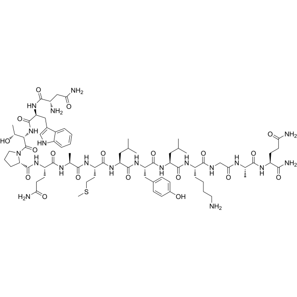
Spexin (Neuropeptide Q) is a selective agonist of galanin receptors GAL2 and GAL3, and is a conserved peptide that functions as a neurotransmitter/neuromodulator and endocrine factor. Spexin can function through both central and peripheral actions. Spexin upregulates Beclin 1 to inhibit ferroptosis induced by excessive autophagy, reduces the uptake of long-chain fatty acids by adipocytes, and regulates energy metabolism by increasing lipid oxidation (e.g., reducing the respiratory exchange ratio in rodents). Spexin improves cardiac function in the Doxorubicin hydrochloride (HY-15142)-induced cardiotoxicity model, protects mitochondrial membrane potential, and reduces iron accumulation and lipid peroxidation. Spexin can be used to study obesity and its related metabolic disorders, cardiovascular diseases (e.g., cardioprotection), and side effects of tumor chemotherapy.
For research use only. We do not sell to patients.
- Purity : 99.12%
- CAS No.: 1370290-58-6
- Formula: C74H114N20O19S
- Molecular Weight:1619.88
-
Storage:
Sealed storage, away from moisture and light.
Powder -80°C, 2 years , -20°C, 1 year* In solvent : -80°C, 6 months; -20°C, 1 month (sealed storage, away from moisture and light)

To place orders, for customer services and technical support, please contact: MedChemExpress USA
Tel: 609-228-6898 E-mail: [email protected] [email protected]


-
 Biological Activity
-
Chemical Information
-
Solvent & Solubility
- Protocols
- Purity & Documentation
- References
-
 Help & FAQs


-
HY-101986
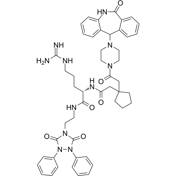
BIIE-0246NPY2R Antagonist -
HY-107725
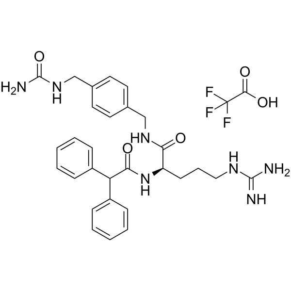
BIBO3304NPY Y1 Receptor Antagonist -
HY-14450
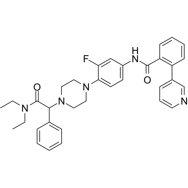
JNJ-31020028Y2 Receptor Antagonist -
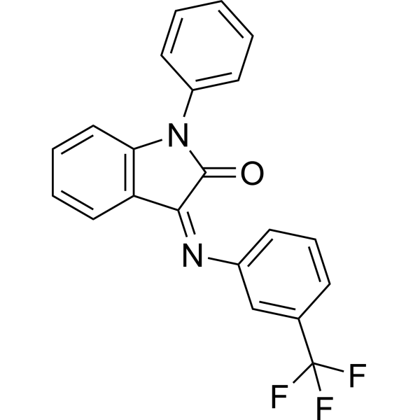
HY-100717
HT-2157Gal3 Antagonist -
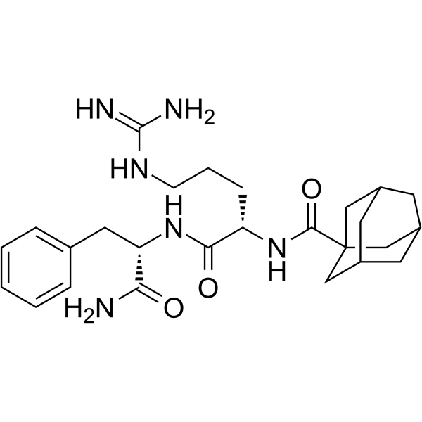
HY-107382
RF9Neuropeptide Y Receptor Antagonist -
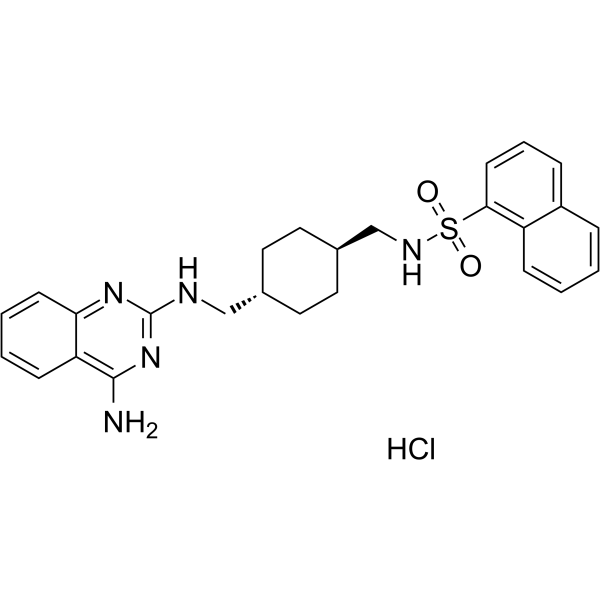
HY-107723
CGP71683 hydrochlorideNeuropeptide Receptor Antagonist -
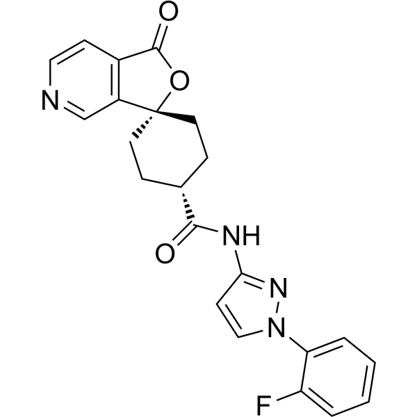
HY-15411
MK-0557Neuropeptide Y Receptor Antagonist -
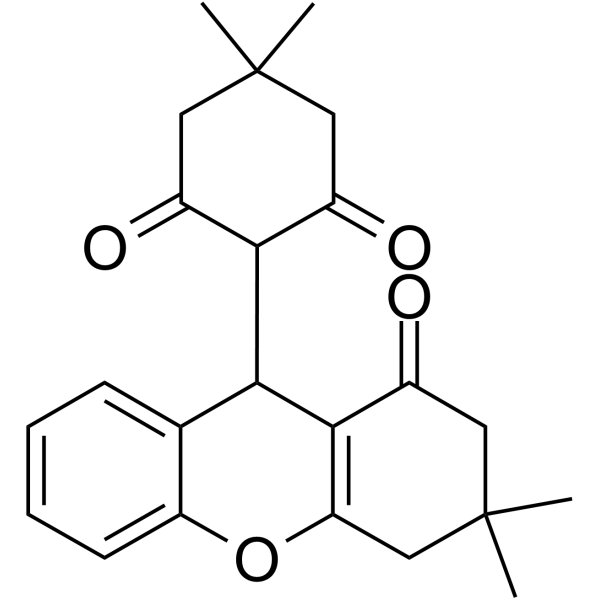
HY-107734
L 152804NPY Y5 Receptor Antagonist -
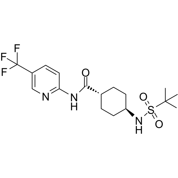
HY-14423
VelneperitNeuropeptide Y Receptor Antagonist -
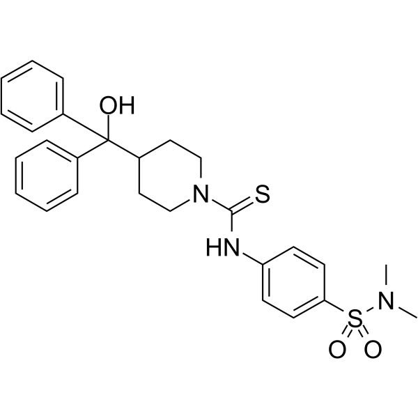
HY-107735
CYM 9484NPYY2 Receptor Antagonist -
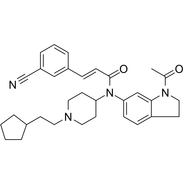
HY-107732
JNJ-5207787Neuropeptide Y Y2 Receptor -
HY-110107
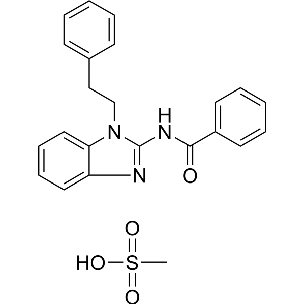
GW438014ANPY-Y5 receptor Antagonist
Publications Citing Use of MedChemExpress (MCE) Spexin
MoreAll Neuropeptide Y Receptor Isoforms
More

Biological Activity
Description
IC50 & Target
|
GAL2 |
GAL3 |
In Vitro
Spexin (1 μM; 24 h) increases viability and reduces apoptosis in neonatal rat cardiomyocytes (NRCs) treated with Doxorubicin (HY-15142A), reverses the loss of mitochondrial membrane potential and upregulates GPX4/SLC7A11[2].
Spexin (20 ng/mL; 2 h) inhibits long-chain fatty acid (LCFA) uptake in isolated mouse adipocytes[3].
MedChemExpress (MCE) has not independently confirmed the accuracy of these methods. They are for reference only.
-
Cell Line:Neonatal rat cardiomyocytes (NRCs)
-
Concentration:1 μM
-
Incubation Time:24 h; with or without 0.5 μM Doxorubicin
-
Result:Showed a 35% increase in viability compared to Doxorubicin-treated controls, with reduced LDH release.
In Vivo
Spexin (35 μg/kg/day; subcutaneous injection; 19 days) reduces food intake and weight gain in male Sprague-Dawley rats[3].
Spexin (25 μg/kg/day; ip; 10 days) significantly reduces nocturnal respiratory exchange ratio (RER) and increases exercise capacity in mice[3].
Spexin (70 μg/kg/day; ip; single dose) does not induce aversive effects in a conditioned taste aversion test in female rats[3].
MedChemExpress (MCE) has not independently confirmed the accuracy of these methods. They are for reference only.
-
Animal Model:C57BL/6 male mice (8 weeks old, 20-25 g) with Doxorubicin-induced cardiotoxicity model[2]
-
Dosage:50 μg/kg
-
Administration:Intraperitoneal injection (i.p.), daily for 4 weeks, starting 24 h after Doxorubicin hydrochloride injection (5 mg/kg/weeks, i.p., 4 weeks; with injectionson days 0, 7, 14 and 28)
-
Result:Cardiac Function: Echocardiography showed increased ejection fraction (EF) from 45 ± 5% to 58 ± 4% and fractional shortening (FS) from 22 ± 3% to 30 ± 3% in Spexin-treated group compared to doxorubicin control.
Oxidative Stress: Cardiac tissue showed reduced MDA levels (2.5 ± 0.3 nmol/mg protein vs. 3.8 ± 0.4 nmol/mg protein in control) and increased SOD activity (120 ± 10 U/mg protein vs. 90 ± 8 U/mg protein in control).
Histology: H&E staining revealed less myocardial damage and fibrosis in Spexin-treated hearts.
- Mitochondrial membrane-potential fluorescent assay
- Large-size fat particle sorting
- Fluorescent plasma-membrane potential dye assay
- Autophagy
- Mitochondrial membrane-potential and mitochondrial mass staining
- Ferroptosis Solutions
- Lysosome and acidic-vesicle live-cell staining
- Macroautophagy Solutions
- Research Protocol for Metabolic Diseases


Chemical Information
-
CAS No. 1370290-58-6
-
Appearance Solid
-
Molecular Weight 1619.88
-
Formula C74H114N20O19S
-
Color White to off-white
-
Synonyms
Neuropeptide Q
-
Sequence Shortening
NWTPQAMLYLKGAQ-NH2
-
Shipping
Room temperature in continental US; may vary elsewhere.
-
Storage
Sealed storage, away from moisture and light
Powder -80°C 2 years -20°C 1 year * In solvent : -80°C, 6 months; -20°C, 1 month (sealed storage, away from moisture and light)


Publications (2)
-
Journal Impact Factor
-
Most Recent
-
Br J Pharmacol
Spexin inhibits excessive autophagy-induced ferroptosis to alleviate doxorubicin-induced cardiotoxicity by upregulating Beclin 1. [Abstract]2024 Nov;181(21):4195-4213. PMID: 38961632 -
Front Med
Exogenous spexin aggravates renal ischemia reperfusion injury and triggers toxicity in healthy kidneys. [Abstract]2025 Oct;19(5):842-854. PMID: 41016963


Solvent & Solubility
In Vitro:
H2O : 25 mg/mL (15.43 mM; Need ultrasonic)
Please refer to the solubility information to select the appropriate solvent. Once prepared, please aliquot and store the solution to prevent product inactivation from repeated freeze-thaw cycles.
Storage method and period of stock solution: -80°C, 6 months; -20°C, 1 month (sealed storage, away from moisture and light). When stored at -80°C, please use it within 6 months. When stored at -20°C, please use it within 1 month.
* Note: If you choose water as the stock solution, please dilute it to the working solution, then filter and sterilize it with a 0.22 μm filter before use.
Please refer to the solubility information to select the appropriate solvent. Once prepared, please aliquot and store the solution to prevent product inactivation from repeated freeze-thaw cycles.
Storage method and period of stock solution: -80°C, 6 months; -20°C, 1 month (sealed storage, away from moisture and light). When stored at -80°C, please use it within 6 months. When stored at -20°C, please use it within 1 month.
* Note: If you choose water as the stock solution, please dilute it to the working solution, then filter and sterilize it with a 0.22 μm filter before use.
Concentration (start) × Volume (start) = Concentration (final) × Volume (final)


Purity & Documentation
-
Data Sheet (282 KB)
-
SDS (252 KB)
- English - EN (252 KB)
- Français - FR (252 KB)
- Deutsch - DE (252 KB)
- Norwegian - NO (252 KB)
- Español - ES (252 KB)
- Swedish - SV (252 KB)
- Italian - IT (252 KB)
- Korean - KR (252 KB)
- Portuguese - PT (252 KB)
-
Handling Instructions (2659 KB)
References
[1]. Porzionato A, et al. Spexin expression in normal rat tissues. J Histochem Cytochem. 2010;58(9):825-837. [Content Brief]
[2]. Ou W, et al. Spexin inhibits excessive autophagy-induced ferroptosis to alleviate doxorubicin-induced cardiotoxicity by upregulating Beclin 1. Br J Pharmacol. 2024 Nov;181(21):4195-4213. [Content Brief]
[3]. Walewski JL, et al. Spexin is a novel human peptide that reduces adipocyte uptake of long chain fatty acids and causes weight loss in rodents with diet-induced obesity. Obesity (Silver Spring). 2014 Jul;22(7):1643-52. [Content Brief]


Complete Stock Solution Preparation Table
Please refer to the solubility information to select the appropriate solvent. Once prepared, please aliquot and store the solution to prevent product inactivation from repeated freeze-thaw cycles.
Storage method and period of stock solution: -80°C, 6 months; -20°C, 1 month (sealed storage, away from moisture and light). When stored at -80°C, please use it within 6 months. When stored at -20°C, please use it within 1 month.
| Optional Solvent | Concentration Solvent Mass | 1 mg | 5 mg | 10 mg | 25 mg |
|---|---|---|---|---|---|
| H2O | 1 mM | 0.6173 mL | 3.0866 mL | 6.1733 mL | 15.4332 mL |
| 5 mM | 0.1235 mL | 0.6173 mL | 1.2347 mL | 3.0866 mL | |
| 10 mM | 0.0617 mL | 0.3087 mL | 0.6173 mL | 1.5433 mL | |
| 15 mM | 0.0412 mL | 0.2058 mL | 0.4116 mL | 1.0289 mL |
* Note: If you choose water as the stock solution, please dilute it to the working solution, then filter and sterilize it with a 0.22 μm filter before use.


Spexin Related Classifications
HY-P1723 Related Classifications
 Powered by Bioz