



Sensitive ECL Kit
Based on 8 publication(s) in Google Scholar
Sensitive ECL Kit enables low picogram detection of antigen by oxidizing luminol in the presence of HRP and peroxide. This reaction produces a prolonged chemiluminescence which can be visualized on X-ray film or digital imaging systems. The 100 mL is defined as the base specification. All larger sizes correspond to incremental volumes of this base.
-
Storage :
4°C, 1 year.
Protect from light.

To place orders, for customer services and technical support, please contact: MedChemExpress USA
Tel: 609-228-6898 E-mail: [email protected] [email protected]



-
HY-K1005
Ultra High Sensitivity ECL Kit -
HY-K2005
High Sensitivity ECL Kit -

HY-15371
ForskolinAdenylate Cyclase Activator -
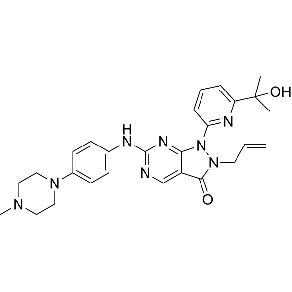
HY-10993
AdavosertibWee1 Inhibitor -
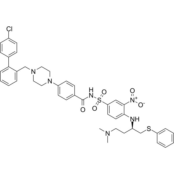
HY-50907
ABT-737Bcl-2 Family Inhibitor/BH3 Mimetic -
HY-K1027
Rapid Blocking Buffer (TBS-T) Powder (100 mL of 1×) -
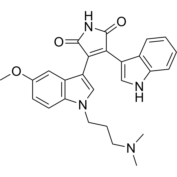
HY-13689
Go 6983PKC Inhibitor -
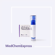
HY-K1100
SDS-PAGE Sample Loading Buffer (4×) -
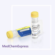
HY-K0022
Phosphatase Inhibitor Cocktail Ⅱ (100× in ddH2O) -
HY-K1022
PBS-T Powder (1 L of 1×) -
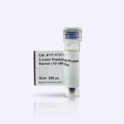
HY-K1011
3-Color Prestained Protein Marker (10-190 kDa)


Description & Advantages
MCE Sensitive ECL Kit enables low picogram detection of antigen by oxidizing luminol in the presence of HRP and peroxide. This reaction produces a prolonged chemiluminescence which can be visualized on X-ray film or digital imaging systems.
The Kit produces a strong, long-lived signal (6-8 hours), which, combined with very low background levels, allows for long exposure times and enables the detection of low-abundance proteins. In the Western Blot experiment, the storage solution of primary antibody (1 mg/mL) can be diluted at a ratio of 1:1,000-1:4,000 (the final concentration is about 0.25-1 μg/mL). The secondary antibody (1 mg/mL) storage solution can be diluted at a ratio of 1:5,000-1:10,000 (the final concentration is about 0.1-0.2 μg/mL).


Protocolo
1. Take the High Sensitivity ECL Kit out of the refrigerator and allow it to equilibrate at room temperature for about 20 minutes.
2. Prepare the ECL working solution.
Note: Approximately 0.125 mL of High Sensitivity ECL Reagent is required per cm2 of membrane area.
a). Mix solutions A and B in a 1:1 ratio. Use a in sufficient volume to ensure that the blot is completely wetted with the ECL working solution.
b). It is recommended to prepare the working solution just before use, although the mixed reagent solution is stable for several hours at room temperature.
3. Place the blot, protein-side up, in a clean container, and add ECL working solution.
4. Incubate the blot for 3-5 minutes at room temperature.
5. Drain the excess substrate and place the blot in a plastic membrane protector. Remove all air bubbles between the blot and the surface of the membrane protector.
6. Visualize the blot using X-ray film or a CCD-based imaging system.


Publications
-
Journal Impact Factor
-
Most Recent
-
Insect Biochem Mol Biol2026 Mar:188:104495. PMID: 41534801
Almacenamiento
4°C, 1 year.
Protect from light.


Componentes
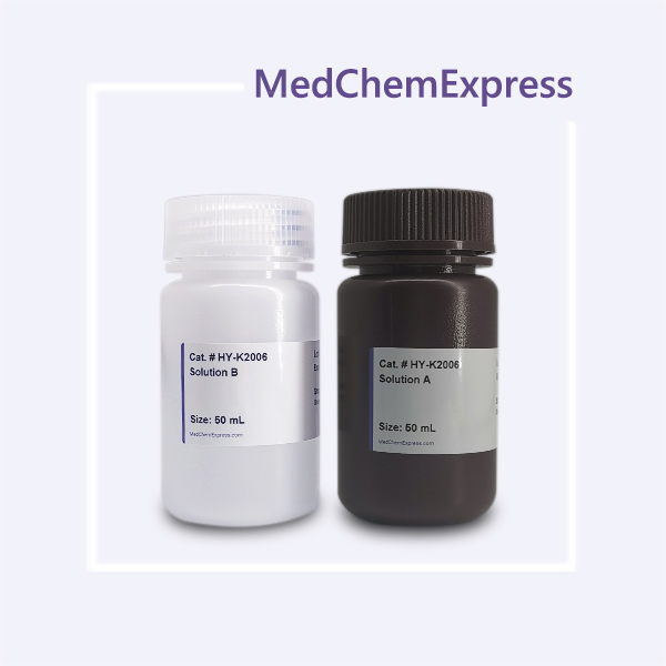
| Components | HY-K2006-100 mL | HY-K2006-500 mL |
|---|---|---|
| Solution A | 50 mL | 50 mL × 5 |
| Solution B | 50 mL | 50 mL × 5 |


 Please enter the correct Lot Number
