



MN58b
Based on 1 publication(s) in Google Scholar
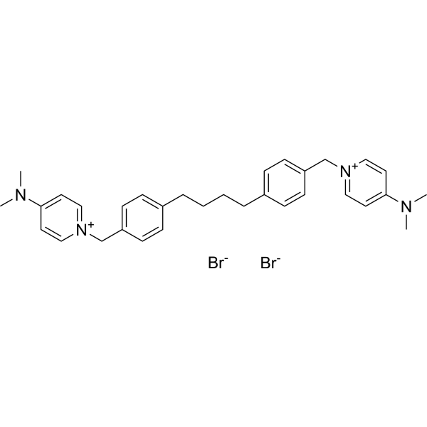
MN58b is a selective choline kinase α (CHKα) inhibitor, and results in inhibition of phosphocholine synthesis. MN58b reduces cell growth through the induction of apoptosis, and also has antitumoral activity.
Para uso exclusivo en investigación. No vendemos a pacientes.
- Pureza : 99.78%
- No. CAS: 203192-01-2
- Fòrmula: C32H40Br2N4
- Peso molecular:640.49
-
Almacenamiento:
-20°C, sealed storage, away from moisture
* In solvent : -80°C, 6 months; -20°C, 1 month (sealed storage, away from moisture)

To place orders, for customer services and technical support, please contact: MedChemExpress USA
Tel: 609-228-6898 E-mail: [email protected] [email protected]


-
 Actividad biológica
-
Chemical Information
-
Solvente y solubilidad
- Protocolo
- Pureza y Documentación
- Referencias
-
 Help & FAQs


-
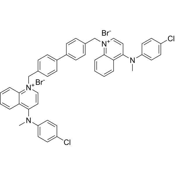
HY-101144
RSM-932AChoKα Inhibitor -
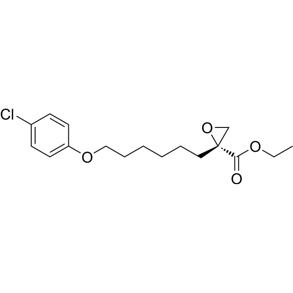
HY-50202
EtomoxirCPT1a Inhibitor -
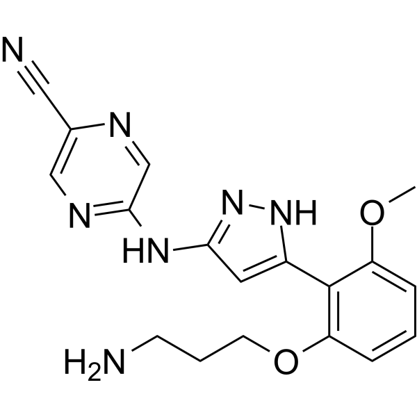
HY-18174
PrexasertibCHK1 Inhibitor -
HY-10992
AZD-7762Chk Inhibitor -
HY-14720
RabusertibCHK1 Inhibitor -
HY-18958
CCT245737CHK1 Inhibitor -
HY-10032
PF 477736Chk1 Inhibitor -
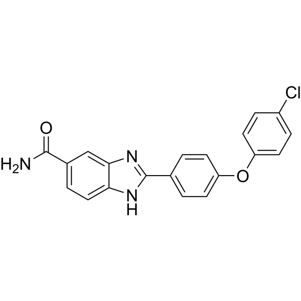
HY-13946
BML-277 -
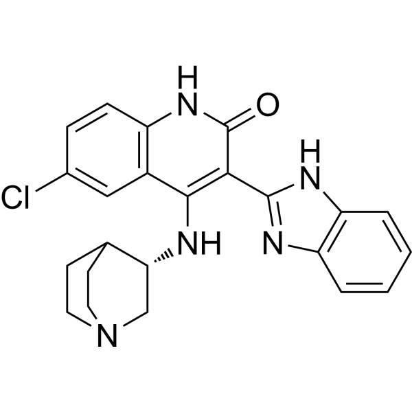
HY-13263
CHIR-124Checkpoint Kinase (Chk) Inhibitor -
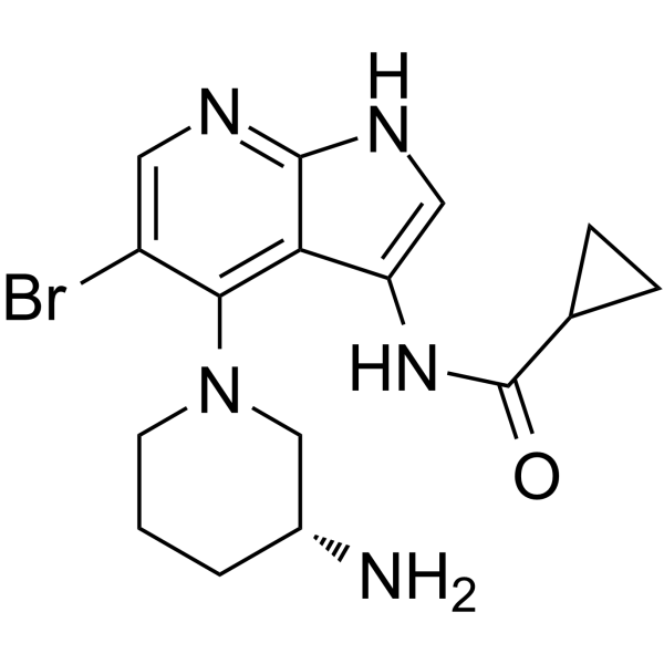
HY-112167
GDC-0575Chk1 Inhibitor -
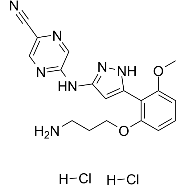
HY-18174A
Prexasertib dihydrochlorideCHK1 Inhibitor -
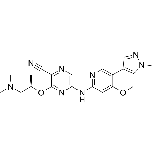
HY-18175
CCT244747
Publications Citing Use of MedChemExpress (MCE) MN58b
More

Actividad biológica
Descripciòn
IC50 & Target
Choline kinase α (CHKα)[1]
Cellular Effect
|
Cell Line

|
Type | Value | Description | References |
|---|---|---|---|---|
| CCRF-CEM | GI50 |
0.21 μM
Compound: MN58b
|
Antiproliferative activity against human CCRF-CEM cells assessed as inhibition of cell proliferation
Antiproliferative activity against human CCRF-CEM cells assessed as inhibition of cell proliferation
|
[PMID: 32977218] |
| HeLa | GI50 |
1.9 μM
Compound: MN58b
|
Antiproliferative activity against human HeLa cells assessed as inhibition of cell proliferation
Antiproliferative activity against human HeLa cells assessed as inhibition of cell proliferation
|
[PMID: 32977218] |
| HT-29 | GI50 |
1.9 μM
Compound: MN58b
|
Antiproliferative activity against human HT-29 cells assessed as inhibition of cell proliferation
Antiproliferative activity against human HT-29 cells assessed as inhibition of cell proliferation
|
[PMID: 32977218] |
| RS4-11 | GI50 |
1 μM
Compound: MN58b
|
Antiproliferative activity against human RS4-11 cells assessed as inhibition of cell proliferation
Antiproliferative activity against human RS4-11 cells assessed as inhibition of cell proliferation
|
[PMID: 32977218] |
In Vitro
The IC50s of MN58b for parental and Gemcitabine-resistant Suit2 007 cells are 3.14 μM and 0.77 μM, respectively[1].
?
MN58b (1-5 μM; 72 hours; SK-PC-1, Suit2 008, IMIM-PC2, and RWP-1 cells) has a marked effect on colony formation at 1 μM, and growth is completely abolished at 5 μM in all the cell lines[1].
?
MN58b ((1-10 μM; 24-48 hours; SK-PC-1, Suit2 008, IMIM-PC2, and RWP-1 cells) induces apoptosis and this response correlates with CHKα expression[1].
MedChemExpress (MCE) has not independently confirmed the accuracy of these methods. They are for reference only.
-
Cell Line:SK-PC-1, Suit2 008, IMIM-PC2, and RWP-1 cells
-
Concentration:1 µM, 5 µM
-
Incubation Time:72 hours
-
Result:Inhibited cells growth.
-
Cell Line:SK-PC-1, Suit2 008, IMIM-PC2, and RWP-1 cells
-
Concentration:1 µM, 2 µM, 5 µM, 10 µM
-
Incubation Time:24 and 48 hours
-
Result:Induced cell apoptosis.
In Vivo
MedChemExpress (MCE) has not independently confirmed the accuracy of these methods. They are for reference only.
-
Animal Model:MF-1 nude mice with HT29 or MDA-MB-231 cells[2]
-
Dosage:4 mg/kg
-
Administration:Intraperitoneal injection; once a day; for 5 days
-
Result:Phosphomonoesters decreased significantly.
- Detection of Apoptosis
- TUNEL staining for apoptotic DNA fragmentation
- Annexin V plus membrane-impermeant dye apoptosis staining
- Protocol for Cell Counting and Cell Density Analysis
- Somatic Cell Culture
- Apoptosis Solutions
- CCK-8/WST-8 Cell Proliferation Assay
- Cell Counting-Based Growth Curve Assay
- MTT Cell Proliferation Assay
- Cell Viability Determination by MTT Colorimetric Assay


Chemical Information
-
No. CAS 203192-01-2
-
Appearance Solid
-
Peso molecular 640.49
-
Fòrmula C32H40Br2N4
-
Color White to off-white
-
SMILES
CN(C)C1=CC=[N+](C=C1)CC2=CC=C(CCCCC3=CC=C(C[N+]4=CC=C(N(C)C)C=C4)C=C3)C=C2.[Br-].[Br-]
-
Envío
Room temperature in continental US; may vary elsewhere.
-
Almacenamiento
-20°C, sealed storage, away from moisture
* In solvent : -80°C, 6 months; -20°C, 1 month (sealed storage, away from moisture)


Publications (1)
-
Journal Impact Factor
-
Most Recent
-
Nat Commun
2025 Mar 13;16(1):2221. PMID: 40082403


Solvente y solubilidad
In Vitro:
DMSO : 14.71 mg/mL (22.97 mM; Need ultrasonic; Hygroscopic DMSO has a significant impact on the solubility of product, please use newly opened DMSO)
Please refer to the solubility information to select the appropriate solvent. Once prepared, please aliquot and store the solution to prevent product inactivation from repeated freeze-thaw cycles.
Storage method and period of stock solution: -80°C, 6 months; -20°C, 1 month (sealed storage, away from moisture). When stored at -80°C, please use it within 6 months. When stored at -20°C, please use it within 1 month.
Please refer to the solubility information to select the appropriate solvent. Once prepared, please aliquot and store the solution to prevent product inactivation from repeated freeze-thaw cycles.
Storage method and period of stock solution: -80°C, 6 months; -20°C, 1 month (sealed storage, away from moisture). When stored at -80°C, please use it within 6 months. When stored at -20°C, please use it within 1 month.
Concentration (start) × Volume (start) = Concentration (final) × Volume (final)
In Vivo:
Select the appropriate dissolution method based on your experimental animal and administration route.
- For the following dissolution methods, please ensure to first prepare a clear stock solution using an In Vitro approach and then sequentially add co-solvents:
- To ensure reliable experimental results, the clarified stock solution can be appropriately stored based on storage conditions. As for the working solution for In Vivo experiments, it is recommended to prepare freshly and use it on the same day.
- The percentages shown for the solvents indicate their volumetric ratio in the final prepared solution. If precipitation or phase separation occurs during preparation, heat and/or sonication can be used to aid dissolution.
Add each solvent one by one: 10% DMSO 40% PEG300 5% Tween-80 45% Saline
Solubility: ≥ 1.47 mg/mL (2.30 mM); Clear solution
This protocol yields a clear solution of ≥ 1.47 mg/mL (saturation unknown).
Taking 1 mL working solution as an example, add 100 μL DMSO stock solution (14.7 mg/mL) to 400 μL PEG300, and mix evenly; then add 50 μL Tween-80 and mix evenly; then add 450 μL Saline to adjust the volume to 1 mL.
Preparation of Saline: Dissolve 0.9 g sodium chloride in ddH₂O and dilute to 100 mL to obtain a clear Saline solution.
Add each solvent one by one: 10% DMSO 90% (20% SBE-β-CD in Saline)
Solubility: ≥ 1.47 mg/mL (2.30 mM); Clear solution
This protocol yields a clear solution of ≥ 1.47 mg/mL (saturation unknown).
Taking 1 mL working solution as an example, add 100 μL DMSO stock solution (14.7 mg/mL) to 900 μL 20% SBE-β-CD in Saline, and mix evenly.
Preparation of 20% SBE-β-CD in Saline (4°C, storage for one week): 2 g SBE-β-CD powder is dissolved in 10 mL Saline, completely dissolve until clear.
In Vivo Dissolution Calculator
Please enter the basic information of animal experiments:
-
-
-
-
Recommended: Prepare an additional quantity of animals to account for potential losses during experiments.
Please enter your animal formula composition:
-
%DMSO +
Recommended: Keep the proportion of DMSO in working solution below 2% if your animal is weak.
-
%+
-
+%Tween-80 + +
-
%Saline +
The co-solvents required include: DMSO, . All of co-solvents are available by MedChemExpress (MCE). , Tween 80. All of co-solvents are available by MedChemExpress (MCE).
Working solution concentration: 0.22 mg/mL
Method for preparing stock solution: mg drug dissolved in μL DMSO. Stock solution concentration: mg/mL. * In solvent : -80°C, 6 months; -20°C, 1 month (sealed storage, away from moisture)
1. Take μL DMSO stock solution;
2. Add μL .
μL , mix evenly;
3. Then add μL Tween 80, mix evenly;
4. Then add μL
Please ensure that the stock solution in the first step is dissolved to a clear state, and add co-solvents in sequence. You can use ultrasonic heating (ultrasonic cleaner, recommended frequency 20-40 kHz), vortexing, etc. to assist dissolution.


Pureza y Documentación
-
Ficha de datos (272 KB)
-
SDS (393 KB)
- English - EN (393 KB)
- Français - FR (393 KB)
- Deutsch - DE (393 KB)
- Norwegian - NO (393 KB)
- Español - ES (393 KB)
- Swedish - SV (393 KB)
- Italian - IT (393 KB)
- Korean - KR (393 KB)
- Portuguese - PT (393 KB)
-
Instrucciones de manejo (2659 KB)
Referencias
[1]. Mazarico JM, et al. Choline Kinase Alpha (CHKα) as a Therapeutic Target in Pancreatic Ductal Adenocarcinoma: Expression, Predictive Value, and Sensitivity to Inhibitors. Mol Cancer Ther. 2016 Feb;15(2):323-33. [Content Brief]
[2]. Al-Saffar NM, et al. Noninvasive magnetic resonance spectroscopic pharmacodynamic markers of the choline kinase inhibitor MN58b in human carcinoma models. Cancer Res. 2006 Jan 1;66(1):427-34. [Content Brief]


Complete Stock Solution Preparation Table
Please refer to the solubility information to select the appropriate solvent. Once prepared, please aliquot and store the solution to prevent product inactivation from repeated freeze-thaw cycles.
Storage method and period of stock solution: -80°C, 6 months; -20°C, 1 month (sealed storage, away from moisture). When stored at -80°C, please use it within 6 months. When stored at -20°C, please use it within 1 month.
| Optional Solvent | Concentration Solvent Mass | 1 mg | 5 mg | 10 mg | 25 mg |
|---|---|---|---|---|---|
| DMSO | 1 mM | 1.5613 mL | 7.8065 mL | 15.6130 mL | 39.0326 mL |
| 5 mM | 0.3123 mL | 1.5613 mL | 3.1226 mL | 7.8065 mL | |
| 10 mM | 0.1561 mL | 0.7807 mL | 1.5613 mL | 3.9033 mL | |
| 15 mM | 0.1041 mL | 0.5204 mL | 1.0409 mL | 2.6022 mL | |
| 20 mM | 0.0781 mL | 0.3903 mL | 0.7807 mL | 1.9516 mL |


MN58b Related Classifications
HY-108431 Related Classifications
 Powered by Bioz