



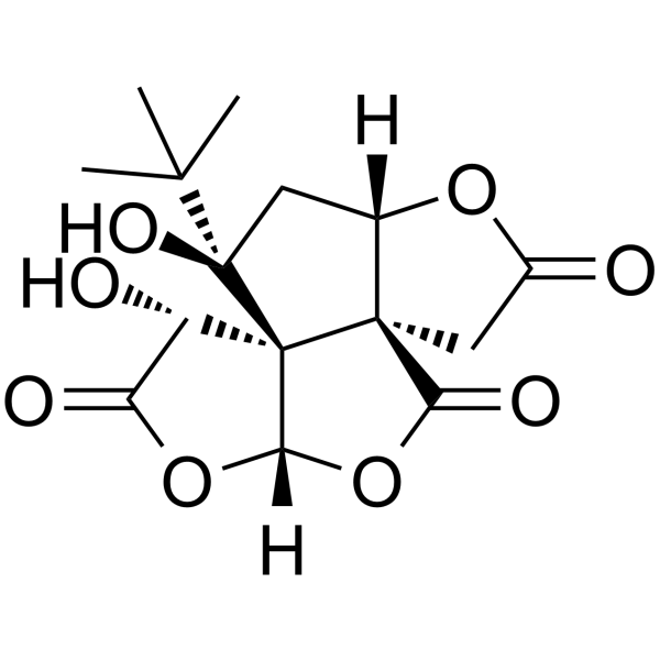
Bilobalide
Based on 3 publication(s) in Google Scholar
Bilobalide, a sesquiterpene trilactone constituent of Ginkgo biloba, inhibits the NMDA-induced efflux of choline with an IC50 value of 2.3 μM. Bilobalide prevents apoptosis through activation of the PI3K/Akt pathway in SH-SY5Y cells. Exerts protective and trophic effects on neurons.
For research use only. We do not sell to patients.
- Purity : 99.92%
- CAS No.: 33570-04-6
- Formula: C15H18O8
- Molecular Weight:326.30
-
Storage:Powder -20°C, 3 years , 4°C, 2 years ; In solvent -80°C, 2 years , -20°C, 1 year

To place orders, for customer services and technical support, please contact: MedChemExpress USA
Tel: 609-228-6898 E-mail: [email protected] [email protected]


-
 Biological Activity
-
Chemical Information
-
Solvent & Solubility
- Protocols
- Purity & Documentation
- References
-
 Help & FAQs


-
HY-N0784
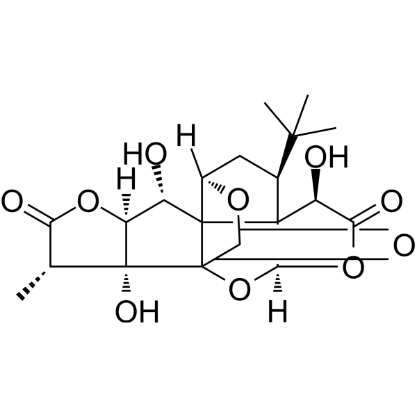
Ginkgolide BPlatelet Activating Factor Antagonist -
HY-B0355
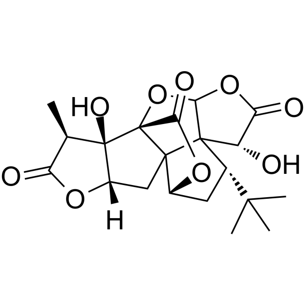
Ginkgolide AGABA Antagonist -

HY-N0076R
Bilobalide (Standard)Neuroprotective Sesquiterpene -
HY-100218A
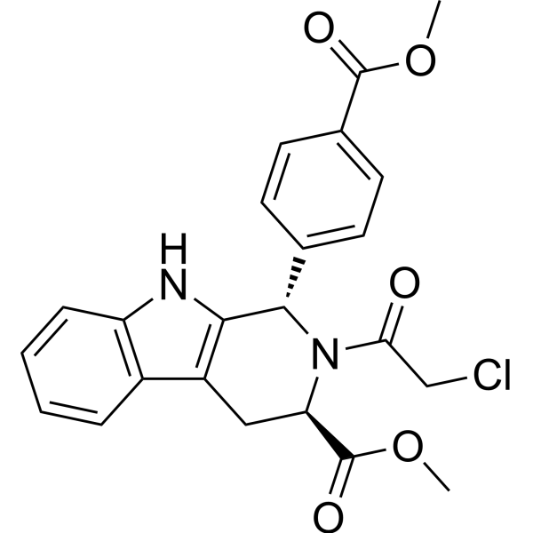
RSL3GPX4 Inhibitor -
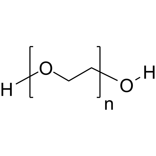
HY-Y0873
PEG300Neutral Polymer -
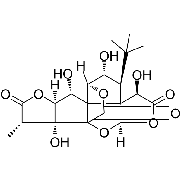
HY-N0785
Ginkgolide CFlavone -
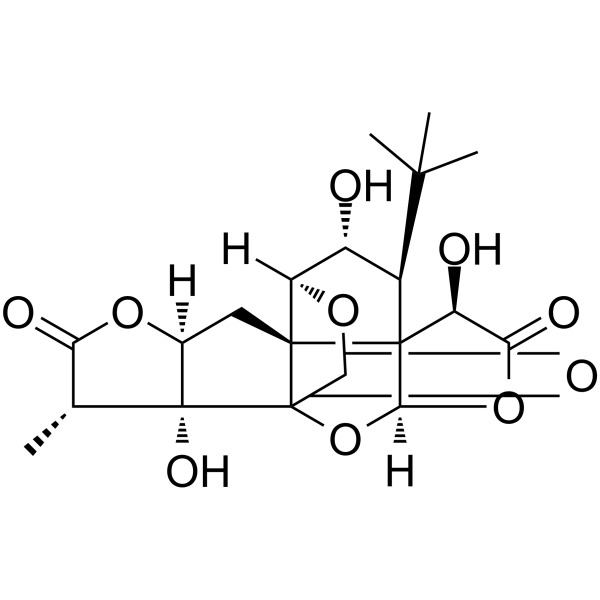
HY-N0786
Ginkgolide JNeuroprotective Agent -
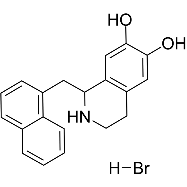
HY-15477
YS-49PI3K/Akt Activator -
HY-N0036
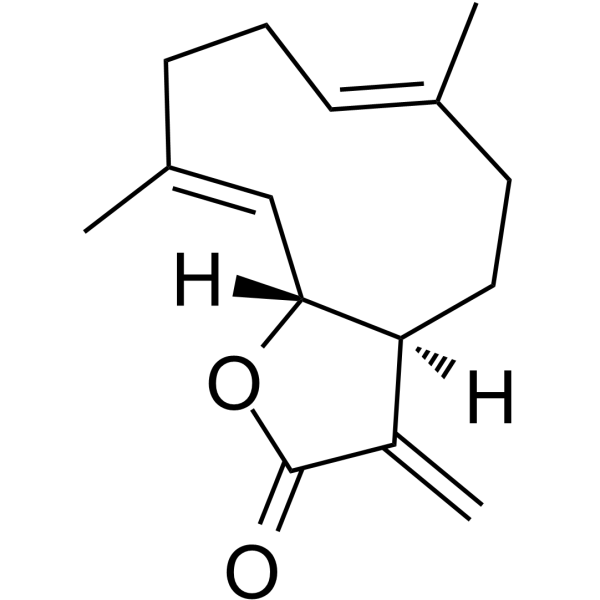
CostunolideApoptosis inducer -
HY-N0271
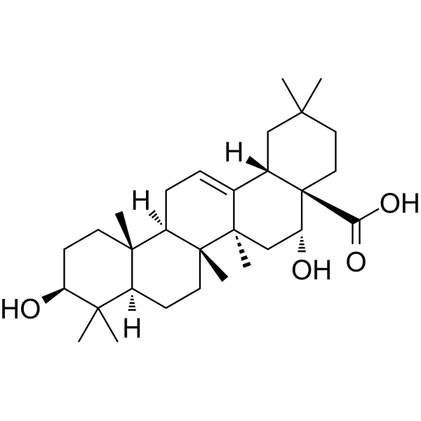
Echinocystic acidAntioxidant/Anti-inflammatory Agent -

HY-N0036R
Costunolide (Standard)Apoptosis inducer
Publications Citing Use of MedChemExpress (MCE) Bilobalide
More Customer Validation & Images

-
RT-PCR
-
WB
-
IF
-
Cell Proliferation/Viability Assay
-
ELISA
All Endogenous Metabolite Isoforms
More

Biological Activity
Description
IC50 & Target
|
Human Endogenous Metabolite |
In Vitro
Bilobalide (1-100 μM) completely suppresses the NMDA-evoked release of choline in a concentration-dependent manner with IC50 value of 2.3 μM[1].
Bilobalide (1, 5 and 10 μM) alone for 24 h does not affect cell viability of SH-SY5Y cells. Pre-treatment of cells with Bilobalide concentration-dependently prevents Aβ 1-42-, H2O2- and serum deprivation-induced decrease of cell viability, with the best protective effect obtained at 10 μM[2].
Bilobalide (5 and 10 μM; 24 h) treatment dose-dependently increases levels of p-Akt (Ser473 and Thr308) in SH-SY5Y cells[2].
MedChemExpress (MCE) has not independently confirmed the accuracy of these methods. They are for reference only.
-
Cell Line:SH-SY5Y cells
-
Concentration:5 and 10 μM
-
Incubation Time:24 hours
-
Result:Induced a significant increase in levels of p-Akt (Ser473 and Thr308).
In Vivo
MedChemExpress (MCE) has not independently confirmed the accuracy of these methods. They are for reference only.
-
Animal Model:Male Wistar rats (250-350 g)[1]
-
Dosage:20 mg/kg
-
Administration:I.p. injection 60 min before NMDA infusion
-
Result:Lowered basal choline efflux only slightly (by 7%) but fully antagonized the NMDA-induced increase of choline release. The convulsive effect of NMDA was almost completely suppressed.


Chemical Information
-
CAS No. 33570-04-6
-
Appearance Solid
-
Molecular Weight 326.30
-
Formula C15H18O8
-
Color White to off-white
-
SMILES
O=C(O1)C[C@@]2([C@]1([H])C[C@]3(C(C)(C)C)O)[C@@]34[C@](OC([C@@H]4O)=O)([H])OC2=O
-
Synonyms
(-)-Bilobalide
-
Structure Classification
-
Initial Source
-
Shipping
Room temperature in continental US; may vary elsewhere.
-
Storage
Powder -20°C 3 years 4°C 2 years In solvent -80°C 2 years -20°C 1 year


Publications (3)
-
Journal Impact Factor
-
Most Recent
-
-
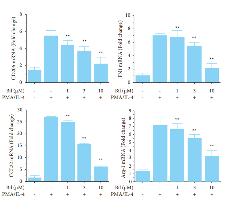
Bilobalide purchased from MedChemExpress. Usage Cited in: Eur J Cancer Care (Engl). 2024 Apr 4.
Bilobalide (1, 3, 10 μM) signifcantly reduced IL-10 and Arg-1 expression in a dose-dependent manner. Similar to the RT-qPCR analysis.
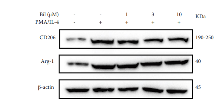
Bilobalide purchased from MedChemExpress. Usage Cited in: Eur J Cancer Care (Engl). 2024 Apr 4.
Bilobalide (1, 3, 10 μM) signifcantly decreased the levels of M2 markers by Western blotting.
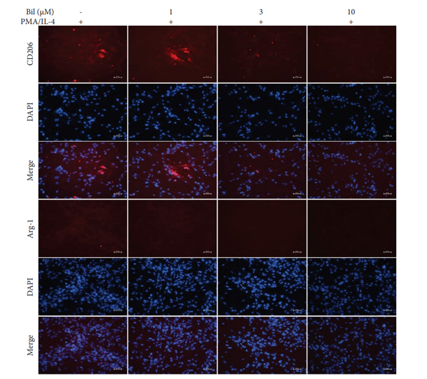
Bilobalide purchased from MedChemExpress. Usage Cited in: Eur J Cancer Care (Engl). 2024 Apr 4.
Bilobalide (1, 3, 10 μM) signifcantly decreased the levels of M2 markers by immunofuorescence.
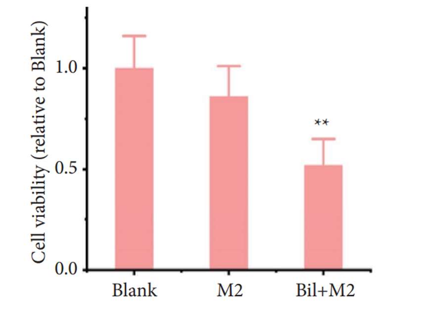
Bilobalide purchased from MedChemExpress. Usage Cited in: Eur J Cancer Care (Engl). 2024 Apr 4.
The viability of the CRC organoids was signifcantly reduced in the Bilobalide (10 μM)-treated group compared to the blank or M2 groups.
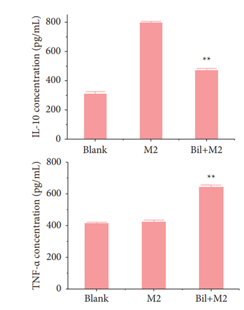
Bilobalide purchased from MedChemExpress. Usage Cited in: Eur J Cancer Care (Engl). 2024 Apr 4.
Bilobalide (10 μM) treatment (Bil + M2 group) caused a decrease and increase in IL-10 and TNF-α concentration, respectively by ELISA.
-


Solvent & Solubility
In Vitro:
DMSO : ≥ 100 mg/mL (306.47 mM; Hygroscopic DMSO has a significant impact on the solubility of product, please use newly opened DMSO)
* "≥" means soluble, but saturation unknown.
Please refer to the solubility information to select the appropriate solvent. Once prepared, please aliquot and store the solution to prevent product inactivation from repeated freeze-thaw cycles.
Storage method and period of stock solution: -80°C, 2 years; -20°C, 1 year. When stored at -80°C, please use it within 2 years. When stored at -20°C, please use it within 1 year.
Please refer to the solubility information to select the appropriate solvent. Once prepared, please aliquot and store the solution to prevent product inactivation from repeated freeze-thaw cycles.
Storage method and period of stock solution: -80°C, 2 years; -20°C, 1 year. When stored at -80°C, please use it within 2 years. When stored at -20°C, please use it within 1 year.
Concentration (start) × Volume (start) = Concentration (final) × Volume (final)
In Vivo:
Select the appropriate dissolution method based on your experimental animal and administration route.
- For the following dissolution methods, please ensure to first prepare a clear stock solution using an In Vitro approach and then sequentially add co-solvents:
- To ensure reliable experimental results, the clarified stock solution can be appropriately stored based on storage conditions. As for the working solution for In Vivo experiments, it is recommended to prepare freshly and use it on the same day.
- The percentages shown for the solvents indicate their volumetric ratio in the final prepared solution. If precipitation or phase separation occurs during preparation, heat and/or sonication can be used to aid dissolution.
Add each solvent one by one: 10% DMSO 40% PEG300 5% Tween-80 45% Saline
Solubility: ≥ 2.08 mg/mL (6.37 mM); Clear solution
This protocol yields a clear solution of ≥ 2.08 mg/mL (saturation unknown).
Taking 1 mL working solution as an example, add 100 μL DMSO stock solution (20.8 mg/mL) to 400 μL PEG300, and mix evenly; then add 50 μL Tween-80 and mix evenly; then add 450 μL Saline to adjust the volume to 1 mL.
Preparation of Saline: Dissolve 0.9 g sodium chloride in ddH₂O and dilute to 100 mL to obtain a clear Saline solution.
Add each solvent one by one: 10% DMSO 90% (20% SBE-β-CD in Saline)
Solubility: ≥ 2.08 mg/mL (6.37 mM); Clear solution
This protocol yields a clear solution of ≥ 2.08 mg/mL (saturation unknown).
Taking 1 mL working solution as an example, add 100 μL DMSO stock solution (20.8 mg/mL) to 900 μL 20% SBE-β-CD in Saline, and mix evenly.
Preparation of 20% SBE-β-CD in Saline (4°C, storage for one week): 2 g SBE-β-CD powder is dissolved in 10 mL Saline, completely dissolve until clear.
In Vivo Dissolution Calculator
Please enter the basic information of animal experiments:
-
-
-
-
Recommended: Prepare an additional quantity of animals to account for potential losses during experiments.
Please enter your animal formula composition:
-
%DMSO +
Recommended: Keep the proportion of DMSO in working solution below 2% if your animal is weak.
-
%+
-
+%Tween-80 + +
-
%Saline +
The co-solvents required include: DMSO, . All of co-solvents are available by MedChemExpress (MCE). , Tween 80. All of co-solvents are available by MedChemExpress (MCE).
Working solution concentration: 0.22 mg/mL
Method for preparing stock solution: mg drug dissolved in μL DMSO. Stock solution concentration: mg/mL.
1. Take μL DMSO stock solution;
2. Add μL .
μL , mix evenly;
3. Then add μL Tween 80, mix evenly;
4. Then add μL
Please ensure that the stock solution in the first step is dissolved to a clear state, and add co-solvents in sequence. You can use ultrasonic heating (ultrasonic cleaner, recommended frequency 20-40 kHz), vortexing, etc. to assist dissolution.


Purity & Documentation
-
Data Sheet (285 KB)
-
SDS (393 KB)
- English - EN (393 KB)
- Français - FR (393 KB)
- Deutsch - DE (393 KB)
- Norwegian - NO (393 KB)
- Español - ES (393 KB)
- Swedish - SV (393 KB)
- Italian - IT (393 KB)
- Korean - KR (393 KB)
- Portuguese - PT (393 KB)
-
Handling Instructions (2659 KB)
References
[1]. O Weichel, et al. Bilobalide, a constituent of Ginkgo biloba, inhibits NMDA-induced phospholipase A2 activation and phospholipid breakdown in rat hippocampus. Naunyn Schmiedebergs Arch Pharmacol. 1999 Dec;360(6):609-15. [Content Brief]
[2]. Chun Shi, et al. Bilobalide prevents apoptosis through activation of the PI3K/Akt pathway in SH-SY5Y cells. Apoptosis. 2010 Jun;15(6):715-27. [Content Brief]


Complete Stock Solution Preparation Table
Please refer to the solubility information to select the appropriate solvent. Once prepared, please aliquot and store the solution to prevent product inactivation from repeated freeze-thaw cycles.
Storage method and period of stock solution: -80°C, 2 years; -20°C, 1 year. When stored at -80°C, please use it within 2 years. When stored at -20°C, please use it within 1 year.
| Optional Solvent | Concentration Solvent Mass | 1 mg | 5 mg | 10 mg | 25 mg |
|---|---|---|---|---|---|
| DMSO | 1 mM | 3.0647 mL | 15.3233 mL | 30.6466 mL | 76.6166 mL |
| 5 mM | 0.6129 mL | 3.0647 mL | 6.1293 mL | 15.3233 mL | |
| 10 mM | 0.3065 mL | 1.5323 mL | 3.0647 mL | 7.6617 mL | |
| 15 mM | 0.2043 mL | 1.0216 mL | 2.0431 mL | 5.1078 mL | |
| 20 mM | 0.1532 mL | 0.7662 mL | 1.5323 mL | 3.8308 mL | |
| 25 mM | 0.1226 mL | 0.6129 mL | 1.2259 mL | 3.0647 mL | |
| 30 mM | 0.1022 mL | 0.5108 mL | 1.0216 mL | 2.5539 mL | |
| 40 mM | 0.0766 mL | 0.3831 mL | 0.7662 mL | 1.9154 mL | |
| 50 mM | 0.0613 mL | 0.3065 mL | 0.6129 mL | 1.5323 mL | |
| 60 mM | 0.0511 mL | 0.2554 mL | 0.5108 mL | 1.2769 mL | |
| 80 mM | 0.0383 mL | 0.1915 mL | 0.3831 mL | 0.9577 mL | |
| 100 mM | 0.0306 mL | 0.1532 mL | 0.3065 mL | 0.7662 mL |


Bilobalide Related Classifications
HY-N0076 Related Classifications
 Powered by Bioz