



BI-0474

Based on 1 Customer Validation
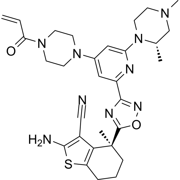
BI-0474 is a potent KRASG12C inhibitor with an IC50 value of 7.0 nM for the GDP-KRAS::SOS1 protein-protein interaction. BI-0474 exhibits good anti-proliferative activity against NCI-H358 cells carrying the G12C mutation. BI-0474 also shows good anti-tumour activity in non-small cell lung cancer xenograft models.
For research use only. We do not sell to patients.
The BI-0474 was designed by Boehringer Ingelheim and could be obtained free of charge through the Boehringer Ingelheim open innovation portal opnMe.com, associated with its negative control.
- Purity : 99.83%
- CAS No.: 2750570-55-7
- Formula: C30H37N9O2S
- Molecular Weight:587.74
-
Storage:Powder -20°C, 3 years , 4°C, 2 years ; In solvent -80°C, 6 months , -20°C, 1 month

To place orders, for customer services and technical support, please contact: MedChemExpress USA
Tel: 609-228-6898 E-mail: [email protected] [email protected]


-
 Biological Activity
-
Chemical Information
-
Solvent & Solubility
- Protocols
- Purity & Documentation
- References
-
 Help & FAQs


-
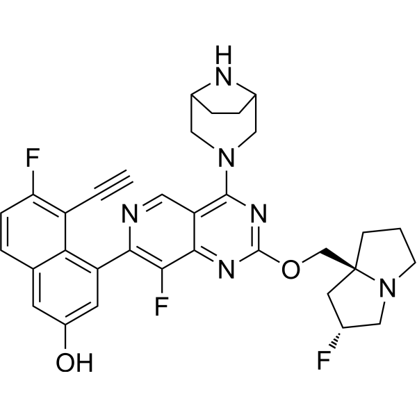
HY-134813
MRTX1133KRAS G12D Inhibitor -
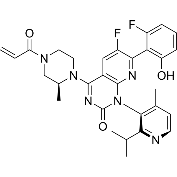
HY-114277
SotorasibKRAS G12C Inhibitor -
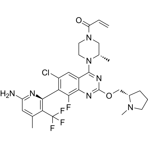
HY-145928
DivarasibKRAS G12C Inhibitor -
HY-148439
DaraxonrasibRAS(ON) Inhibitor -
HY-153724
BI-2865Pan-KRAS Inhibitor -
HY-132966
ASP2453Ras Inhibitor -
HY-153346
ElironrasibKRAS-G12C(ON) Inhibitor -
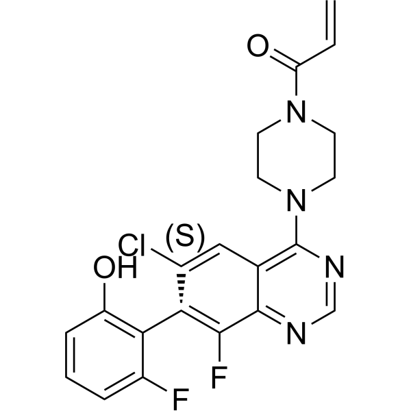
HY-U00418
ARS-1620 -
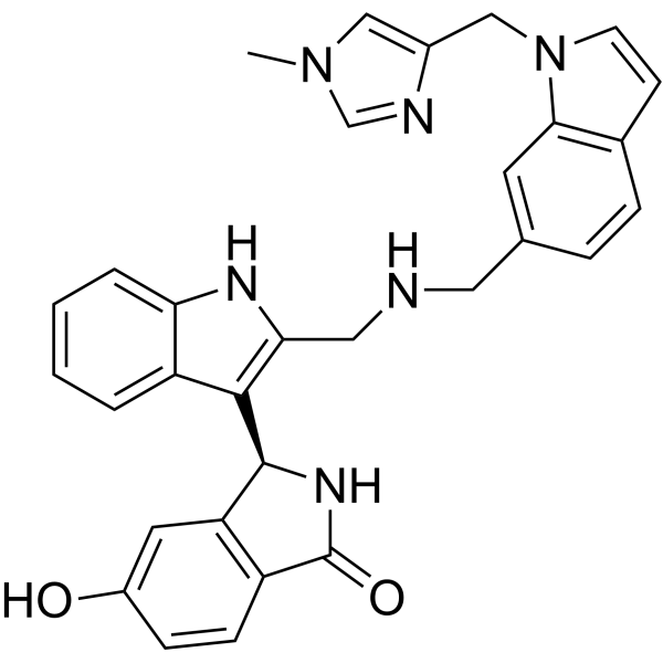
HY-126247
BI-2852KRAS Switch I/II Pocket Inhibitor -
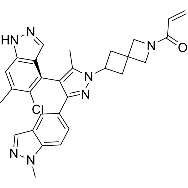
HY-139612
OpnurasibKRAS G12C Inhibitor -
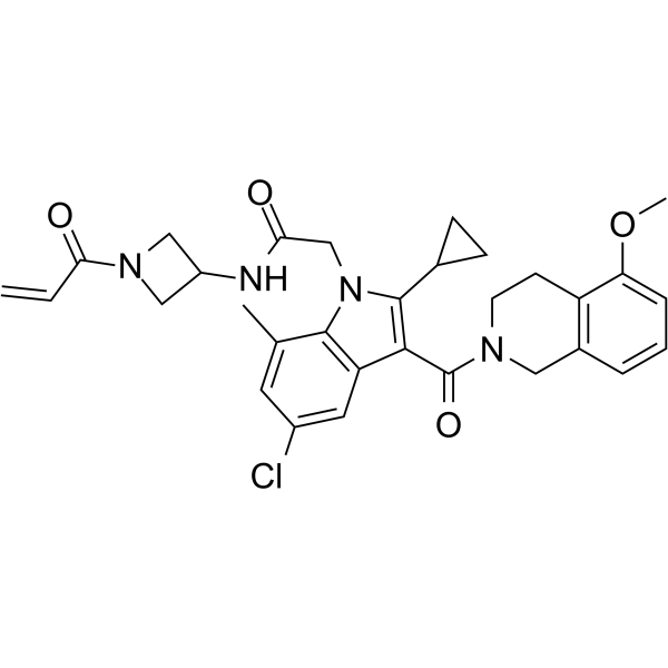
HY-128771
K-Ras G12C-IN-4Ras Inhibitor -

HY-114436
MRTX-1257KRAS G12C Inhibitor


Biological Activity
Description
IC50 & Target
|
KRAS-SOS1 7.0 nM (IC50) |
Cellular Effect
|
Cell Line

|
Type | Value | Description | References |
|---|---|---|---|---|
| LS-513 | EC50 |
>4 μM
Compound: 23; BI-0474
|
Antiproliferative activity against human LS-513 cells harboring KRAS G12D mutant incubated for 5 days by celltiter glo luminescent assay
Antiproliferative activity against human LS-513 cells harboring KRAS G12D mutant incubated for 5 days by celltiter glo luminescent assay
|
[PMID: 36300829] |
| NCI-H358 | EC50 |
26 nM
Compound: 23; BI-0474
|
Antiproliferative activity against human NCI-H358 cells harboring KRAS G12C mutant incubated for 3 days by celltiter glo luminescent assay
Antiproliferative activity against human NCI-H358 cells harboring KRAS G12C mutant incubated for 3 days by celltiter glo luminescent assay
|
[PMID: 36300829] |
In Vitro
BI-0474 (1-10,000 nM; 3 days) shows potent antiproliferative activity of 26 nM on NCI-H358 cells carrying a G12C mutation[1].
MedChemExpress (MCE) has not independently confirmed the accuracy of these methods. They are for reference only. Further protocols information, click here.
-
Cell Line:NCI-H358 cells (carrying a G12C mutation)
-
Concentration:1-10,000 nM
-
Incubation Time:3 days
-
Result:Inhibited proliferation of NCI-H358 cells with an EC50 of 26 nM.
In Vivo
MedChemExpress (MCE) has not independently confirmed the accuracy of these methods. They are for reference only.
-
Animal Model:NMRI nude mice (NCI-H358 cell line-derived non-small cell lung cancer xenograft model)[1].
-
Dosage:40 mg/kg
-
Administration:Intraperitoneal administration; single daily for 3 days
-
Result:Led to induction of programmed cell death in this xenograft model.


Chemical Information
-
CAS No. 2750570-55-7
-
Appearance Solid
-
Molecular Weight 587.74
-
Formula C30H37N9O2S
-
Color White to off-white
-
SMILES
N#CC1=C(N)SC2=C1[C@@](C)(C3=NC(C4=NC(N5[C@@H](C)CN(C)CC5)=CC(N6CCN(C(C=C)=O)CC6)=C4)=NO3)CCC2
-
Shipping
Room temperature in continental US; may vary elsewhere.
-
Storage
Powder -20°C 3 years 4°C 2 years In solvent -80°C 6 months -20°C 1 month


Solvent & Solubility
In Vitro:
DMSO : 100 mg/mL (170.14 mM; Need ultrasonic; Hygroscopic DMSO has a significant impact on the solubility of product, please use newly opened DMSO)
Please refer to the solubility information to select the appropriate solvent. Once prepared, please aliquot and store the solution to prevent product inactivation from repeated freeze-thaw cycles.
Storage method and period of stock solution: -80°C, 6 months; -20°C, 1 month. When stored at -80°C, please use it within 6 months. When stored at -20°C, please use it within 1 month.
Please refer to the solubility information to select the appropriate solvent. Once prepared, please aliquot and store the solution to prevent product inactivation from repeated freeze-thaw cycles.
Storage method and period of stock solution: -80°C, 6 months; -20°C, 1 month. When stored at -80°C, please use it within 6 months. When stored at -20°C, please use it within 1 month.
Concentration (start) × Volume (start) = Concentration (final) × Volume (final)
In Vivo:
Select the appropriate dissolution method based on your experimental animal and administration route.
- For the following dissolution methods, please ensure to first prepare a clear stock solution using an In Vitro approach and then sequentially add co-solvents:
- To ensure reliable experimental results, the clarified stock solution can be appropriately stored based on storage conditions. As for the working solution for In Vivo experiments, it is recommended to prepare freshly and use it on the same day.
- The percentages shown for the solvents indicate their volumetric ratio in the final prepared solution. If precipitation or phase separation occurs during preparation, heat and/or sonication can be used to aid dissolution.
Add each solvent one by one: 10% DMSO 40% PEG300 5% Tween-80 45% Saline
Solubility: 2.5 mg/mL (4.25 mM); Clear solution; Need ultrasonic
This protocol yields a clear solution of 2.5 mg/mL.
Taking 1 mL working solution as an example, add 100 μL DMSO stock solution (25.0 mg/mL) to 400 μL PEG300, and mix evenly; then add 50 μL Tween-80 and mix evenly; then add 450 μL Saline to adjust the volume to 1 mL.
Preparation of Saline: Dissolve 0.9 g sodium chloride in ddH₂O and dilute to 100 mL to obtain a clear Saline solution.
In Vivo Dissolution Calculator
Please enter the basic information of animal experiments:
-
-
-
-
Recommended: Prepare an additional quantity of animals to account for potential losses during experiments.
Please enter your animal formula composition:
-
%DMSO +
Recommended: Keep the proportion of DMSO in working solution below 2% if your animal is weak.
-
%+
-
+%Tween-80 + +
-
%Saline +
The co-solvents required include: DMSO, . All of co-solvents are available by MedChemExpress (MCE). , Tween 80. All of co-solvents are available by MedChemExpress (MCE).
Working solution concentration: 0.22 mg/mL
Method for preparing stock solution: mg drug dissolved in μL DMSO. Stock solution concentration: mg/mL.
1. Take μL DMSO stock solution;
2. Add μL .
μL , mix evenly;
3. Then add μL Tween 80, mix evenly;
4. Then add μL
Please ensure that the stock solution in the first step is dissolved to a clear state, and add co-solvents in sequence. You can use ultrasonic heating (ultrasonic cleaner, recommended frequency 20-40 kHz), vortexing, etc. to assist dissolution.

Protocols
-
Immunoprecipitation
Immunoprecipitation (IP) is an experimental method that uses the principle of antibody specific binding to purify and enrich target proteins.
-
Protocol for Bimolecular Fluorescence Complementation (BiFC) Assay
Bimolecular fluorescence complementation detects protein-protein proximity in living or fixed cells by fusing two candidate interaction partners to nonfluorescent N- and C-terminal fragments of a fluorescent protein; when the partners interact or remain close enough, the fluorescent fragments complement, mature, and generate a fluorescent signal at the site of the protein complex. The BiFC readout is fluorescence intensity and subcellular localization of the reconstituted fluorophore, which reflects formation or stabilization of a protein complex rather than direct biochemical binding kinetics; BiFC is therefore useful for mapping where interactions occur in cancer cells, neurons, macrophages, organoid-derived cells, or drug-screening systems, but results should be validated by independent assays such as co-IP or Western blot. BiFC signal formation is delayed by fluorophore maturation and can stabilize otherwise transient complexes, so it is not a real-time reversible interaction assay
-
Cell Viability Determination by MTT Colorimetric Assay
The following protocol uses the MTT colorimetric assay as a classic literature-established method for assessing cell viability/metabolic activity in cultured mammalian cells. MTT[3-(4,5-dimethylthiazol-2-yl)-2,5-diphenyltetrazolium bromide] is reduced by metabolically active cells to a colored formazan product; the amount of formazan is quantified spectrophotometrically and provides an indirect measure of metabolically active viable cells. Importantly, MTT reduction reflects cellular oxidoreductase/metabolic activity rather than an absolute direct count of living cells, so changes in cellular metabolism can alter the signal independently of cell number.
-
Cell Cytotoxicity Assay
Cytotoxicity assays are usually based on the assessment of cell membrane damage, which can also be indirectly detected by measuring cell viability. Detection methods include MTT assay, CKK-8 assay, LDH assay and ATP assay, etc.
-
Pull-down
The pull-down assay is an in vitro technique used to detect physical interactions between two or more proteins and an invaluable tool for confirming a predicted protein-protein interaction or identifying novel interacting partners. This method typically involves the use of affinity purification with various wash and elution steps.
-
Patient-Derived Xenograft (PDX)
Patient-derived xenograft (PDX) models are generated by engrafting primary human tumor tissue directly into immunodeficient mice, allowing in vivo propagation of patient tumor biology without initial in vitro adaptation. These models are used to preserve key histopathological and molecular characteristics of the original tumor and enable assessment of tumor growth dynamics and therapeutic response in a living organism. The biological readout is tumor engraftment and subsequent growth in the murine host, which reflects the ability of human tumor cells to survive, vascularize, and expand in an immunocompromised microenvironment.
-
Splenic/Portal-Vein Liver Metastasis Xenograft
Splenic and portal-vein liver metastasis xenograft models deliver tumor cells into the portal circulation so that cells reach the liver first and form hepatic metastatic lesions; splenic injection uses the spleen as an access route to the portal system, while direct portal-vein injection introduces cells into the portal vein without requiring splenectomy. The assay detects liver colonization, intrahepatic tumor growth, tumor distribution, treatment response, survival, and liver-metastasis microenvironment changes; readouts include bioluminescence or fluorescence imaging, gross liver nodule counts, liver weight or tumor burden, histology, and survival.
-
Subcutaneous Cell-Line-Derived Xenograft
Subcutaneous cell-line-derived xenograft (CDX) models are established by implanting cultured human cancer cell lines into immunodeficient mice, where the injected cells form localized tumors that can be monitored in vivo as a measure of tumorigenic potential, growth kinetics, and treatment response. These models are widely used in oncology research because they allow reproducible tumor formation and enable comparative assessment of tumor growth between different cell lines or genetic manipulations in a controlled in vivo microenvironment. Subcutaneous implantation of cancer cells in immunodeficient mice is a standard approach for evaluating tumor growth behavior and therapeutic response across multiple cancer types, including prostate, esophageal, pancreatic, and colon cancer models.
-
Genotoxicity/Mutagenicity Study
The bacterial reverse mutation assay detects point mutations that restore amino-acid prototrophy in auxotrophic Salmonella typhimurium or Escherichia coli tester strains; after exposure to a test article, mutagenic activity is read out as an increased number of revertant colonies on minimal agar compared with the vehicle control. The assay uses tester strains with different mutation targets so that base-substitution and frameshift mutagens can be detected, and testing is performed with and without exogenous mammalian metabolic activation because some chemicals require biotransformation to become mutagenic.
-
Orthotopic Cell-Line Xenograft
Orthotopic cell-line xenograft models involve implantation of human cancer cell lines into the anatomically corresponding organ of immunodeficient mice to reproduce tumor growth within a native microenvironment, enabling more clinically relevant tumor behavior compared with subcutaneous models. These models are widely used because orthotopic placement better recapitulates tumor progression, including invasion and metastatic spread, which are often underrepresented in heterotopic implantation systems. Compared with conventional xenografts, orthotopic implantation is described as more technically complex but provides improved simulation of tumor-microenvironment interactions and metastatic behavior, making it particularly valuable for translational oncology research. Surgical orthotopic implantation approaches have been emphasized as enabling faithful reproduction of clinical cancer features, including metastasis and disease progression patterns that align with the tumor’s organ of origi
-
Co-Immunoprecipitation
Co-immunoprecipitation technology can verify protein interaction based on the specific immune reaction between antibodies and antigens.
-
Protocol for Yeast Two-Hybrid (Y2H) Assay
The yeast two-hybrid assay detects binary protein-protein interactions by separating a transcription factor into a DNA-binding domain fused to a "bait" protein and a transcriptional activation domain fused to a "prey" protein; if bait and prey interact in yeast, the transcription factor is reconstituted and activates reporter genes such as HIS3, ADE2, lacZ, MEL1, or other selectable/readable reporters. The readout is yeast growth on selective medium and/or reporter activity, which reflects proximity-dependent transcriptional activation in the yeast nucleus rather than direct biochemical binding in the original mammalian, tumor, neuronal, macrophage, or organoid context. Because yeast two-hybrid can generate false positives and false negatives, interaction claims should be validated using independent assays such as co-immunoprecipitation, Western blot, immunofluorescence colocalization, BiFC, pull-down, or mammalian two-hybrid assays.
-
Intraperitoneal/Peritoneal Dissemination Xenograft
Intraperitoneal (IP) or peritoneal dissemination xenograft models are based on the introduction of human cancer cells into the peritoneal cavity of immunodeficient mice, where they attach to peritoneal surfaces, form multicellular aggregates or spheroids, and progressively generate disseminated tumor nodules that mimic advanced peritoneal metastatic disease. These models are widely used to study ovarian cancer progression, tumor-microenvironment interactions, and intraperitoneal therapeutic responses, often incorporating bioluminescence or fluorescence imaging to longitudinally monitor tumor burden in vivo. The biological principle relies on the capacity of tumor cells such as SKOV3 or related ovarian carcinoma lines to survive in suspension, aggregate within ascites-like fluid, adhere to mesothelial surfaces, and invade peritoneal organs, thereby recapitulating human peritoneal carcinomatosis patterns observed in advanced disease.


Purity & Documentation
-
Data Sheet (282 KB)
-
SDS (251 KB)
- English - EN (251 KB)
- Français - FR (251 KB)
- Deutsch - DE (251 KB)
- Norwegian - NO (251 KB)
- Español - ES (251 KB)
- Swedish - SV (251 KB)
- Italian - IT (251 KB)
- Korean - KR (251 KB)
- Portuguese - PT (251 KB)
-
Handling Instructions (2659 KB)
References


Complete Stock Solution Preparation Table
Please refer to the solubility information to select the appropriate solvent. Once prepared, please aliquot and store the solution to prevent product inactivation from repeated freeze-thaw cycles.
Storage method and period of stock solution: -80°C, 6 months; -20°C, 1 month. When stored at -80°C, please use it within 6 months. When stored at -20°C, please use it within 1 month.
| Optional Solvent | Concentration Solvent Mass | 1 mg | 5 mg | 10 mg | 25 mg |
|---|---|---|---|---|---|
| DMSO | 1 mM | 1.7014 mL | 8.5072 mL | 17.0143 mL | 42.5358 mL |
| 5 mM | 0.3403 mL | 1.7014 mL | 3.4029 mL | 8.5072 mL | |
| 10 mM | 0.1701 mL | 0.8507 mL | 1.7014 mL | 4.2536 mL | |
| 15 mM | 0.1134 mL | 0.5671 mL | 1.1343 mL | 2.8357 mL | |
| 20 mM | 0.0851 mL | 0.4254 mL | 0.8507 mL | 2.1268 mL | |
| 25 mM | 0.0681 mL | 0.3403 mL | 0.6806 mL | 1.7014 mL | |
| 30 mM | 0.0567 mL | 0.2836 mL | 0.5671 mL | 1.4179 mL | |
| 40 mM | 0.0425 mL | 0.2127 mL | 0.4254 mL | 1.0634 mL | |
| 50 mM | 0.0340 mL | 0.1701 mL | 0.3403 mL | 0.8507 mL | |
| 60 mM | 0.0284 mL | 0.1418 mL | 0.2836 mL | 0.7089 mL | |
| 80 mM | 0.0213 mL | 0.1063 mL | 0.2127 mL | 0.5317 mL | |
| 100 mM | 0.0170 mL | 0.0851 mL | 0.1701 mL | 0.4254 mL |


BI-0474 Related Classifications
HY-148278 Related Classifications
 Powered by Bioz