



Tyramide Amplification Buffer
Based on 1 publication(s) in Google Scholar
Tyramide Amplification Buffer is a ready-to-use buffer primarily intended for immunostaining of cells and tissues by the technique of tyramide signal amplification (TSA). Tyramide Amplification Buffer enables the reaction between tyramide-labeled antibodies and fluorescently labeled tyramide substitutes (tyramide) using peroxidase, resulting in a highly amplified fluorescent signal. Tyramide Amplification Buffer can be used for signal enhancement in detection methods such as immunofluorescence (IF), immunohistochemistry (IHC), or in situ hybridization (FISH).
For research use only. We do not sell to patients.
-
Storage:
4°C, protect from light
* In solvent : -80°C, 6 months; -20°C, 1 month (protect from light)

To place orders, for customer services and technical support, please contact: MedChemExpress USA
Tel: 609-228-6898 E-mail: [email protected] [email protected]


-
 Biological Activity
-
Chemical Information
- Protocols
- Purity & Documentation
- References
-
 Help & FAQs


-

HY-D1837
Vari Fluor 488 TSA(200×)Fluorescent Dye -

HY-D1836
Vari Fluor 555 TSA (200x)Fluorescent Probe -

HY-D1835
Vari Fluor 594 TSA (200×)Fluorescent Dye -

HY-D1834
Vari Fluor 640 TSA (200×)Fluorescent Dye -
HY-125658
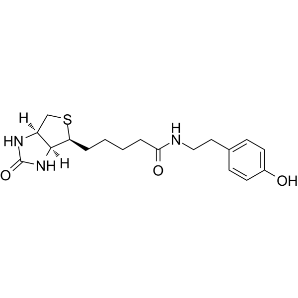
Biotinyl tyramideBiotin Derivative/Reagent -

HY-P83652
Polymer HRP-conjugated Goat Anti-Mouse/Rabbit lgG(H&L) (Ready to use) -

HY-D1833
Vari Fluor 680 TSA (200×)Fluorescent Dye -

HY-D1831
Vari Fluor 620 TSA (200×)Fluorescent Dye -

HY-D1832
Vari Fluor 532 TSA (200×)Fluorescent Dye
Publications Citing Use of MedChemExpress (MCE) Tyramide Amplification Buffer
More

Biological Activity
Description
In Vitro
Guide (The following is our recommended solution. This solution is merely a guideline and should be modified according to your specific needs.)
The following scheme is applicable to fixed cells in a 96-well plate or tissue sections of approximately 10 mm × 10 mm. It can be scaled proportionally according to the size of the sample [1].
Perform the corresponding fixation, blocking and antibody labeling steps using the samples and antibodies. The samples must be labeled with antibodies or HRP conjugates coupled with streptavidin.
After the antibody (streptavidin)-HRP conjugate is labeled, the sample is washed three times with the 1×PBS washing buffer, for 5 minutes each time.
3. Prepare the working solution of the dye-tyramide probe using 1× Tyramide Amplification Buffer. The final concentration of the dye-tyramide probe can be adjusted according to specific applications. Add 100 μL of the working solution to each well of a 96-well plate or to each tissue section. The working solution can be stored under light protection at room temperature for up to 24 hours.
4. Incubate at room temperature in the dark for 10 minutes.
5. Wash the samples three times with the washing buffer 1×PBS, for 5 minutes each time.
6. Optional: If using Biotin-acylamide, fluorescence staining can be performed using fluorescently-labeled streptavidin, or streptavidin labeled with HRP can be used and then stained with DAB.
7. Microscopy imaging. For tissue samples on the slide, cover the slide with a cover glass and seal it, then perform microscopy imaging.
MedChemExpress (MCE) has not independently confirmed the accuracy of these methods. They are for reference only.
- Ki-67 Immunostaining Proliferation Assay
- Polymer-Based Two-Step IHC Detection
- Immunohistochemistry-Frozen
- Avidin-Biotin/Streptavidin-Biotin IHC
- Double or multiplex chromogenic IHC
- Protocol for Fluorescence In Situ Hybridization (FISH)
- Antibody-based immunofluorescence/immunocytochemistry staining
- Immunocytochemistry/Immunofluorescence
- In situ hybridization


Chemical Information
-
Appearance Liquid
-
Color Colorless to light yellow
-
SMILES
[Tyramide Amplification Buffer]
-
Shipping
Room temperature in continental US; may vary elsewhere.
-
Storage
4°C, protect from light
* In solvent : -80°C, 6 months; -20°C, 1 month (protect from light)


Publications (1)
-
Journal Impact Factor
-
Most Recent
-
Anal Chem
Chemiluminescence-Driven Tyramide Amplification for High-Sensitivity and Long-Term Immunofluorescence Imaging. [Abstract]2026 Jan 13;98(1):1185-1198. PMID: 41480935


Purity & Documentation
-
Data Sheet (267 KB)
-
SDS (252 KB)
- English - EN (252 KB)
- Français - FR (252 KB)
- Deutsch - DE (252 KB)
- Norwegian - NO (252 KB)
- Español - ES (252 KB)
- Swedish - SV (252 KB)
- Italian - IT (252 KB)
- Korean - KR (252 KB)
- Portuguese - PT (252 KB)
-
Handling Instructions (2659 KB)
References

Calculators
Concentration (start) × Volume (start) = Concentration (final) × Volume (final)


Tyramide Amplification Buffer Related Classifications
HY-D1840 Related Classifications
 Powered by Bioz