



Succinyl phosphonate
Based on 14 publication(s) in Google Scholar
Succinyl phosphonate is a α-Ketoglutarate Dehydrogenase Complex (KGDHC) modulator with neuroprotective activity. Succinyl phosphonate protects this complex, reduces cellular succinyl-CoA concentration, downregulates protein succinylation levels, and inhibits the activity of the α-ketoglutarate dehydrogenase complex. Succinyl phosphonate corrects hypoxic or ethanol-induced behavioral impairments, modulates exploratory behavior and emotional stress responses, and improves hypoxia tolerance. Succinyl phosphonate reduces glutamate excitotoxicity, restores the activity of the α-ketoglutarate dehydrogenase complex, reverses the changes in glutamate dehydrogenase and glutamine synthetase activities induced by β-amyloid (Amyloid-β), modulates cognitive function, and prevents β-amyloid-induced neuronal damage. Succinyl phosphonate improves microglial senescence, alleviates neuroinflammation, reactive oxygen species (ROS) production, lipid peroxidation, and the expression of proinflammatory cytokines. Succinyl phosphonate can be used in the research of Alzheimer's disease, aging-related neuroinflammation, and Parkinson's disease.
연구목적의 판매만을 진행합니다. 환자를 대상으로 한 판매는 하지 않습니다.
- CAS No.: 26647-82-5
- 화학식: C4H7O6P
- 분자량:182.07
-
보관:Pure form -20°C, 3 years , 4°C, 2 years ; In solvent -80°C, 6 months , -20°C, 1 month

To place orders, for customer services and technical support, please contact: MedChemExpress USA
Tel: 609-228-6898 E-mail: [email protected] [email protected]


-
 Biological Activity
-
Chemical Information
- Protocol
- 순도&문서
- References
-
 Help & FAQs


Publications Citing Use of MedChemExpress (MCE) Succinyl phosphonate
More- Cancer Res. 2019 Jul 1;79(13):3281-3293. [Abstract]
- Redox Biol. 2023 Jun:62:102669. [Abstract]
- Cell Death Dis. 2021 Oct 25;12(11):999. [Abstract]
- Free Radic Biol Med. 2016 Jul:96:22-33. [Abstract]
- Planta. 2018 Oct;248(4):963-979. [Abstract]
- Plant Signal Behav. 2019;14(7):1604015. [Abstract]
- J Cell Sci. 2026 Mar 16:jcs.264420. [Abstract]
- ACS Chem Biol. 2020 Aug 21;15(8):2041-2047. [Abstract]
- Exp Cell Res. 2019 Sep 15;382(2):111483. [Abstract]
- Clin Rheumatol. 2025 Nov 20. [Abstract]
- Radiat Med Prot. 2026 May 20.
- bioRxiv. 2024 Jan 15.
- Research Square Preprint. 2022.
- Research Square Print. 2022 May.
 Customer Validation & Images

-
Cell Proliferation/Viability Assay
-
Bio/Physico-chemical Assay
-
IP
-
Cell Proliferation/Viability Assay
-
RT-PCR


Biological Activity
제품 설명
In Vitro
Succinyl phosphonate (10 μM; 72 h) reduces succinylation levels and succinyl-CoA concentrations, mitigates LPS (HY-D1056)-induced reactive oxygen species production, lipid accumulation, lipid peroxidation, and pro-inflammatory cytokine expression in BV2 cells and primary mouse microglia, and reduces senescence in VP-16 (HY-13629)-induced aged BV2 cells[3].
Succinyl phosphonate upregulates active OGDHC quantity in cultured neurons, but this upregulation is impaired by co-treatment with ethanol[4].
MedChemExpress (MCE) has not independently confirmed the accuracy of these methods. They are for reference only. Further protocols information, click here.
In Vivo
Succinyl phosphonate (10 nM; intra-CA1; 30 minutes after first Aβ25-35 injection) can block the damaging effects induced by Aβ and neuronal damage, improve the spatial learning and memory abilities of rats, and reverse the decrease in α-KGDHC enzyme activity[2].
Succinyl phosphonate (1 mg/kg; i.c.v.; single injection) reduces LPS (HY-D1056)-induced hippocampal mitochondrial protein succinylation, including succinylation of SDHA and ECHA, in male C57BL/6J mice[3].
MedChemExpress (MCE) has not independently confirmed the accuracy of these methods. They are for reference only.
-
Animal Model:Wistar rats (adult male, 230-250 g, Alzheimer’s disease model via bilateral microinjection of aggregated amyloid-beta25-35 into dorsal CA1 area for 4 consecutive days)[2]
-
Dosage:10 nM
-
Administration:intra-CA1; single dose; 30 minutes after first Aβ25-35 injection
-
Result:Prevented Aβ25-35-induced spatial learning deficits, as shown by significantly reduced AUC of escape latencies and improved escape latencies in specific trials.
Reversed Aβ25-35-induced spatial memory deficits, with time spent in the MWM target quadrant not differing from controls.
Restored hippocampal α-KGDHC activity to control levels.
Reduced elevated GDH activity to control levels.
Prevented the Aβ-induced reduction in GS activity.
Reduced Aβ-induced neuronal damage in the hippocampal CA1, CA3, and DG regions, with significantly more surviving neurons compared to the Aβ-only group.
-
Animal Model:LPS-induced C57BL/6J (2-4 month-old male, wild type)[3]
-
Dosage:1 mg/kg
-
Administration:i.c.v.; single injection
-
Result:Reversed the LPS-induced increase in hippocampal mitochondrial protein succinylation, restoring it to near control levels.
Significantly reduced the LPS-elevated succinylation levels of succinate dehydrogenase (SDHA) and trifunctional enzyme subunit alpha (ECHA) in mouse hippocampus.


Chemical Information
-
CAS No. 26647-82-5
-
Appearance Oil
-
분자량 182.07
-
화학식 C4H7O6P
-
Color Colorless to light yellow
-
SMILES
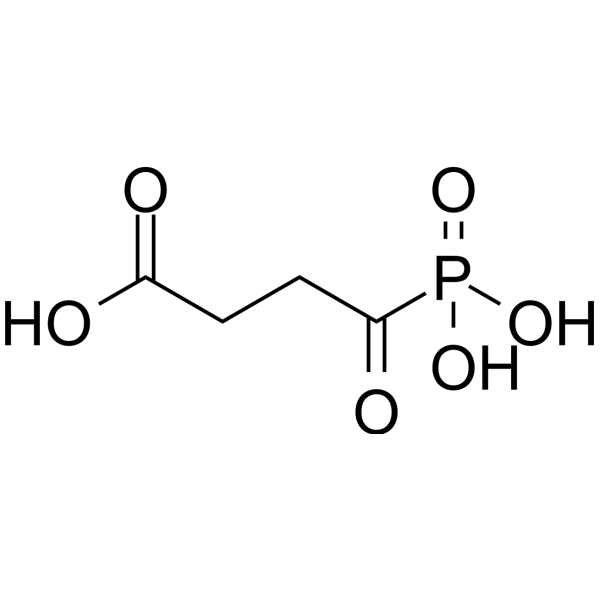
O=C(O)CCC(P(O)(O)=O)=O
-
Structure Classification
-
Initial Source
-
선적
Room temperature in continental US; may vary elsewhere.
-
보관
Pure form -20°C 3 years 4°C 2 years In solvent -80°C 6 months -20°C 1 month


Publications (14)
-
Journal Impact Factor
-
Most Recent
-
Cancer Res
APC/C-CDH1-Regulated IDH3β Coordinates with the Cell Cycle to Promote Cell Proliferation. [Abstract]2019 Jul 1;79(13):3281-3293. PMID: 31053633 -
Redox Biol
2023 Jun:62:102669. PMID: 36933393 -
Cell Death Dis
2021 Oct 25;12(11):999. PMID: 34697294 -
Free Radic Biol Med
Reactive oxygen species production in cardiac mitochondria after complex I inhibition: Modulation by substrate-dependent regulation of the NADH/NAD(+) ratio. [Abstract]2016 Jul:96:22-33. PMID: 27068062 -
Planta
Inhibition of α-ketoglutarate dehydrogenase activity affects adventitious root growth in poplar via changes in GABA shunt. [Abstract]2018 Oct;248(4):963-979. PMID: 29982922 -
Plant Signal Behav
2019;14(7):1604015. PMID: 30999791 -
J Cell Sci
α-Ketoglutarate dehydrogenase complex activity modulates glutamate excitotoxicity via metabotropic regulation of NMDA receptors in primary cultures. [Abstract]2026 Mar 16:jcs.264420. PMID: 41834724
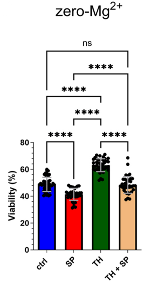
Succinyl phosphonate purchased from MedChemExpress. Usage Cited in: J Cell Sci. 2026 Mar 16:jcs.264420. [Abstract]
Bar graphs (depicting mean±s.d. together with the individual data points) of neuronal viability determined after exposure to zero-Mg2+-induced excitotoxicity of untreated cultures (ctrl, n=45), cultures pretreated for 36 to 48 h with Succinyl phosphonate trisodium salt (SP, 200 µM) (n=36), with TH (n=42) or with both compounds (TH+SP, n=41) (from three independent culture preparations).
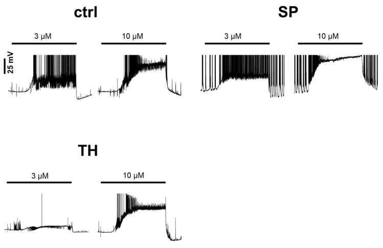
Succinyl phosphonate purchased from MedChemExpress. Usage Cited in: J Cell Sci. 2026 Mar 16:jcs.264420. [Abstract]
Depiction of representative voltage responses to application of 3 µM and 10 µM glutamate of control neurons, Succinyl phosphonate trisodium salt (SP)-treated neurons and TH-treated neurons. Responses were characterized by triggering of or increase in action potential discharge, and at 10 µM glutamate, by a transition into depolarisation block, which manifests itself in a cessation of discharge activity during sustained depolarization. Action potentials are truncated at −10 mV.
-
ACS Chem Biol
2020 Aug 21;15(8):2041-2047. PMID: 32633484 -
Exp Cell Res
OGDH mediates the inhibition of SIRT5 on cell proliferation and migration of gastric cancer. [Abstract]2019 Sep 15;382(2):111483. PMID: 31247190 -
Clin Rheumatol
High glucose-induced mitophagy accelerates premature aging of T cells in patients with rheumatoid arthritis. [Abstract]2025 Nov 20. PMID: 41261244
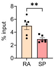
Succinyl phosphonate purchased from MedChemExpress. Usage Cited in: Clin Rheumatol. 2025 Nov 20. [Abstract]
ZNF76 activity quantified upon Succinyl phosphonate trisodium salt (SP) (50 μM) treatment. The results showed that the application of the OGDH inhibitor SP significantly reduced the binding of ZNF76 to the promoter region of the DRP1 gene.
-
-
-
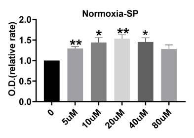
Succinyl phosphonate purchased from MedChemExpress. Usage Cited in: Research Square Preprint. 2022.
Proliferation assay of EPCs treated with vehicle or various concentration of Succinyl phosphonate trisodium salt (SP, 5-80 μM; 3 days) under normoxia (n = 6).
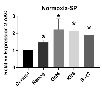
Succinyl phosphonate purchased from MedChemExpress. Usage Cited in: Research Square Preprint. 2022.
qRT-PCR showed higher expression of EPC stemness markers in the presence of 20 uM Succinyl phosphonate trisodium salt (SP) than with vehicle control under normoxia (n=5).
-

Protocol
-
RNA extraction experimental
By lysing cells, releasing RNA, and removing impurities such as proteins and DNA, high-purity RNA products are finally obtained. The commonly used traditional method is the guanidine isothiocyanate/phenol/chloroform method (Trizol), which is suitable for a variety of animal materials including animal tissues, microorganisms, cultured cells, etc., and most plant materials.
-
Protocol for Open Field Test (OF)
The Open Field Test is a rodent behavioral assay that measures spontaneous locomotion, exploratory behavior, and anxiety-like behavior when an animal is placed in a novel open arena. The main readouts are total distance traveled, movement time, velocity, center-zone entries, center-zone time, peripheral-zone time, and thigmotaxis. The assay is based on the conflict between exploration of a novel environment and avoidance of exposed open areas; higher center exploration is commonly interpreted as lower anxiety-like behavior, whereas increased wall-following or peripheral occupancy is interpreted as higher anxiety-like behavior.
-
ROS/oxidative-stress fluorescent staining
ROS/oxidative-stress fluorescent staining uses cell-permeant fluorogenic probes that become fluorescent after oxidation inside cells or tissues; commonly used examples include DCFH-DA/DCFDA for broad cellular oxidant detection, DHE for superoxide-related signal detection, MitoSOX for mitochondrial superoxide-related signal detection, and CellROX probes for oxidative-stress-associated fluorescence readouts. The assay detects probe oxidation rather than a single ROS species unless the probe and analysis method have been chemically validated for that species. DCFH-DA enters cells, is deacetylated by intracellular esterases to DCFH, and produces fluorescent DCF after oxidation, so the readout is used as an operational measure of total cellular oxidative stress rather than a species-specific ROS measurement. DHE and MitoSOX can report superoxide-related oxidation, but red fluorescence alone can include non-specific ethidium-like oxidation products; HPLC or optimized spectral approaches are
-
Human pluripotent stem cell midbrain dopaminergic neuron differentiation
Human pluripotent stem cells are directed toward midbrain dopaminergic neurons by first inducing a neural floor-plate-like progenitor state, then patterning cells with ventralizing SHH signaling and midbrain/WNT-FGF cues, and finally maturing progenitors into neurons expressing dopaminergic markers such as TH, NURR1/NR4A2, PITX3, DAT/SLC6A3, VMAT2/SLC18A2, GIRK2/KCNJ6, FOXA2, LMX1A, and EN1. The main readouts are loss of pluripotency, acquisition of FOXA2+/LMX1A+ midbrain floor-plate progenitors, emergence of βIII-tubulin+/MAP2+ neurons, and production of TH+ dopaminergic neurons with molecular, dopamine-release, and electrophysiological features of midbrain dopaminergic identity.
-
Protocol for Water Maze
The Morris Water Maze is a rodent spatial learning and memory assay in which a mouse or rat swims in opaque water to find an escape platform; in the hidden-platform version, the animal cannot see the platform and must use distal extra-maze cues to learn its fixed spatial location. The assay primarily measures hippocampus-dependent spatial learning during acquisition trials and spatial reference memory during probe trials after platform removal; readouts include escape latency, swim path length, swim speed, quadrant occupancy, platform-site crossings, and proximity to the former platform location.
-
Ferroptosis Solutions
Ferroptosis is an iron-dependent, non-apoptotic form of regulated cell death characterized by lethal lipid peroxidation and sensitivity to suppression by iron chelators or lipophilic radical-trapping antioxidants. The core pathway links cystine uptake through system Xc−, glutathione availability, GPX4-dependent detoxification of phospholipid hydroperoxides, iron-dependent oxidative reactions, and polyunsaturated-phospholipid metabolism into a cell-death program that is biochemically and morphologically distinct from apoptosis, necrosis, and autophagy. The ferroptosis pathway is experimentally linked to phenotype through chemical and genetic perturbation. Erastin induces ferroptosis by inhibiting cystine uptake through system Xc− and weakening antioxidant defenses, while GPX4 inhibition or depletion causes lipid peroxide accumulation and ferroptotic cancer-cell death. ACSL4 and oxidizable arachidonoyl- or adrenoyl-containing phosphatidylethanolamines shape ferroptosis sensitivity by con
-
Research Protocol for Neurological Diseases
PINK1/Parkin-mediated mitophagy pathway is a mitochondrial quality-control signaling axis in which mitochondrial depolarization stabilizes PINK1 on damaged mitochondria, activates Parkin recruitment and E3 ubiquitin ligase activity, promotes ubiquitination of outer mitochondrial membrane proteins, recruits selective autophagy adaptors, and drives lysosomal degradation of damaged mitochondria. In neurological disease research, this pathway is experimentally important because neurons, especially dopaminergic neurons, are highly dependent on mitochondrial integrity, and defective mitochondrial turnover can lead to mitochondrial dysfunction, oxidative stress, impaired neuronal survival, α-synuclein accumulation, and neuroinflammatory damage-associated signals. The genetic disease link is strongest in Parkinson’s disease because mutations in PRKN/parkin cause autosomal recessive juvenile parkinsonism, mutations in PINK1 cause hereditary early-onset Parkinson’s disease, and Drosophila studie
-
Amyloid: Congo Red Amyloid Staining
Congo red amyloid staining is a histochemical method used to detect extracellular amyloid deposits in tissue sections based on the affinity of Congo red dye for β-pleated sheet-rich protein aggregates. When bound to amyloid, Congo red produces characteristic apple-green birefringence under polarized light microscopy, which is widely regarded as a diagnostic feature of amyloid deposition in histopathology. The diagnostic principle relies on the combination of dye binding (congophilia) and optical anisotropy under polarized illumination, which distinguishes amyloid from most non-amyloid eosinophilic extracellular deposits in routine histological evaluation. Amyloid identification by Congo red staining remains a cornerstone in diagnostic pathology despite the availability of adjunct methods such as immunohistochemistry and mass spectrometry, particularly because of its ability to localize deposits directly within tissue architecture. The specificity of Congo red-positive deposits is incre
-
Senescence-associated β-galactosidase staining
Senescence-associated β-galactosidase staining detects β-galactosidase activity that is histochemically visible at pH 6. 0 in senescent cells, where X-gal cleavage produces an insoluble blue precipitate observable by bright-field microscopy. This activity reflects increased lysosomal β-galactosidase/lysosomal mass rather than a senescence-essential enzyme, because GLB1 depletion or genetic lysosomal β-galactosidase deficiency can abolish SA-β-gal staining while cells still undergo senescence. SA-β-gal was originally reported in senescent but not presenescent fibroblasts and keratinocytes, absent from quiescent fibroblasts and terminally differentiated keratinocytes, and increased with donor age in human skin samples. Because SA-β-gal can also appear in some non-senescent or tissue-specific contexts, interpretation should be paired with experimental controls and, when possible, independent senescence markers.
-
Alzheimer’s Disease Modeling
Alzheimer’s Disease (AD) is a neurodegenerative disorder characterized by a progressive decline in cognitive functions and loss of specific types of neurons and synapses. Alzheimer's symptoms can be simulated in mice by injecting drugs (such as Aβ) or genetically modified.


순도&문서
-
Data Sheet (277 KB)
-
SDS (252 KB)
- Français - FR (252 KB)
- Deutsch - DE (252 KB)
- Norwegian - NO (252 KB)
- Español - ES (252 KB)
- Swedish - SV (252 KB)
- Italian - IT (252 KB)
- Korean - KR (252 KB)
- Portuguese - PT (252 KB)
-
Handling Instructions (2659 KB)
References
[2]. Sayehmiri F, et al. Phosphonate analog of 2-oxoglutarate regulates glutamate-glutamine homeostasis and counteracts amyloid beta induced learning and memory deficits in rats. Exp Gerontol. 2022;168:111944. [Content Brief]
[3]. Zhao X, et al. Up-regulated succinylation modifications induce a senescence phenotype in microglia by altering mitochondrial energy metabolism. J Neuroinflammation. 2024;21(1):296. Published 2024 Nov 14. [Content Brief]
[4]. Artiukhov AV, et al. Phosphonate Inhibitors of Pyruvate Dehydrogenase Perturb Homeostasis of Amino Acids and Protein Succinylation in the Brain. Int J Mol Sci. 2022;23(21):13186. Published 2022 Oct 29. [Content Brief]

Calculators
Concentration (start) × Volume (start) = Concentration (final) × Volume (final)


Succinyl phosphonate Related Classifications
HY-12688 Related Classifications
 Powered by Bioz
Keywords
- Succinyl phosphonate
- 26647-82-5
- Mitochondrial Metabolism
- Reactive Oxygen Species (ROS)
- Amyloid-β
- primary mouse microglia
- protein succinylation
- glutamine synthetase
- male Wistar rats
- BV2 cells
- glutamate dehydrogenase
- amyloid-beta
- male C57BL/6J mice
- succinyl-CoA
- 2-oxoglutarate dehydrogenase complex
- Inhibitor
- inhibitor
- inhibit