



Triamcinolone acetonide
Based on 7 publication(s) in Google Scholar
Triamcinolone acetonide inhibits basic fibroblast growth factor (bFGF) induced proliferation of retinal endothelial cells. Triamcinolone acetonide reduces chondrocyte viability and leads to cartilage destruction. Triamcinolone acetonide activates macrophage with anti-inflammatory characteristics. Triamcinolone acetonide can be used in the study of diseases such as atopic dermatitis.
연구목적의 판매만을 진행합니다. 환자를 대상으로 한 판매는 하지 않습니다.
- Purity : 99.95%
- CAS No.: 76-25-5
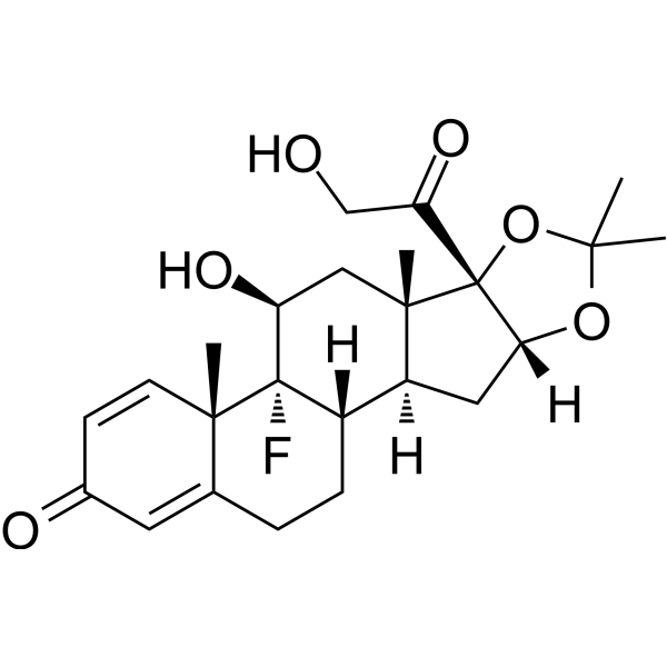
- 화학식: C24H31FO6
- 분자량:434.50
-
보관:Powder -20°C, 3 years , 4°C, 2 years ; In solvent -80°C, 6 months , -20°C, 1 month

To place orders, for customer services and technical support, please contact: MedChemExpress USA
Tel: 609-228-6898 E-mail: [email protected] [email protected]


-
 Biological Activity
-
Chemical Information
-
용액&용해도
- Protocol
- 순도&문서
- References
-
 Help & FAQs


-

HY-14648
DexamethasoneGR Agonist -
HY-B0154
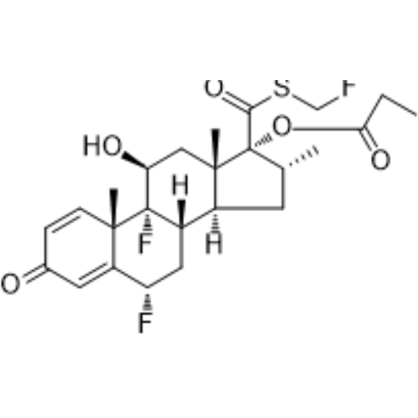
Fluticasone propionateGlucocorticoid Receptor Agonist -
HY-13580
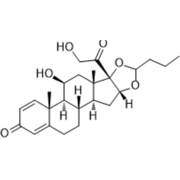
BudesonideGlucocorticoid Receptor Agonist -
HY-13570
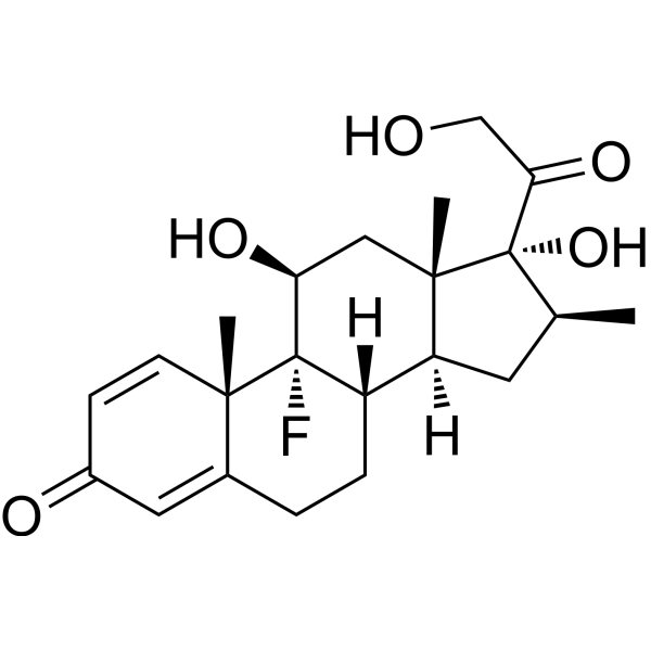
BetamethasoneGlucocorticoid Receptor Agonist -
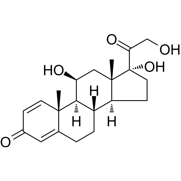
HY-17463
PrednisoloneGlucocorticoid -
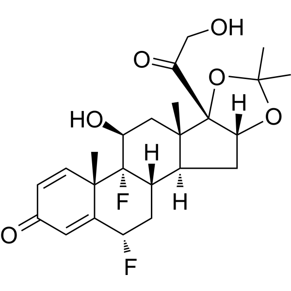
HY-B0415
Fluocinolone (Acetonide)Glucocorticoid Receptor Agonist -
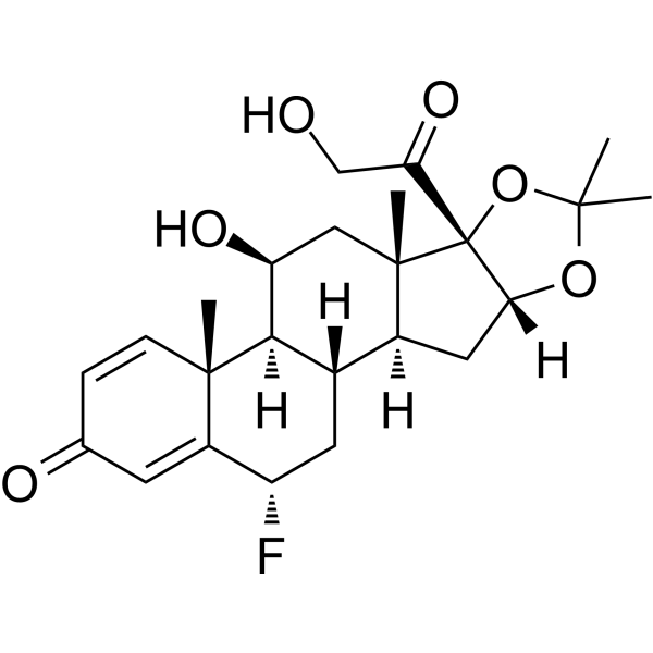
HY-B1121
FlunisolideGlucocorticoid Receptor Activator -
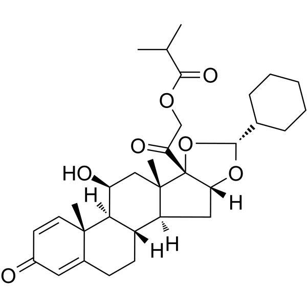
HY-B0625
CiclesonideGlucocorticoid -
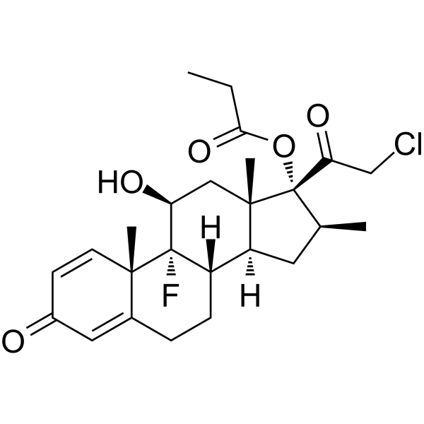
HY-13600
Clobetasol propionateCYP3A5 Inhibitor -
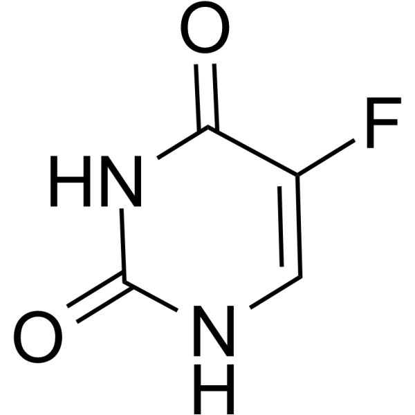
HY-90006
5-FluorouracilThymidylate Synthase Inhibitor -
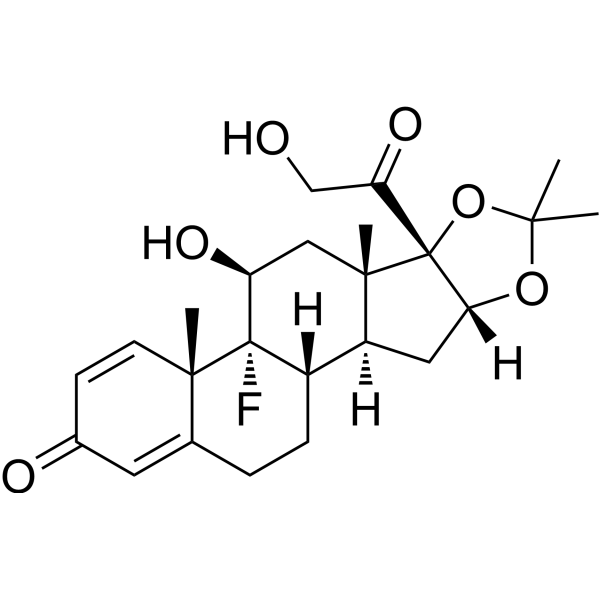
HY-B0636R
Triamcinolone acetonide (Standard) -

HY-10227
BortezomibProteasome Inhibitor
Publications Citing Use of MedChemExpress (MCE) Triamcinolone acetonide
More Customer Validation & Images

-
Histological Imaging/Staining
-
Histological Imaging/Staining
-
IF
-
IHC
-
In Vivo Efficacy Study


Biological Activity
제품 설명
Cellular Effect
|
Cell Line

|
Type | Value | Description | References |
|---|---|---|---|---|
| NIH3T3 | IC50 |
2.5 μM
Compound: 2 ; Triamcinolone acetonide (TA)
|
Inhibition of NIH 3T3 cell growth required to induce 50% of maximum response using antiproliferative assay
Inhibition of NIH 3T3 cell growth required to induce 50% of maximum response using antiproliferative assay
|
[PMID: 11855992] |
In Vitro
Triamcinolone acetonide (0.05-3 mg/mL, 48-60 h) decreases the proliferation of BRECs with increasing concentration[1].
Triamcinolone acetonide (0.04-5 mg/mL, 24 h) reduces chondrocyte viability in both normal and osteoarthritic (OA) chondrocytes in a concentration-dependent manner[2].
Triamcinolone acetonide (0.04-5 mg/mL, 24 h) increases the severity of cartilage structural damage, chondrocyte loss and cluster formation, and proteoglycan loss in OA cartilage[2].
Triamcinolone acetonide (100 nM, 7 days) strongly induces monocyte differentiation towards an M2 and anti-inflammatory macrophage phenotype[3].
MedChemExpress (MCE) has not independently confirmed the accuracy of these methods. They are for reference only. Further protocols information, click here.
-
Cell Line:Chondrocyte
-
Concentration:0.04, 0.08, 0.16, 0.31, 0.63, 1.25, 2.5, and 5 mg/ml
-
Incubation Time:24 h
-
Result:Reduced cell viability with the value of IC50 was 2.23 mg/mL in normal chondrocytes and 1.14 mg/mL in OA chondrocytes.
In Vivo
MedChemExpress (MCE) has not independently confirmed the accuracy of these methods. They are for reference only.
-
Animal Model:Severe OA rat model [3]
-
Dosage:1.43 mg/mL
-
Administration:Intraperitoneal injection (i.p.)
-
Result:Decreased body weight during OA induction. Showed more macrophage activation and minimal or no osteophyte formation when injected knee joints.
Clinical Trial
| NCT Number | Sponsor | Condition | Start Date |
Phase
|
|---|---|---|---|---|
| NCT01329991 | Plexxikon| | 2011-05 | PHASE1 |


Chemical Information
-
CAS No. 76-25-5
-
Appearance Solid
-
분자량 434.50
-
화학식 C24H31FO6
-
Color White to off-white
-
SMILES
OCC([C@]12[C@@]3([C@@]([C@@]4([H])[C@]([C@H](C3)O)([C@@]5(C(CC4)=CC(C=C5)=O)C)F)([H])C[C@@]1([H])OC(C)(O2)C)C)=O
-
선적
Room temperature in continental US; may vary elsewhere.
-
보관
Powder -20°C 3 years 4°C 2 years In solvent -80°C 6 months -20°C 1 month


Publications (7)
-
Journal Impact Factor
-
Most Recent
-
Proc Natl Acad Sci U S A
NSUN5/TET2-directed chromatin-associated RNA modification of 5-methylcytosine to 5-hydroxymethylcytosine governs glioma immune evasion. [Abstract]2024 Apr 2;121(14):e2321611121. PMID: 38547058 -
Biochim Biophys Acta Mol Cell Res
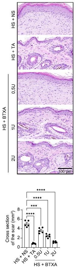
Botulinum toxin A prevents hypertrophic scarring by suppressing PARP14/SOCS2-mediated M2 polarization of macrophages. [Abstract]2025 Oct;1872(7):120003. PMID: 40505894
Triamcinolone acetonide purchased from MedChemExpress. Usage Cited in: Biochim Biophys Acta Mol Cell Res. 2025 Oct;1872(7):120003. [Abstract]
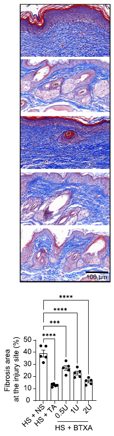
Triamcinolone acetonide (TA) (0.05 mL/g) treatment reduced dermal thickness and epidermal hyperplasia, decreased cell numbers, increased the number of dermal appendages, and significantly decreased the scar cross-sectional area in HS mice.
Triamcinolone acetonide purchased from MedChemExpress. Usage Cited in: Biochim Biophys Acta Mol Cell Res. 2025 Oct;1872(7):120003. [Abstract]
Triamcinolone acetonide (TA) (0.05 mL/g) reduced collagen deposition in the scars and diminished the fibrotic area at the injury site in HS mice.
Triamcinolone acetonide purchased from MedChemExpress. Usage Cited in: Biochim Biophys Acta Mol Cell Res. 2025 Oct;1872(7):120003. [Abstract]
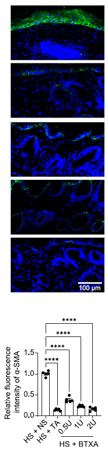
Triamcinolone acetonide (TA) (0.05 mL/g) significantly reduced the relative fluorescence intensity of α-SMA, a marker of myofibroblasts and indicative of collagen matrix contraction ability in the scar tissues of HS mice.
Triamcinolone acetonide purchased from MedChemExpress. Usage Cited in: Biochim Biophys Acta Mol Cell Res. 2025 Oct;1872(7):120003. [Abstract]
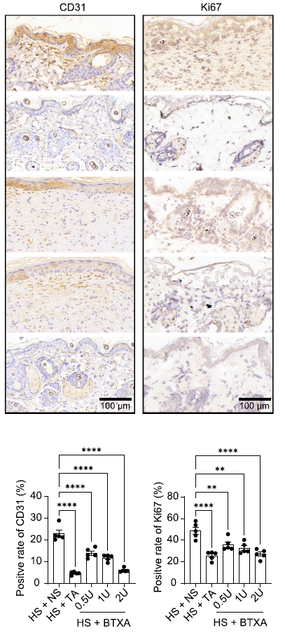
Triamcinolone acetonide (TA) (0.05 mL/g) significantly reduced the levels of CD31 and Ki67, marker proteins of angiogenesis and proliferation, in the scar tissue of HS mice.
-
-
Drug Test Anal
High-throughput liquid chromatography tandem mass spectrometry assay as initial testing procedure for analysis of total urinary fraction. [Abstract]2021 Feb;13(2):283-298. PMID: 32852861 -
Ghent university
Triamcinolone acetonide purchased from MedChemExpress. Usage Cited in: Ghent university
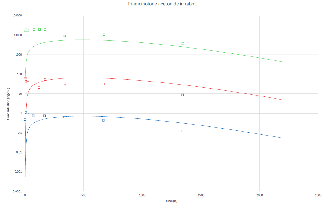
Simulation output of Triamcinolone acetonide (TA) (4 mg; i.v.; 2500 h) ocular rabbit model. Observed (squares) and predicted (solid line) values in VH (green), AH (red), and plasma (blue). Concentrations in ng/mL.
-
-


용액&용해도
In Vitro:
DMSO : 50 mg/mL (115.07 mM; Need ultrasonic; Hygroscopic DMSO has a significant impact on the solubility of product, please use newly opened DMSO)
H2O : < 0.1 mg/mL (insoluble)
Please refer to the solubility information to select the appropriate solvent. Once prepared, please aliquot and store the solution to prevent product inactivation from repeated freeze-thaw cycles.
Storage method and period of stock solution: -80°C, 6 months; -20°C, 1 month. When stored at -80°C, please use it within 6 months. When stored at -20°C, please use it within 1 month.
Please refer to the solubility information to select the appropriate solvent. Once prepared, please aliquot and store the solution to prevent product inactivation from repeated freeze-thaw cycles.
Storage method and period of stock solution: -80°C, 6 months; -20°C, 1 month. When stored at -80°C, please use it within 6 months. When stored at -20°C, please use it within 1 month.
Concentration (start) × Volume (start) = Concentration (final) × Volume (final)
In Vivo:
Select the appropriate dissolution method based on your experimental animal and administration route.
- For the following dissolution methods, please ensure to first prepare a clear stock solution using an In Vitro approach and then sequentially add co-solvents:
- To ensure reliable experimental results, the clarified stock solution can be appropriately stored based on storage conditions. As for the working solution for In Vivo experiments, it is recommended to prepare freshly and use it on the same day.
- The percentages shown for the solvents indicate their volumetric ratio in the final prepared solution. If precipitation or phase separation occurs during preparation, heat and/or sonication can be used to aid dissolution.
Add each solvent one by one: 10% DMSO 40% PEG300 5% Tween-80 45% Saline
Solubility: ≥ 2.08 mg/mL (4.79 mM); Clear solution
This protocol yields a clear solution of ≥ 2.08 mg/mL (saturation unknown).
Taking 1 mL working solution as an example, add 100 μL DMSO stock solution (20.8 mg/mL) to 400 μL PEG300, and mix evenly; then add 50 μL Tween-80 and mix evenly; then add 450 μL Saline to adjust the volume to 1 mL.
Preparation of Saline: Dissolve 0.9 g sodium chloride in ddH₂O and dilute to 100 mL to obtain a clear Saline solution.
Add each solvent one by one: 10% DMSO 90% (20% SBE-β-CD in Saline)
Solubility: ≥ 2.08 mg/mL (4.79 mM); Clear solution
This protocol yields a clear solution of ≥ 2.08 mg/mL (saturation unknown).
Taking 1 mL working solution as an example, add 100 μL DMSO stock solution (20.8 mg/mL) to 900 μL 20% SBE-β-CD in Saline, and mix evenly.
Preparation of 20% SBE-β-CD in Saline (4°C, storage for one week): 2 g SBE-β-CD powder is dissolved in 10 mL Saline, completely dissolve until clear.
In Vivo Dissolution Calculator
Please enter the basic information of animal experiments:
-
-
-
-
Recommended: Prepare an additional quantity of animals to account for potential losses during experiments.
Please enter your animal formula composition:
-
%DMSO +
Recommended: Keep the proportion of DMSO in working solution below 2% if your animal is weak.
-
%+
-
+%Tween-80 + +
-
%Saline +
The co-solvents required include: DMSO, . All of co-solvents are available by MedChemExpress (MCE). , Tween 80. All of co-solvents are available by MedChemExpress (MCE).
Working solution concentration: 0.22 mg/mL
Method for preparing stock solution: mg drug dissolved in μL DMSO. Stock solution concentration: mg/mL.
1. Take μL DMSO stock solution;
2. Add μL .
μL , mix evenly;
3. Then add μL Tween 80, mix evenly;
4. Then add μL
Please ensure that the stock solution in the first step is dissolved to a clear state, and add co-solvents in sequence. You can use ultrasonic heating (ultrasonic cleaner, recommended frequency 20-40 kHz), vortexing, etc. to assist dissolution.

Protocol
-
TPA/Croton Oil Ear Edema and Dermatitis
The TPA (12-O-tetradecanoylphorbol-13-acetate) and croton oil-induced mouse ear edema model is a well-established acute cutaneous inflammation system used to evaluate topical anti-inflammatory activity by measuring edema formation, neutrophil infiltration, vascular permeability, and cytokine-mediated skin responses in vivo. The inflammatory response is triggered by topical application of phorbol esters (TPA) or croton oil constituents, leading to rapid activation of protein kinase C signaling, leukocyte recruitment, and increased vascular permeability, which can be quantified by ear thickness, weight, dye extravasation, and biochemical markers such as myeloperoxidase (MPO) activity and pro-inflammatory mediators in ear tissue homogenates. This model is widely used for screening anti-inflammatory agents, where reductions in edema and inflammatory biomarkers reflect suppression of acute dermal inflammation and immune cell infiltration. Histological evaluation typically confirms epidermal
-
Endothelial Tube Formation Assay
Endothelial tube formation assay evaluates the ability of endothelial cells to attach, migrate, align, and organize into capillary-like networks when cultured on gelled basement membrane extract or Matrigel; the readout is the morphology and quantity of tube-like networks, which reflects an in vitro endothelial morphogenesis step related to angiogenesis. Basement membrane extract/Matrigel provides laminin-rich extracellular matrix cues that support endothelial differentiation into capillary-like structures, but it can contain biologically active growth factors, so growth-factor-reduced matrix is preferred when testing defined angiogenic stimulators or inhibitors.
-
Research Protocol for Inflammation-related Diseases
The NLRP3 inflammasome is a cytosolic innate immune signaling platform that integrates priming signals and danger-signal activation to promote caspase-1 activation, maturation of IL-1β and IL-18, and gasdermin D-mediated pyroptotic cell death. The core experimental logic is to determine whether inflammatory disease phenotypes are driven by increased NLRP3 expression, ASC-containing inflammasome assembly, caspase-1 cleavage, GSDMD cleavage, and extracellular release of IL-1β/IL-18 rather than by nonspecific cell injury alone. The pathway is strongly linked to inflammation-related disease phenotypes because monosodium urate crystals activate NALP3/NLRP3 inflammasome signaling in gout-like crystal inflammation, cholesterol crystals activate NLRP3 inflammasomes in atherogenesis models, and DSS-induced intestinal inflammation has been reported to involve NLRP3 inflammasome activity. However, experimental colitis studies also show context-dependent protective effects of NLRP3 inflammasome co
-
Cell Viability Determination by MTT Colorimetric Assay
The following protocol uses the MTT colorimetric assay as a classic literature-established method for assessing cell viability/metabolic activity in cultured mammalian cells. MTT[3-(4,5-dimethylthiazol-2-yl)-2,5-diphenyltetrazolium bromide] is reduced by metabolically active cells to a colored formazan product; the amount of formazan is quantified spectrophotometrically and provides an indirect measure of metabolically active viable cells. Importantly, MTT reduction reflects cellular oxidoreductase/metabolic activity rather than an absolute direct count of living cells, so changes in cellular metabolism can alter the signal independently of cell number.


순도&문서
-
Data Sheet (284 KB)
-
SDS (419 KB)
- English - EN (419 KB)
- Français - FR (419 KB)
- Deutsch - DE (419 KB)
- Norwegian - NO (419 KB)
- Español - ES (419 KB)
- Swedish - SV (419 KB)
- Italian - IT (419 KB)
- Korean - KR (419 KB)
- Portuguese - PT (419 KB)
-
Handling Instructions (2659 KB)
References


Complete Stock Solution Preparation Table
Please refer to the solubility information to select the appropriate solvent. Once prepared, please aliquot and store the solution to prevent product inactivation from repeated freeze-thaw cycles.
Storage method and period of stock solution: -80°C, 6 months; -20°C, 1 month. When stored at -80°C, please use it within 6 months. When stored at -20°C, please use it within 1 month.
| Optional Solvent | Concentration Solvent Mass | 1 mg | 5 mg | 10 mg | 25 mg |
|---|---|---|---|---|---|
| DMSO | 1 mM | 2.3015 mL | 11.5075 mL | 23.0150 mL | 57.5374 mL |
| 5 mM | 0.4603 mL | 2.3015 mL | 4.6030 mL | 11.5075 mL | |
| 10 mM | 0.2301 mL | 1.1507 mL | 2.3015 mL | 5.7537 mL | |
| 15 mM | 0.1534 mL | 0.7672 mL | 1.5343 mL | 3.8358 mL | |
| 20 mM | 0.1151 mL | 0.5754 mL | 1.1507 mL | 2.8769 mL | |
| 25 mM | 0.0921 mL | 0.4603 mL | 0.9206 mL | 2.3015 mL | |
| 30 mM | 0.0767 mL | 0.3836 mL | 0.7672 mL | 1.9179 mL | |
| 40 mM | 0.0575 mL | 0.2877 mL | 0.5754 mL | 1.4384 mL | |
| 50 mM | 0.0460 mL | 0.2301 mL | 0.4603 mL | 1.1507 mL | |
| 60 mM | 0.0384 mL | 0.1918 mL | 0.3836 mL | 0.9590 mL | |
| 80 mM | 0.0288 mL | 0.1438 mL | 0.2877 mL | 0.7192 mL | |
| 100 mM | 0.0230 mL | 0.1151 mL | 0.2301 mL | 0.5754 mL |


Triamcinolone acetonide Related Classifications
HY-B0636 Related Classifications
 Powered by Bioz