



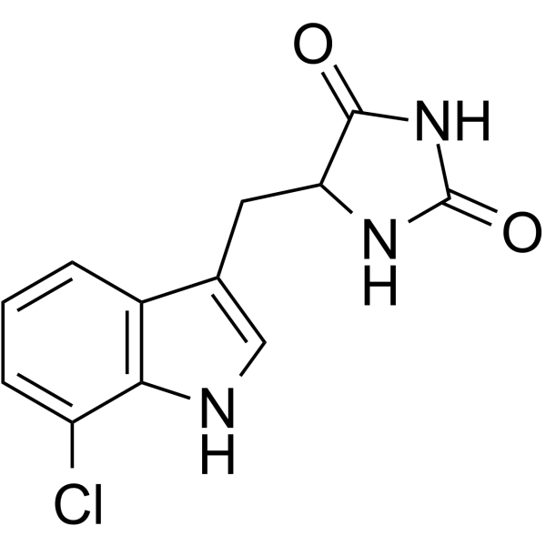
KWCN-41

Based on 1 Customer Validation
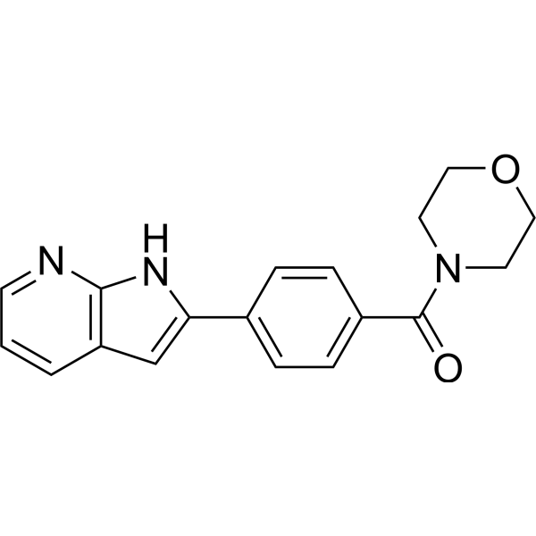
KWCN-41 is a selective and efficient inhibitor of RIPK1 kinase with an IC50 value of 88 nM. KWCN-41 specifically inhibits cell necrosis but does not inhibit apoptosis. KWCN-41 also has anti-inflammatory effects.
For research use only. We do not sell to patients.
- Purity : 99.29%
- CAS No.: 2913223-17-1
- Formula: C18H17N3O2
- Molecular Weight:307.35
-
Storage:Powder -20°C, 3 years , 4°C, 2 years ; In solvent -80°C, 6 months , -20°C, 1 month

To place orders, for customer services and technical support, please contact: MedChemExpress USA
Tel: 609-228-6898 E-mail: [email protected] [email protected]


-
 Biological Activity
-
Chemical Information
-
Solvent & Solubility
- Protocols
- Purity & Documentation
- References
-
 Help & FAQs




Biological Activity
Description
IC50 & Target
[1]|
RIPK1 88 nM (IC50) |
Cellular Effect
|
Cell Line

|
Type | Value | Description | References |
|---|---|---|---|---|
| L929 | CC50 |
>50 μM
Compound: 41; KWCN-41
|
Cytotoxicity against mouse L929 cells by CCK-8 assay
Cytotoxicity against mouse L929 cells by CCK-8 assay
|
[PMID: 36801518] |
| L929 | EC50 |
0.056 μM
Compound: 41; KWCN-41
|
Anti-necroptosis activity against TNFalpha/z-VAD-fmk-induced mouse L929 cell necroptosis model assessed as cell viability pretreated with compound for 1 hr followed by treated with TZ for 6 hrs
Anti-necroptosis activity against TNFalpha/z-VAD-fmk-induced mouse L929 cell necroptosis model assessed as cell viability pretreated with compound for 1 hr followed by treated with TZ for 6 hrs
|
[PMID: 36801518] |
| L929 | EC50 |
56 nM
Compound: KWCN-41; L8
|
Cytotoxicity against mouse L929 cells assessed as cell viability measured after 24 hrs by MTT assay
Cytotoxicity against mouse L929 cells assessed as cell viability measured after 24 hrs by MTT assay
|
[PMID: 38199165] |
In Vitro
KWCN-41 (10, 50, 250 nM; 1 h) inhibits L929 cell necroptosis in a dosedependent manner and increases the number of cells in the viable cell zone[1].
KWCN-41 (10, 50, 250 nM; 0.5, 1.5, 2.5 h) inhibits the phosphorylation of RIPK1/3 and MLKL in L929 cells[1].
KWCN-41 (0-10 μM) inhibits TZ-induced necroptosis of L929 cells in a dose-dependent manner and similarly protects cells from TSZ (TNF-α, Smac mimetic, and z-VAD-FMK)-induced necroptosis of HT-29 and U937 cells[1].
MedChemExpress (MCE) has not independently confirmed the accuracy of these methods. They are for reference only.
-
Cell Line:L929 cells
-
Concentration:10, 50, 250 nM
-
Incubation Time:1 h
-
Result:Inhibited L929 cell necroptosis in a Dosedependent manner and increased the number of cells in the viable cell zone.
In Vivo
MedChemExpress (MCE) has not independently confirmed the accuracy of these methods. They are for reference only.
-
Animal Model:Inflammatory mouse model[1]
-
Dosage:10, 25, 40 mg/kg
-
Administration:i.p.; dissolved in saline containing 10% DMSO and 20% Tween80; 72 h
-
Result:At 25, 40 mg/kg completely inhibited TNF-α induced death in mice, achieving 100% survival compared to 75% survival at 10 mg/kg.


Chemical Information
-
CAS No. 2913223-17-1
-
Appearance Solid
-
Molecular Weight 307.35
-
Formula C18H17N3O2
-
Color Off-white to light yellow
-
SMILES
O=C(N1CCOCC1)C2=CC=C(C3=CC4=C(N3)N=CC=C4)C=C2
-
Shipping
Room temperature in continental US; may vary elsewhere.
-
Storage
Powder -20°C 3 years 4°C 2 years In solvent -80°C 6 months -20°C 1 month


Solvent & Solubility
In Vitro:
DMSO : 100 mg/mL (325.36 mM; Need ultrasonic; Hygroscopic DMSO has a significant impact on the solubility of product, please use newly opened DMSO)
Please refer to the solubility information to select the appropriate solvent. Once prepared, please aliquot and store the solution to prevent product inactivation from repeated freeze-thaw cycles.
Storage method and period of stock solution: -80°C, 6 months; -20°C, 1 month. When stored at -80°C, please use it within 6 months. When stored at -20°C, please use it within 1 month.
Please refer to the solubility information to select the appropriate solvent. Once prepared, please aliquot and store the solution to prevent product inactivation from repeated freeze-thaw cycles.
Storage method and period of stock solution: -80°C, 6 months; -20°C, 1 month. When stored at -80°C, please use it within 6 months. When stored at -20°C, please use it within 1 month.
Concentration (start) × Volume (start) = Concentration (final) × Volume (final)


Purity & Documentation
-
Data Sheet (273 KB)
-
SDS (251 KB)
- English - EN (251 KB)
- Français - FR (251 KB)
- Deutsch - DE (251 KB)
- Norwegian - NO (251 KB)
- Español - ES (251 KB)
- Swedish - SV (251 KB)
- Italian - IT (251 KB)
- Korean - KR (251 KB)
- Portuguese - PT (251 KB)
-
Handling Instructions (2659 KB)
References


Complete Stock Solution Preparation Table
Please refer to the solubility information to select the appropriate solvent. Once prepared, please aliquot and store the solution to prevent product inactivation from repeated freeze-thaw cycles.
Storage method and period of stock solution: -80°C, 6 months; -20°C, 1 month. When stored at -80°C, please use it within 6 months. When stored at -20°C, please use it within 1 month.
| Optional Solvent | Concentration Solvent Mass | 1 mg | 5 mg | 10 mg | 25 mg |
|---|---|---|---|---|---|
| DMSO | 1 mM | 3.2536 mL | 16.2681 mL | 32.5362 mL | 81.3405 mL |
| 5 mM | 0.6507 mL | 3.2536 mL | 6.5072 mL | 16.2681 mL | |
| 10 mM | 0.3254 mL | 1.6268 mL | 3.2536 mL | 8.1340 mL | |
| 15 mM | 0.2169 mL | 1.0845 mL | 2.1691 mL | 5.4227 mL | |
| 20 mM | 0.1627 mL | 0.8134 mL | 1.6268 mL | 4.0670 mL | |
| 25 mM | 0.1301 mL | 0.6507 mL | 1.3014 mL | 3.2536 mL | |
| 30 mM | 0.1085 mL | 0.5423 mL | 1.0845 mL | 2.7113 mL | |
| 40 mM | 0.0813 mL | 0.4067 mL | 0.8134 mL | 2.0335 mL | |
| 50 mM | 0.0651 mL | 0.3254 mL | 0.6507 mL | 1.6268 mL | |
| 60 mM | 0.0542 mL | 0.2711 mL | 0.5423 mL | 1.3557 mL | |
| 80 mM | 0.0407 mL | 0.2034 mL | 0.4067 mL | 1.0168 mL | |
| 100 mM | 0.0325 mL | 0.1627 mL | 0.3254 mL | 0.8134 mL |


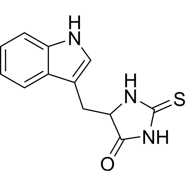
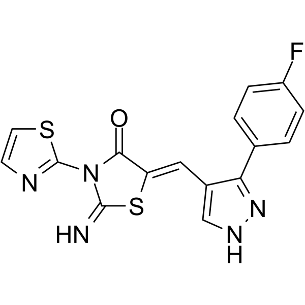
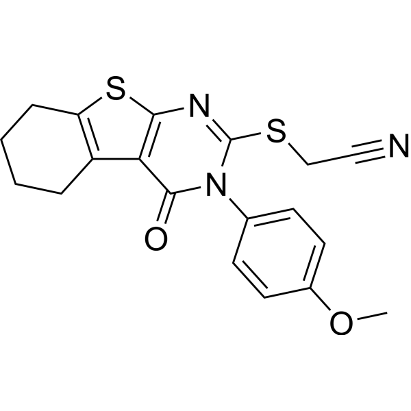
KWCN-41 Related Classifications
HY-149258 Related Classifications
 Powered by Bioz