



Levomepromazine
Based on 1 publication(s) in Google Scholar
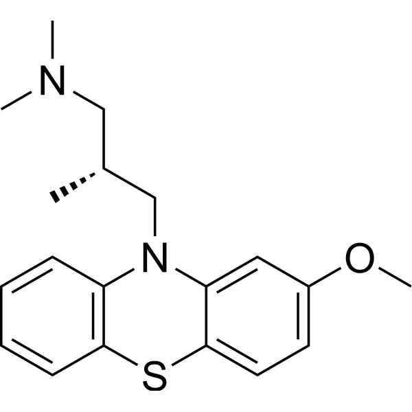
Levomepromazine (Methotrimeprazine) is an orally active antipsychotic compound and Ca2+ release inducer. Levomepromazine inhibits SERCA pump and induces an increase in cytoplasmic Ca2+ levels. Levomepromazine has antagonistic effects on a variety of neurotransmitter receptors, including dopamine, cholinergic, serotonin, and histamine receptors. Levomepromazine can induce adaptive ER stress and autophagy. In addition, Levomepromazine has antiviral, anti-inflammatory, neuroprotective and analgesic, sedative and anti-injurious activities. Levomepromazine can be used in the study psychiatric disorders and relieving nausea and vomiting.
For research use only. We do not sell to patients.
- Purity : 99.94%
- CAS No.: 60-99-1
- Formula: C19H24N2OS
- Molecular Weight:328.47
-
Storage:Powder -20°C, 3 years , 4°C, 2 years ; In solvent -80°C, 6 months , -20°C, 1 month

To place orders, for customer services and technical support, please contact: MedChemExpress USA
Tel: 609-228-6898 E-mail: [email protected] [email protected]


-
 Biological Activity
-
Chemical Information
-
Solvent & Solubility
- Protocols
- Purity & Documentation
- References
-
 Help & FAQs




Publications Citing Use of MedChemExpress (MCE) Levomepromazine
MoreAll 5-HT Receptor Isoforms
MoreAll Dopamine Receptor Isoforms
MoreAll Histamine Receptor Isoforms
MoreAll Calcium Channel Isoforms
More

Biological Activity
Description
Cellular Effect
|
Cell Line

|
Type | Value | Description | References |
|---|---|---|---|---|
| Huh-7 | CC50 |
24.5 μM
Compound: 50
|
Cytotoxicity against human Huh-7 cells
Cytotoxicity against human Huh-7 cells
|
[PMID: 32283298] |
In Vitro
Levomepromazine (10 μM; 24 h) reduces Japanese encephalitis virus (JEV)-induced Neuro2a cell death, reduces JEV RNA levels and viral titers, and significantly inhibits JEV protein translation/replication complex formation and ROS production[1].
Levomepromazine (10 μM; 24 h) decreases the inflammatory response of microglia by inducing autophagy[1].
MedChemExpress (MCE) has not independently confirmed the accuracy of these methods. They are for reference only.
-
Cell Line:Japanese encephalitis virus treated Neuro2a
-
Concentration:10 μM
-
Incubation Time:24 h
-
Result:Reduced the level of JEV RNA.
In Vivo
Levomepromazine (5-20 mg/kg; intraperitoneal injection; single dose) has analgesic, sedative and anti-injurious effects in mice[2].
MedChemExpress (MCE) has not independently confirmed the accuracy of these methods. They are for reference only.
-
Animal Model:Japanese encephalitis virus-S3 infected C57BL/6 mouse[1]
-
Dosage:2 mg/kg
-
Administration:Oral gavage (i.g.); 15 days
-
Result:Delayed virus invasion into the brain, and significantly lower viremia.
Showed complete protection of the barrier.
Reduced levels of multiple pro-inflammatory cytokines and interferon in the brains of infected mice.


Chemical Information
-
CAS No. 60-99-1
-
Appearance Solid
-
Molecular Weight 328.47
-
Formula C19H24N2OS
-
Color White to off-white
-
SMILES
COC1=CC=C2SC3=C(N(C2=C1)C[C@@H](CN(C)C)C)C=CC=C3
-
Synonyms
Methotrimeprazine
-
Shipping
Room temperature in continental US; may vary elsewhere.
-
Storage
Powder -20°C 3 years 4°C 2 years In solvent -80°C 6 months -20°C 1 month


Publications (1)
-
Journal Impact Factor
-
Most Recent
-
Br J Pharmacol
Tricyclic antipsychotics and antidepressants can inhibit α5-containing GABAA receptors by two distinct mechanisms. [Abstract]2022 Jul;179(14):3675-3692. PMID: 35088415


Solvent & Solubility
In Vitro:
DMSO : 41.67 mg/mL (126.86 mM; ultrasonic and warming and heat to 60°C; Hygroscopic DMSO has a significant impact on the solubility of product, please use newly opened DMSO)
Please refer to the solubility information to select the appropriate solvent. Once prepared, please aliquot and store the solution to prevent product inactivation from repeated freeze-thaw cycles.
Storage method and period of stock solution: -80°C, 6 months; -20°C, 1 month. When stored at -80°C, please use it within 6 months. When stored at -20°C, please use it within 1 month.
Please refer to the solubility information to select the appropriate solvent. Once prepared, please aliquot and store the solution to prevent product inactivation from repeated freeze-thaw cycles.
Storage method and period of stock solution: -80°C, 6 months; -20°C, 1 month. When stored at -80°C, please use it within 6 months. When stored at -20°C, please use it within 1 month.
Concentration (start) × Volume (start) = Concentration (final) × Volume (final)
In Vivo:
Select the appropriate dissolution method based on your experimental animal and administration route.
- For the following dissolution methods, please ensure to first prepare a clear stock solution using an In Vitro approach and then sequentially add co-solvents:
- To ensure reliable experimental results, the clarified stock solution can be appropriately stored based on storage conditions. As for the working solution for In Vivo experiments, it is recommended to prepare freshly and use it on the same day.
- The percentages shown for the solvents indicate their volumetric ratio in the final prepared solution. If precipitation or phase separation occurs during preparation, heat and/or sonication can be used to aid dissolution.
Add each solvent one by one: 10% DMSO 40% PEG300 5% Tween-80 45% Saline
Solubility: ≥ 1.43 mg/mL (4.35 mM); Clear solution
This protocol yields a clear solution of ≥ 1.43 mg/mL (saturation unknown).
Taking 1 mL working solution as an example, add 100 μL DMSO stock solution (14.3 mg/mL) to 400 μL PEG300, and mix evenly; then add 50 μL Tween-80 and mix evenly; then add 450 μL Saline to adjust the volume to 1 mL.
Preparation of Saline: Dissolve 0.9 g sodium chloride in ddH₂O and dilute to 100 mL to obtain a clear Saline solution.
Add each solvent one by one: 10% DMSO 90% (20% SBE-β-CD in Saline)
Solubility: ≥ 1.43 mg/mL (4.35 mM); Clear solution
This protocol yields a clear solution of ≥ 1.43 mg/mL (saturation unknown).
Taking 1 mL working solution as an example, add 100 μL DMSO stock solution (14.3 mg/mL) to 900 μL 20% SBE-β-CD in Saline, and mix evenly.
Preparation of 20% SBE-β-CD in Saline (4°C, storage for one week): 2 g SBE-β-CD powder is dissolved in 10 mL Saline, completely dissolve until clear.
In Vivo Dissolution Calculator
Please enter the basic information of animal experiments:
-
-
-
-
Recommended: Prepare an additional quantity of animals to account for potential losses during experiments.
Please enter your animal formula composition:
-
%DMSO +
Recommended: Keep the proportion of DMSO in working solution below 2% if your animal is weak.
-
%+
-
+%Tween-80 + +
-
%Saline +
The co-solvents required include: DMSO, . All of co-solvents are available by MedChemExpress (MCE). , Tween 80. All of co-solvents are available by MedChemExpress (MCE).
Working solution concentration: 0.22 mg/mL
Method for preparing stock solution: mg drug dissolved in μL DMSO. Stock solution concentration: mg/mL.
1. Take μL DMSO stock solution;
2. Add μL .
μL , mix evenly;
3. Then add μL Tween 80, mix evenly;
4. Then add μL
Please ensure that the stock solution in the first step is dissolved to a clear state, and add co-solvents in sequence. You can use ultrasonic heating (ultrasonic cleaner, recommended frequency 20-40 kHz), vortexing, etc. to assist dissolution.


Purity & Documentation
-
Data Sheet (278 KB)
-
SDS (393 KB)
- English - EN (393 KB)
- Français - FR (393 KB)
- Deutsch - DE (393 KB)
- Norwegian - NO (393 KB)
- Español - ES (393 KB)
- Swedish - SV (393 KB)
- Italian - IT (393 KB)
- Korean - KR (393 KB)
- Portuguese - PT (393 KB)
-
Handling Instructions (2659 KB)
References
[1]. Prajapat SK, et al. Methotrimeprazine is a neuroprotective antiviral in JEV infection via adaptive ER stress and autophagy. EMBO Mol Med. 2024 Jan;16(1):185-217. [Content Brief]
[2]. Petts HV, et al. Interactions of morphine and methotrimeprazine in mouse and man with respect to analgesia, respiration and sedation. Br J Anaesth. 1983 May;55(5):437-41. [Content Brief]
[3]. Cox L, et al. Levomepromazine for nausea and vomiting in palliative care. Cochrane Database Syst Rev. 2015 Nov 2;(11):CD009420. [Content Brief]
[4]. Sivaraman P, et al. Levomepromazine for schizophrenia. Cochrane Database Syst Rev. 2010 Oct 6;(10):CD007779. [Content Brief]


Complete Stock Solution Preparation Table
Please refer to the solubility information to select the appropriate solvent. Once prepared, please aliquot and store the solution to prevent product inactivation from repeated freeze-thaw cycles.
Storage method and period of stock solution: -80°C, 6 months; -20°C, 1 month. When stored at -80°C, please use it within 6 months. When stored at -20°C, please use it within 1 month.
| Optional Solvent | Concentration Solvent Mass | 1 mg | 5 mg | 10 mg | 25 mg |
|---|---|---|---|---|---|
| DMSO | 1 mM | 3.0444 mL | 15.2221 mL | 30.4442 mL | 76.1105 mL |
| 5 mM | 0.6089 mL | 3.0444 mL | 6.0888 mL | 15.2221 mL | |
| 10 mM | 0.3044 mL | 1.5222 mL | 3.0444 mL | 7.6110 mL | |
| 15 mM | 0.2030 mL | 1.0148 mL | 2.0296 mL | 5.0740 mL | |
| 20 mM | 0.1522 mL | 0.7611 mL | 1.5222 mL | 3.8055 mL | |
| 25 mM | 0.1218 mL | 0.6089 mL | 1.2178 mL | 3.0444 mL | |
| 30 mM | 0.1015 mL | 0.5074 mL | 1.0148 mL | 2.5370 mL | |
| 40 mM | 0.0761 mL | 0.3806 mL | 0.7611 mL | 1.9028 mL | |
| 50 mM | 0.0609 mL | 0.3044 mL | 0.6089 mL | 1.5222 mL | |
| 60 mM | 0.0507 mL | 0.2537 mL | 0.5074 mL | 1.2685 mL | |
| 80 mM | 0.0381 mL | 0.1903 mL | 0.3806 mL | 0.9514 mL | |
| 100 mM | 0.0304 mL | 0.1522 mL | 0.3044 mL | 0.7611 mL |


Levomepromazine Related Classifications
HY-B1693 Related Classifications
 Powered by Bioz