



Manelimab
Manelimab (BCD-135) is a monoclonal antibody targeting PD-L1 with PD-L1 inhibitory activity. Manelimab blocks the interaction between PD-L1 and PD-1 to abrogate the immunosuppressive signals of T cells in the tumor microenvironment. Manelimab is applicable for tumor-related research.
For research use only. We do not sell to patients.
- CAS No.: 2168561-26-8
-
Storage:
Please store the product under the recommended conditions in the Certificate of Analysis.


To place orders, for customer services and technical support, please contact: MedChemExpress USA
Tel: 609-228-6898 E-mail: [email protected] [email protected]


-
 Biological Activity
-
Chemical Information
- Protocols
- Purity & Documentation
- References
-
 Help & FAQs


-

HY-P9902
PembrolizumabPD-1 Inhibitor -

HY-P9904
AtezolizumabAnti-PD-L1 Antibody -
HY-P9903
NivolumabPD-1 Inhibitor -

HY-P9919
DurvalumabPD-L1 Blocking Antibody -

HY-P99052
TislelizumabPD-1 Inhibitor -

HY-P9978
ToripalimabPD-1 Inhibitor -

HY-108730
AvelumabAnti-PD-L1 Antibody -

HY-P99048
SintilimabPD-1/PD-L1 Inhibitor -
HY-P9919A
Durvalumab (anti-PD-L1)PD-1/PD-L1 Inhibitor -

HY-P9971
CamrelizumabPD-1 Neutralizing Antibody -

HY-P99203
CemiplimabPD-1 Inhibitor -

HY-P99785
OpucolimabAnti-PD-L1 Antibody


Biological Activity
Description
Isotype
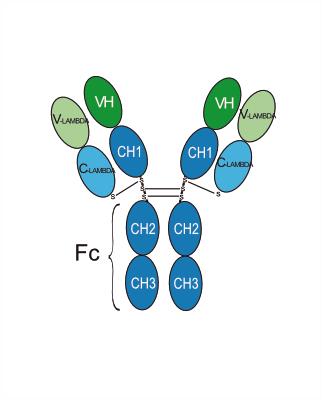
Human IgG1 lambda2
Recommend Isotype Controls
Species Reactivity
Human
IC50 & Target
B7-H1/PD-L1/CD274
In Vitro
MedChemExpress (MCE) has not independently confirmed the accuracy of these methods. They are for reference only.
Clinical Trial
| NCT Number | Sponsor | Condition | Start Date |
Phase

|
|---|---|---|---|---|
| NCT01329991 | Plexxikon| | 2011-05 | PHASE1 |
Gene ID
Accession
Conjugated
Unconjugated
Reconsititution
The product can be reconstituted/diluted with sterile PBS or saline.
Format
-
Human IgG1 lambda2
Application
ELISA, FACS, Functional assay


Chemical Information
-
CAS No. 2168561-26-8
-
SMILES
[Manelimab]
-
Synonyms
BCD-135
-
Formulation
Please refer to the lot-specific COA for specific buffer information.
-
Storage
Please store the product under the recommended conditions in the Certificate of Analysis.


Purity & Documentation
References

Calculators
Concentration (start) × Volume (start) = Concentration (final) × Volume (final)


Manelimab Related Classifications
HY-P99723 Related Classifications
 Powered by Bioz