



MIF-IN-5
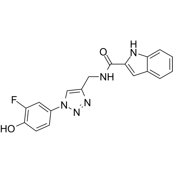
MIF-IN-5 (compound 1d) is a potent and reversible macrophage migration inhibitory factor (MIF) competitive inhibitor with an IC50 of 4.8 μM and a Ki value of 3.3 μM, respectively.
For research use only. We do not sell to patients.
- CAS No.: 2582755-31-3
- Formula: C18H14FN5O2
- Molecular Weight:351.33
-
Storage:
Please store the product under the recommended conditions in the Certificate of Analysis.

To place orders, for customer services and technical support, please contact: MedChemExpress USA
Tel: 609-228-6898 E-mail: [email protected] [email protected]


-
 Biological Activity
-
Chemical Information
- Protocols
- Purity & Documentation
- References
-
 Help & FAQs




Biological Activity
Description
IC50 & Target


Chemical Information
-
CAS No. 2582755-31-3
-
Molecular Weight 351.33
-
Formula C18H14FN5O2
-
SMILES
O=C(NCC1=CN(N=N1)C2=CC(F)=C(C=C2)O)C3=CC4=CC=CC=C4N3
-
Shipping
Room temperature in continental US; may vary elsewhere.
-
Storage
Please store the product under the recommended conditions in the Certificate of Analysis.

Protocols
-
Phagocytosis Functional Assay
A phagocytosis functional assay measures the ability of phagocytic cells, such as neutrophils, macrophages, monocytes, or microglia/macrophages, to bind and internalize particulate targets including bacteria, yeast particles, beads, or myelin particles. Fluorescent flow-cytometry assays detect target uptake as fluorescence associated with gated phagocytes, while pH-sensitive dyes such as pHrodo increase signal in acidic phagosomal compartments and therefore preferentially report internalized particles rather than particles remaining outside the cell. Microscopy or high-content imaging can be used to confirm intracellular localization and, in some protocols, to follow uptake kinetics.
-
Multiplex immunofluorescence IHC
Multiplex immunofluorescence IHC detects multiple protein biomarkers in one tissue section by sequential antibody staining, HRP-mediated tyramide fluorophore deposition, heat-mediated antibody stripping, nuclear counterstaining, multispectral imaging, spectral unmixing, and digital cell phenotyping; TSA deposits fluorophore near the antigen so the fluorescence signal remains after primary and secondary antibodies are removed, enabling repeated staining cycles, including with antibodies from the same host species. Classic FFPE tumor immune-profiling applications use panels such as CD3, CD8, CD68/CD163, FOXP3, PD-1, PD-L1, pancytokeratin, Ki67, and DAPI to identify tumor cells, immune-cell subsets, checkpoint-marker expression, co-expression phenotypes, cell density, and spatial relationships in the tumor microenvironment.
-
Cell migration
Cell migration is a method that plays an important role in wound healing, cell differentiation, embryonic development, etc.
-
Multiplex immunohistochemistry
Multiplex immunohistochemistry (mIHC), also known as tyramide dignal amplification (TSA), is an enzymatic detection method that uses horseradish peroxidase (HRP) to perform high-density in-situ labeling of target proteins or nucleic acids.


Purity & Documentation
References

Calculators
Concentration (start) × Volume (start) = Concentration (final) × Volume (final)


MIF-IN-5 Related Classifications
HY-146318 Related Classifications
 Powered by Bioz