



ML00253764

Based on 1 Customer Validation
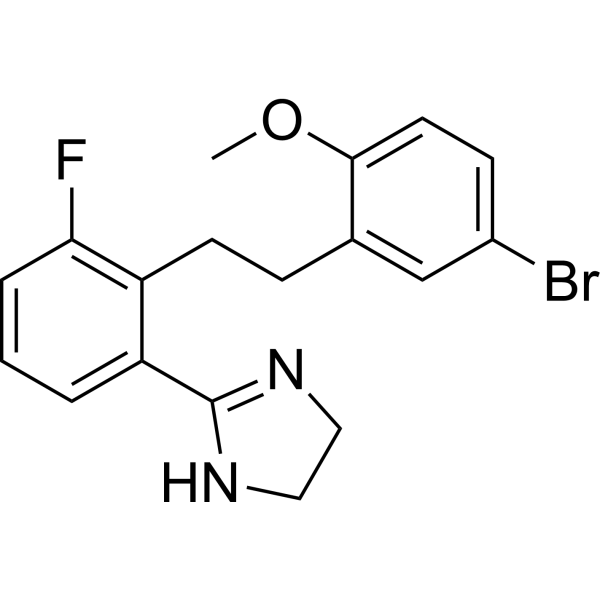
ML00253764 is a selective melanocortin receptor 4 (MC4R) antagonist, can induce apoptosis by inhibiting ERK1/2 and Akt phosphorylation, and has anticancer activity.
For research use only. We do not sell to patients.
- Purity : 99.71%
- CAS No.: 681847-92-7
- Formula: C18H18BrFN2O
- Molecular Weight:377.25
-
Storage:Powder -20°C, 3 years , 4°C, 2 years ; In solvent -80°C, 6 months , -20°C, 1 month

To place orders, for customer services and technical support, please contact: MedChemExpress USA
Tel: 609-228-6898 E-mail: [email protected] [email protected]


-
 Biological Activity
-
Chemical Information
-
Solvent & Solubility
- Protocols
- Purity & Documentation
- References
-
 Help & FAQs


-
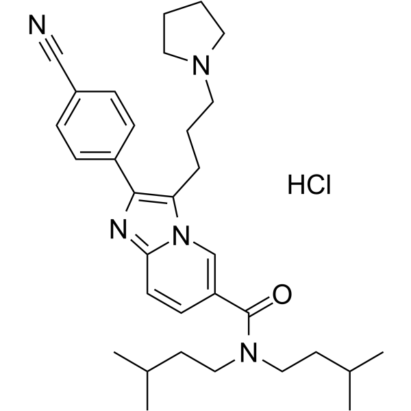
HY-11029
SNT-207707MC-4 Receptor Antagonist -
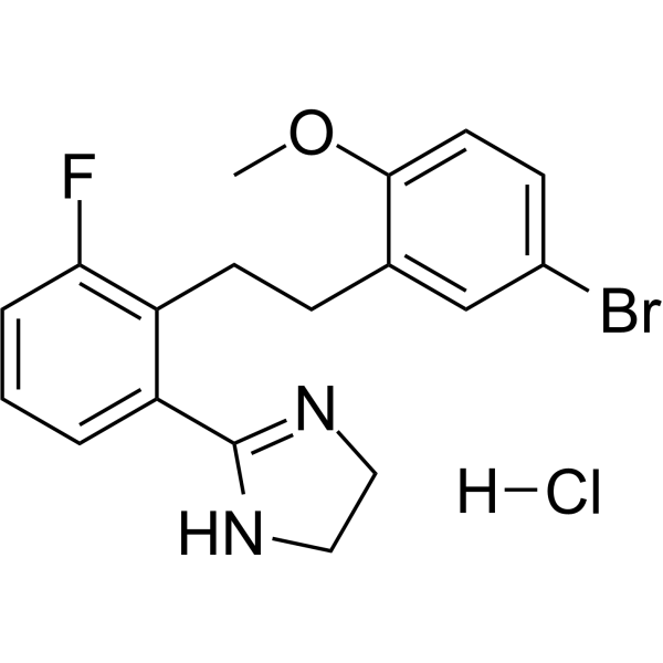
HY-110123
ML-00253764 hydrochlorideMC4R Antagonist -
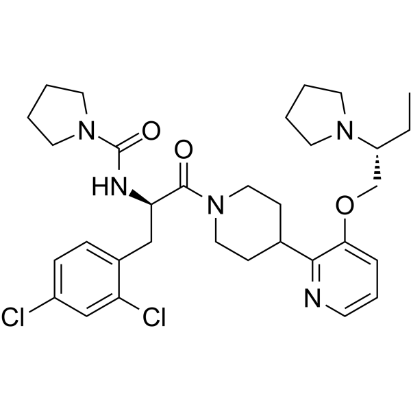
HY-11030A
SNT-207858 free baseMelanocortin Receptor Antagonist -
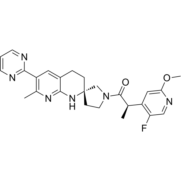
HY-148349
PF-07258669MC4 Antagonist -
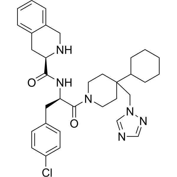
HY-10624
THIQMC4R Agonist -
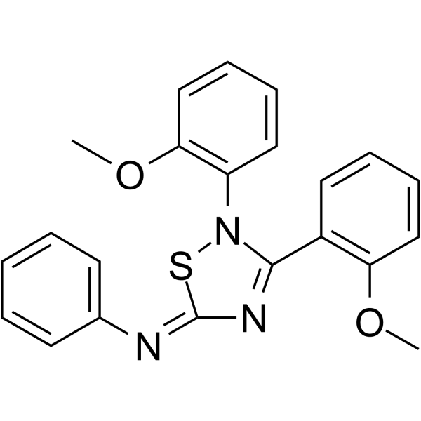
HY-107139
JNJ-10229570Melanocortin Receptor Antagonist -
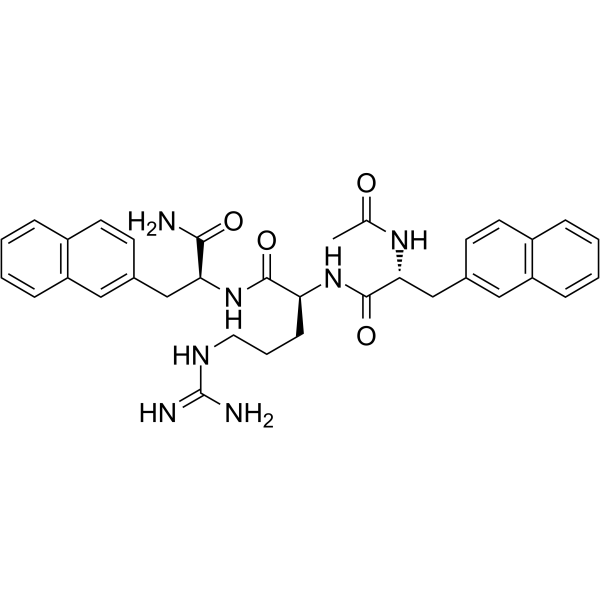
HY-107627
MCL0020MC4 Receptor Antagonist -
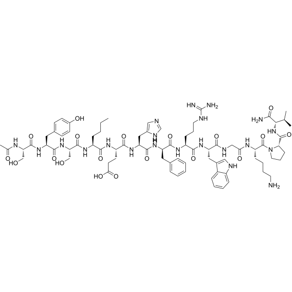
HY-N2466
Melanotan IMelanocortin Receptor Activator -
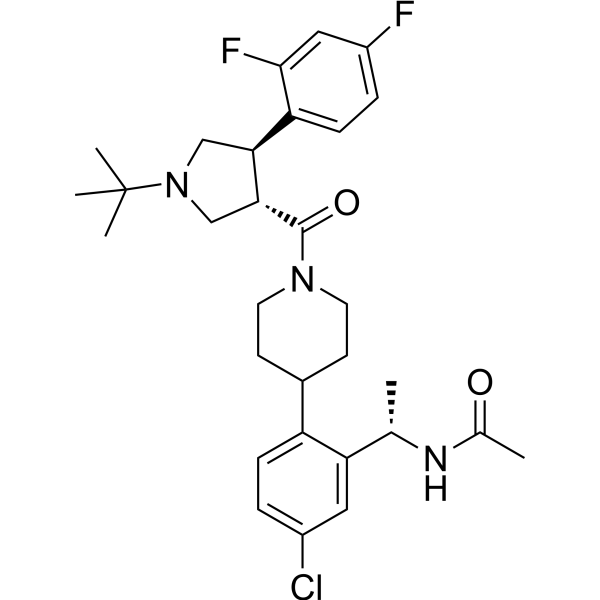
HY-118930
MK-0493MC4R Agonist -
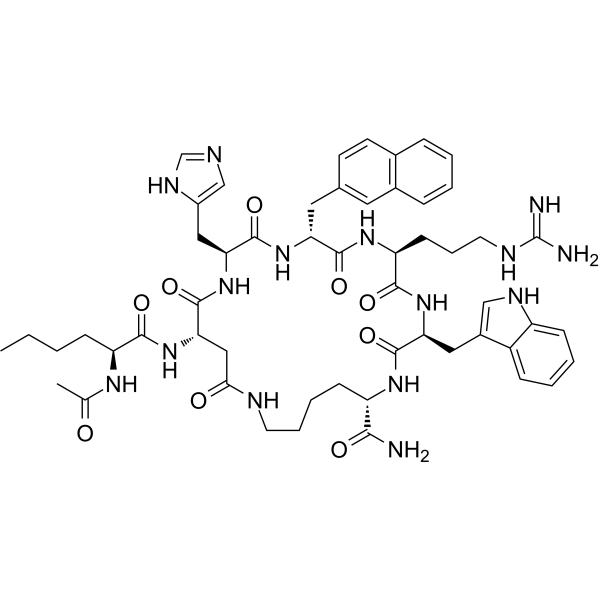
HY-P0227
SHU 9119MC3/4R Antagonist -

HY-19870
SetmelanotideMC4R Agonist -
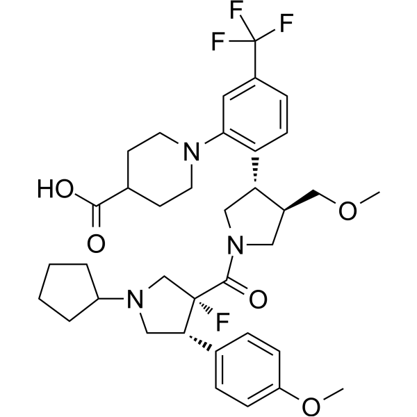
HY-109114
DersimelagonMC1R Agonist


Biological Activity
Description
Cellular Effect
|
Cell Line

|
Type | Value | Description | References |
|---|---|---|---|---|
| HEK293 | IC50 |
1030 nM
Compound: 5
|
Antagonist activity at the mutant MC4R Y268F expressed in HEK293 cells by cAMP accumulation
Antagonist activity at the mutant MC4R Y268F expressed in HEK293 cells by cAMP accumulation
|
[PMID: 16451057] |
| HEK293 | IC50 |
160 nM
Compound: 5
|
Antagonist activity at the mutant MC4R I125A expressed in HEK293 cells by cAMP accumulation
Antagonist activity at the mutant MC4R I125A expressed in HEK293 cells by cAMP accumulation
|
[PMID: 16451057] |
| HEK293 | IC50 |
2708 nM
Compound: 5
|
Antagonist activity at the mutant MC4R Y268A expressed in HEK293 cells by cAMP accumulation
Antagonist activity at the mutant MC4R Y268A expressed in HEK293 cells by cAMP accumulation
|
[PMID: 16451057] |
| HEK293 | IC50 |
2860 nM
Compound: 5
|
Antagonist activity at the mutant MC4R F184L expressed in HEK293 cells by cAMP accumulation
Antagonist activity at the mutant MC4R F184L expressed in HEK293 cells by cAMP accumulation
|
[PMID: 16451057] |
| HEK293 | IC50 |
2900 nM
Compound: 5
|
Antagonist activity at the mutant MC4R Y287A expressed in HEK293 cells by cAMP accumulation
Antagonist activity at the mutant MC4R Y287A expressed in HEK293 cells by cAMP accumulation
|
[PMID: 16451057] |
| HEK293 | IC50 |
355 nM
Compound: 5
|
Antagonist activity at the mutant MC4R Y287F expressed in HEK293 cells by cAMP accumulation
Antagonist activity at the mutant MC4R Y287F expressed in HEK293 cells by cAMP accumulation
|
[PMID: 16451057] |
| HEK293 | IC50 |
3770 nM
Compound: 5
|
Antagonist activity at the mutant MC4R F184A expressed in HEK293 cells by cAMP accumulation
Antagonist activity at the mutant MC4R F184A expressed in HEK293 cells by cAMP accumulation
|
[PMID: 16451057] |
| HEK293 | IC50 |
560 nM
Compound: 5
|
Antagonist activity at the human wild type MC4R expressed in HEK293 cells by cAMP accumulation
Antagonist activity at the human wild type MC4R expressed in HEK293 cells by cAMP accumulation
|
[PMID: 16451057] |
| HEK293 | IC50 |
6190 nM
Compound: 5
|
Antagonist activity at the mutant MC4R I125L expressed in HEK293 cells by cAMP accumulation
Antagonist activity at the mutant MC4R I125L expressed in HEK293 cells by cAMP accumulation
|
[PMID: 16451057] |
| HEK293 | IC50 |
708 nM
Compound: 5
|
Antagonist activity at the mutant MC4R I291A expressed in HEK293 cells by cAMP accumulation
Antagonist activity at the mutant MC4R I291A expressed in HEK293 cells by cAMP accumulation
|
[PMID: 16451057] |
In Vitro
ML00253764 (0.001-50 μM, 24 h or 72 h) has significant time- and concentration-dependent inhibitory activity on the proliferation of human glioblastoma cells, and the IC50 value for U-118 cells is 6.56 μM, which can induce U -87 cells apoptosis and show significant inhibition of ERK1/2 phosphorylation in both cell lines[1].
MedChemExpress (MCE) has not independently confirmed the accuracy of these methods. They are for reference only. Further protocols information, click here.
In Vivo
MedChemExpress (MCE) has not independently confirmed the accuracy of these methods. They are for reference only.


Chemical Information
-
CAS No. 681847-92-7
-
Appearance Solid
-
Molecular Weight 377.25
-
Formula C18H18BrFN2O
-
Color White to off-white
-
SMILES
FC1=C(CCC2=CC(Br)=CC=C2OC)C(C3=NCCN3)=CC=C1
-
Shipping
Room temperature in continental US; may vary elsewhere.
-
Storage
Powder -20°C 3 years 4°C 2 years In solvent -80°C 6 months -20°C 1 month


Solvent & Solubility
In Vitro:
DMSO : 66.67 mg/mL (176.73 mM; Need ultrasonic; Hygroscopic DMSO has a significant impact on the solubility of product, please use newly opened DMSO)
Please refer to the solubility information to select the appropriate solvent. Once prepared, please aliquot and store the solution to prevent product inactivation from repeated freeze-thaw cycles.
Storage method and period of stock solution: -80°C, 6 months; -20°C, 1 month. When stored at -80°C, please use it within 6 months. When stored at -20°C, please use it within 1 month.
Please refer to the solubility information to select the appropriate solvent. Once prepared, please aliquot and store the solution to prevent product inactivation from repeated freeze-thaw cycles.
Storage method and period of stock solution: -80°C, 6 months; -20°C, 1 month. When stored at -80°C, please use it within 6 months. When stored at -20°C, please use it within 1 month.
Concentration (start) × Volume (start) = Concentration (final) × Volume (final)

Protocols
-
Kinase activity and phosphorylation assays
Kinase activity assays measure the ability of kinases to transfer phosphate groups from ATP to specific substrates, while phosphorylation assays detect the presence and levels of phosphorylated proteins. Common methods include radiolabeled ATP incorporation (e. g. ,), ADP release detection via bioluminescence (e. g. ,[3]), enzyme-linked immunosorbent assays (ELISA) for phospho-specific epitopes (e. g. ,[6]), and microtiter-based formats for high-throughput screening (e. g. ,[8]). The ADP-Glo assay quantifies kinase activity by measuring ADP produced during phosphorylation using a luciferase-based system. Radiometric assays involve autoradiography or scintillation counting after incorporation of 32P-labeled ATP into substrate proteins. ELISA-based approaches rely on phospho-specific antibodies to detect activated kinases in cell lysates or purified samples.
-
Western Blot
Western blotting (WB) is a commonly used experimental method in molecular biology, biochemistry, and immunogenetics for identifying and quantifying target proteins. It combines gel electrophoresis with immunoassay, enabling researchers to analyze protein expression, post-translational modifications, and molecular weight.
-
Apoptosis
Apoptosis, also called programmed cell death, is generally characterized by distinct morphological characteristics.
-
TUNEL staining for apoptotic DNA fragmentation
TUNEL staining detects DNA strand breaks by using terminal deoxynucleotidyl transferase to add labeled nucleotides to exposed 3′-OH DNA termini, generating either microscopic staining in fixed cells or tissue sections, or fluorescence/cytometric signal in cell suspensions. TUNEL positivity reflects DNA fragmentation but should not be interpreted alone as definitive apoptosis, because TUNEL can also label necrotic, autolytic, mechanically damaged, or DNA-repair-associated DNA breaks.
-
Annexin V plus membrane-impermeant dye apoptosis staining
Annexin V-based apoptosis assays rely on the detection of phosphatidylserine (PS) externalization from the inner leaflet of the plasma membrane to the outer leaflet, an early biochemical hallmark of apoptosis. Fluorescently labeled Annexin V binds PS in a calcium-dependent manner, enabling identification of early apoptotic cells by flow cytometry or fluorescence microscopy. When combined with a membrane-impermeant DNA-binding dye (e. g. , propidium iodide), this approach allows discrimination between viable (Annexin V−/dye−), early apoptotic (Annexin V+/dye−), and late apoptotic or necrotic (Annexin V+/dye+) cell populations by assessing membrane integrity and PS exposure.
-
Apoptosis Solutions
Apoptosis is a regulated, generally non-lytic cell-death pathway that removes unwanted, damaged, infected, or abnormal cells through coordinated morphological changes, caspase activation, DNA fragmentation, and membrane remodeling. The intrinsic apoptosis pathway is controlled mainly by mitochondrial outer membrane permeabilization, BCL-2 family proteins, cytochrome c release, apoptosome formation, caspase-9 activation, and downstream executioner caspase-3/7 activation. The extrinsic apoptosis pathway is initiated by death receptors such as Fas, TNFR, and TRAIL receptors, which recruit adaptor proteins and activate caspase-8 before engaging executioner caspases or mitochondrial amplification through BID cleavage. Apoptosis is linked to many phenotypes, including cancer cell killing, tissue homeostasis, immune regulation, neurodegeneration, infection response, and treatment-induced cytotoxicity; unresolved questions include how apoptosis interacts with necroptosis, pyroptosis, ferroptos
-
Protocol for Kinase activity and phosphorylation assays
Kinase activity assays measure transfer of phosphate from ATP to a protein or peptide substrate, generating phosphorylated substrate, ADP, or incorporated radiolabeled phosphate as the readout; phosphorylation assays measure site-specific phosphorylation in cells or tissues as a proxy for kinase-pathway activation, inhibition, or substrate regulation. Phosphorylation can be detected by phospho-specific Western blot, immunoprecipitation kinase assay, phospho-immunofluorescence, phospho-flow cytometry, luminescent ADP detection, radiolabeled ATP incorporation, or reporter-based pathway assays, and these readouts can be applied to cancer cells, primary neurons, mouse tumors, organoids, inflammatory macrophages, ferroptosis studies, and mitophagy studies when the kinase target is biologically relevant.


Purity & Documentation
-
Data Sheet (275 KB)
-
SDS (251 KB)
- English - EN (251 KB)
- Français - FR (251 KB)
- Deutsch - DE (251 KB)
- Norwegian - NO (251 KB)
- Español - ES (251 KB)
- Swedish - SV (251 KB)
- Italian - IT (251 KB)
- Korean - KR (251 KB)
- Portuguese - PT (251 KB)
-
Handling Instructions (2659 KB)
References


Complete Stock Solution Preparation Table
Please refer to the solubility information to select the appropriate solvent. Once prepared, please aliquot and store the solution to prevent product inactivation from repeated freeze-thaw cycles.
Storage method and period of stock solution: -80°C, 6 months; -20°C, 1 month. When stored at -80°C, please use it within 6 months. When stored at -20°C, please use it within 1 month.
| Optional Solvent | Concentration Solvent Mass | 1 mg | 5 mg | 10 mg | 25 mg |
|---|---|---|---|---|---|
| DMSO | 1 mM | 2.6508 mL | 13.2538 mL | 26.5076 mL | 66.2691 mL |
| 5 mM | 0.5302 mL | 2.6508 mL | 5.3015 mL | 13.2538 mL | |
| 10 mM | 0.2651 mL | 1.3254 mL | 2.6508 mL | 6.6269 mL | |
| 15 mM | 0.1767 mL | 0.8836 mL | 1.7672 mL | 4.4179 mL | |
| 20 mM | 0.1325 mL | 0.6627 mL | 1.3254 mL | 3.3135 mL | |
| 25 mM | 0.1060 mL | 0.5302 mL | 1.0603 mL | 2.6508 mL | |
| 30 mM | 0.0884 mL | 0.4418 mL | 0.8836 mL | 2.2090 mL | |
| 40 mM | 0.0663 mL | 0.3313 mL | 0.6627 mL | 1.6567 mL | |
| 50 mM | 0.0530 mL | 0.2651 mL | 0.5302 mL | 1.3254 mL | |
| 60 mM | 0.0442 mL | 0.2209 mL | 0.4418 mL | 1.1045 mL | |
| 80 mM | 0.0331 mL | 0.1657 mL | 0.3313 mL | 0.8284 mL | |
| 100 mM | 0.0265 mL | 0.1325 mL | 0.2651 mL | 0.6627 mL |


ML00253764 Related Classifications
HY-124740 Related Classifications
 Powered by Bioz