



Notoginsenoside R1
Based on 4 publication(s) in Google Scholar
Notoginsenoside R1 (Sanchinoside R1), a saponin, is isolated from P. notoginseng. Notoginsenoside R1 exhibits anti-oxidation, anti-inflammatory, anti-angiogenic, and anti-apoptosis activities. Notoginsenoside R1 provides cardioprotection against ischemia/reperfusion (I/R) injury. Notoginsenoside R1 also provides neuroprotection in H2O2-induced oxidative damage in PC12 cells.
For research use only. We do not sell to patients.
- Purity : 99.68%
- CAS No.: 80418-24-2
- Formula: C47H80O18
- Molecular Weight:933.13
-
Storage:Powder -20°C, 3 years , 4°C, 2 years ; In solvent -80°C, 2 years , -20°C, 1 year

To place orders, for customer services and technical support, please contact: MedChemExpress USA
Tel: 609-228-6898 E-mail: [email protected] [email protected]


-
 Biological Activity
-
Chemical Information
-
Solvent & Solubility
- Protocols
- Purity & Documentation
- References
-
 Help & FAQs


-
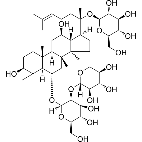
HY-N0045
Ginsenoside Rg1Apoptosis Inducer -
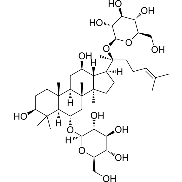
HY-N0431
Astragaloside IVMMP Inhibitor -
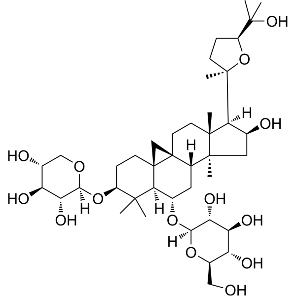
HY-N0039
Ginsenoside Rb1Autophagy Inducer -
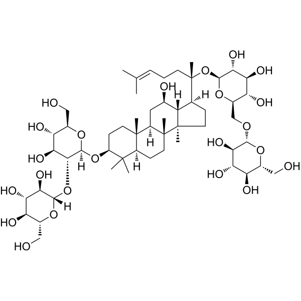
HY-N0135
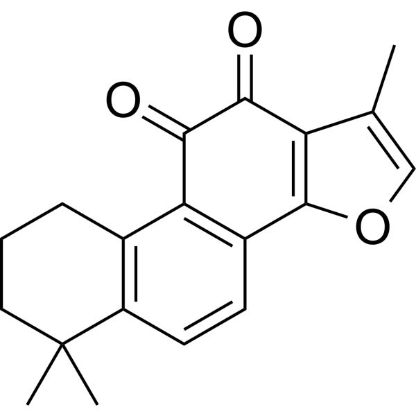
Tanshinone IIAVEGFR Inhibitor -
HY-N0044
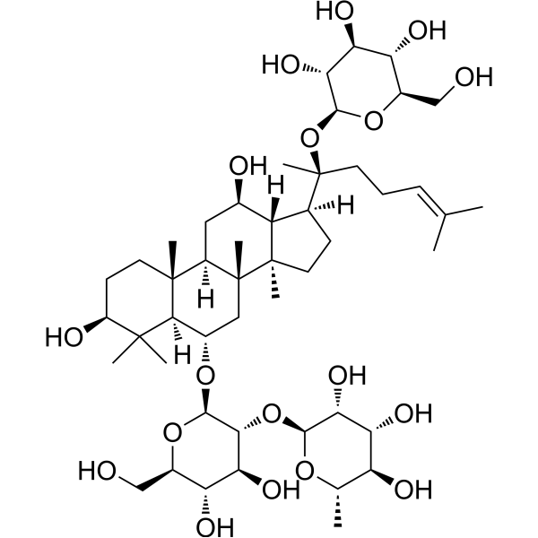
Ginsenoside ReEndogenous Metabolite -
HY-N0145
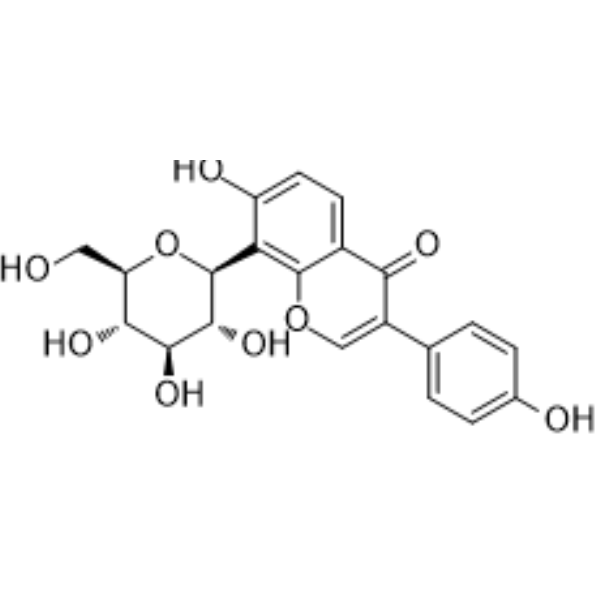
Puerarin5-HT Receptor Antagonist -
HY-N1362
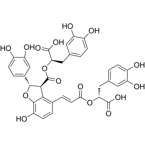
Salvianolic acid BAntioxidant -
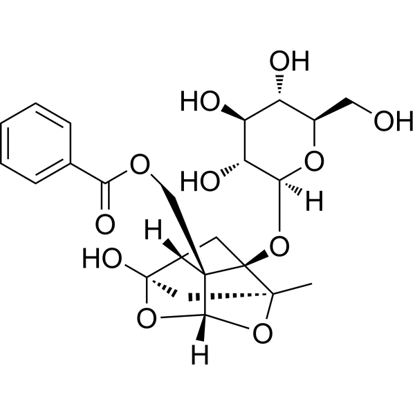
HY-N0293
PaeoniflorinHSP Activator -
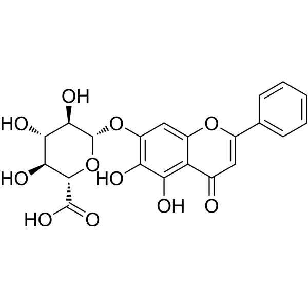
HY-N0197
BaicalinAutophagy Inducer -
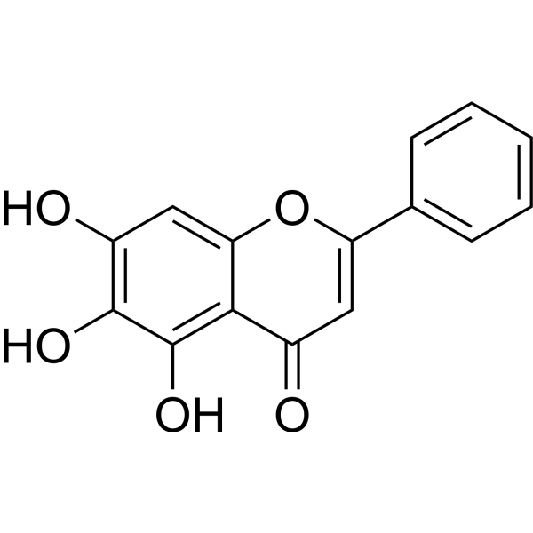
HY-N0196
BaicaleinXanthine Oxidase Inhibitor -
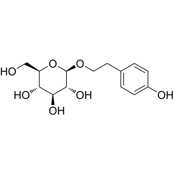
HY-N0109
SalidrosideProlyl Endopeptidase Inhibitor -
HY-N0005
Curcuminp300 Histone Acetylatransferase Inhibitor
Publications Citing Use of MedChemExpress (MCE) Notoginsenoside R1
More Customer Validation & Images

-
Cell Proliferation/Viability Assay
-
Bio/Physico-chemical Assay
-
IF
-
In Vivo Efficacy Study
-
WB


Biological Activity
Description
In Vitro
Notoginsenoside R1 (2.5-80 μM; 24 h) inhibits the hypoxia-reoxygenation (H/R)-induced cell death, intracellular ROS accumulation, and mitochondrial membrane depolarization in H9c2 cardiomyocytes[1].
?
Notoginsenoside R1 (5-20 μM; 24 h) inhibits the H/R-induced H9c2 cardiomyocytes apoptosis in a concentration-dependent manner[1].
?
Notoginsenoside R1 (1-100 μM; 24 h) dose-dependently protects PC12 cells and primary neurons from Aβ-induced cell death and apoptosis[2].
?
Notoginsenoside R1 (10 μM; 24 h) inhibits Aβ25-35-induced ROS production, mitochondrial damage and MAPK activation in PC12 cells[2].
MedChemExpress (MCE) has not independently confirmed the accuracy of these methods. They are for reference only.
In Vivo
MedChemExpress (MCE) has not independently confirmed the accuracy of these methods. They are for reference only.
-
Animal Model:Male Sprague-Dawley (SD) rats (200-250 g)[3]
-
Dosage:5 mg/kg/h
-
Administration:Infused 20 min before LPS infusion via the right jugular vein
-
Result:Ameliorated the LPS-induced reduction in the mesenteric venular shear rate to some extent.
Attenuated the LPS-induced adhesion of leukocytes to the venular wall.
Inhibited mast cell degranulation and cytokine elevation.


Chemical Information
-
CAS No. 80418-24-2
-
Appearance Solid
-
Molecular Weight 933.13
-
Formula C47H80O18
-
Color White to off-white
-
SMILES
C[C@]([C@@](C[C@H]1O)([H])[C@]2(CC[C@@H]3O)C)(C[C@H](O[C@@](O[C@H](CO)[C@@H](O)[C@@H]4O)([H])[C@@H]4O[C@@](OC[C@@H](O)[C@@H]5O)([H])[C@@H]5O)[C@@]2([H])C3(C)C)[C@]6([C@@]1([H])[C@]([C@@](CC/C=C(C)/C)(C)O[C@@H]([C@@H]([C@@H](O)[C@@H]7O)O)O[C@@H]7CO)([H])CC6)C
-
Synonyms
Sanchinoside R1; Sanqi glucoside R1
-
Structure Classification
-
Shipping
Room temperature in continental US; may vary elsewhere.
-
Storage
Powder -20°C 3 years 4°C 2 years In solvent -80°C 2 years -20°C 1 year


Publications (4)
-
Journal Impact Factor
-
Most Recent
-
-
Front Cell Neurosci
Notoginsenoside R1-Induced Neuronal Repair in Models of Alzheimer Disease Is Associated With an Alteration in Neuronal Hyperexcitability, Which Is Regulated by Nav. [Abstract]2020 Sep 4:14:280. PMID: 33088260
Notoginsenoside R1 purchased from MedChemExpress. Usage Cited in: Front Cell Neurosci. 2020 Sep 4:14:280. [Abstract]
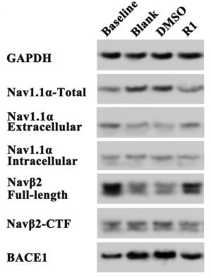
Effect of Notoginsenoside R1 (R1; 5 μM) on the expression, location, and cleavage status of sodium channel proteins after Aβ1-42 treatment.
Notoginsenoside R1 purchased from MedChemExpress. Usage Cited in: Front Cell Neurosci. 2020 Sep 4:14:280. [Abstract]
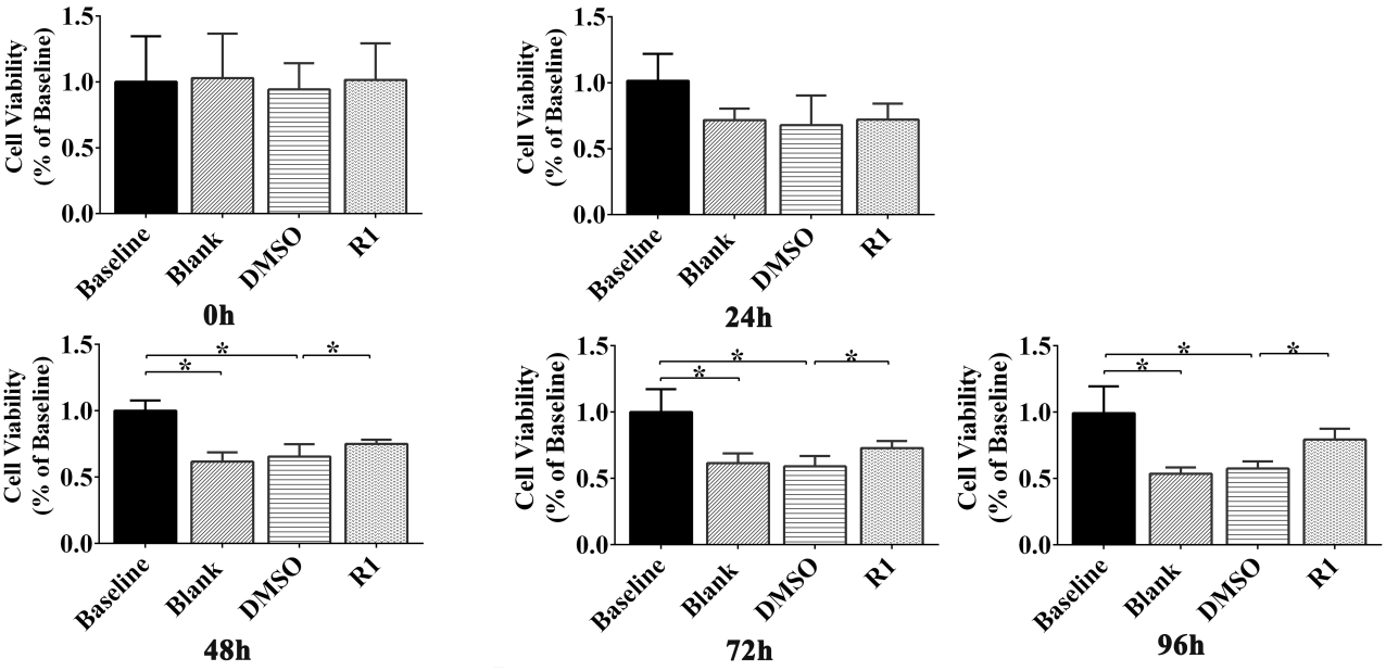
Effect of Notoginsenoside R1 (R1, 5 μM) on Aβ1-42 (5 μM)–induced neuron viability determined using MTT assay. Cell viability of cultured neurons treated with different groups at 0 h, 24 h, 48 h, 72 h, and 96 h.
Notoginsenoside R1 purchased from MedChemExpress. Usage Cited in: Front Cell Neurosci. 2020 Sep 4:14:280. [Abstract]
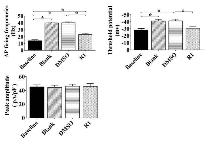
Notoginsenoside R1 (R1, 5 μM) was responsible for reducing abnormal excitability in Aβ1-42–induced neurons.
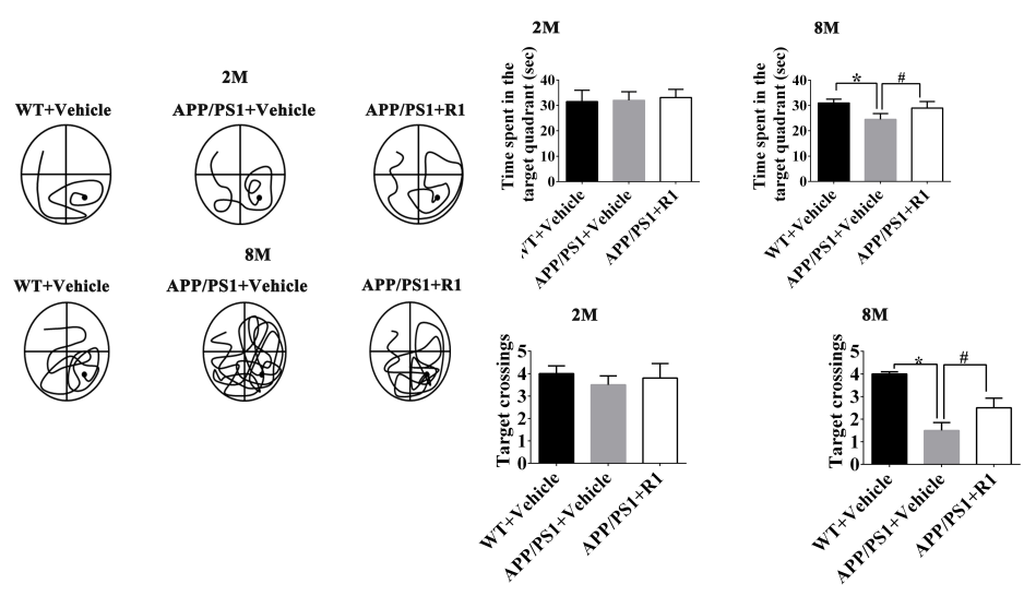
Notoginsenoside R1 purchased from MedChemExpress. Usage Cited in: Front Cell Neurosci. 2020 Sep 4:14:280. [Abstract]
Notoginsenoside R1 (R1, 5 mg/kg; i.g.; once daily for 6 months) partially improved the Marris Water Maze (MWM) test behavior in APP/PS1 mice.
-
Animals (Basel)
Notoginsenoside R1 Inhibits Porcine Deltacoronavirus Infection In Vitro by Restoring SERCA2-Mediated Calcium Homeostasis. [Abstract]2026 Jun 14;16(12):1836. PMID: 42353446 -
Notoginsenoside R1 purchased from MedChemExpress. Usage Cited in: Res Sq. 2024 Jul 12.
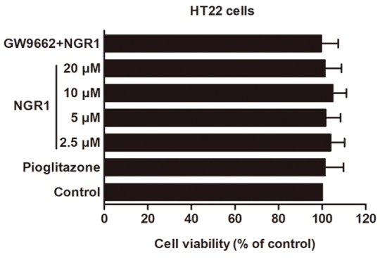
Notoginsenoside R1 (NGR1, 2.5-20 μM; 24 h) treatment did not significantly affect the cell viability of PPARγ/PPRE-transfected HT22 cells.
Notoginsenoside R1 purchased from MedChemExpress. Usage Cited in: Res Sq. 2024 Jul 12.
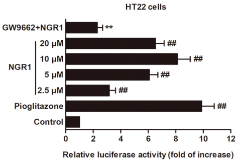
Notoginsenoside R1 (NGR1, 2.5-20 μM; 24 h) treatment notably increased the luciferase activity in the PPARγ/PPRE-transfected HT22 cells.
Notoginsenoside R1 purchased from MedChemExpress. Usage Cited in: Res Sq. 2024 Jul 12.
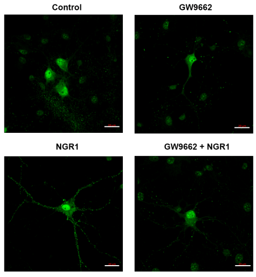
Notoginsenoside R1 (NGR1, 10 µM; 24 h) treatment promoted GLUT4 membrane translocation in primary mouse hippocampal neurons. Scale bar = 20 μm.
Notoginsenoside R1 purchased from MedChemExpress. Usage Cited in: Res Sq. 2024 Jul 12.
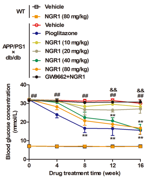
Notoginsenoside R1 (NGR1, 10-80 mg/kg; i.g.; 16 weeks) treatment dramatically decreased the levels of fasting blood glucose in APP/PS1xdb/db mice in a dose-dependent manner.


Solvent & Solubility
In Vitro:
DMSO : 100 mg/mL (107.17 mM; Need ultrasonic; Hygroscopic DMSO has a significant impact on the solubility of product, please use newly opened DMSO)
Please refer to the solubility information to select the appropriate solvent. Once prepared, please aliquot and store the solution to prevent product inactivation from repeated freeze-thaw cycles.
Storage method and period of stock solution: -80°C, 2 years; -20°C, 1 year. When stored at -80°C, please use it within 2 years. When stored at -20°C, please use it within 1 year.
Please refer to the solubility information to select the appropriate solvent. Once prepared, please aliquot and store the solution to prevent product inactivation from repeated freeze-thaw cycles.
Storage method and period of stock solution: -80°C, 2 years; -20°C, 1 year. When stored at -80°C, please use it within 2 years. When stored at -20°C, please use it within 1 year.
Concentration (start) × Volume (start) = Concentration (final) × Volume (final)
In Vivo:
Select the appropriate dissolution method based on your experimental animal and administration route.
- For the following dissolution methods, please ensure to first prepare a clear stock solution using an In Vitro approach and then sequentially add co-solvents:
- To ensure reliable experimental results, the clarified stock solution can be appropriately stored based on storage conditions. As for the working solution for In Vivo experiments, it is recommended to prepare freshly and use it on the same day.
- The percentages shown for the solvents indicate their volumetric ratio in the final prepared solution. If precipitation or phase separation occurs during preparation, heat and/or sonication can be used to aid dissolution.
Add each solvent one by one: 10% DMSO 40% PEG300 5% Tween-80 45% Saline
Solubility: ≥ 2.5 mg/mL (2.68 mM); Clear solution
This protocol yields a clear solution of ≥ 2.5 mg/mL (saturation unknown).
Taking 1 mL working solution as an example, add 100 μL DMSO stock solution (25.0 mg/mL) to 400 μL PEG300, and mix evenly; then add 50 μL Tween-80 and mix evenly; then add 450 μL Saline to adjust the volume to 1 mL.
Preparation of Saline: Dissolve 0.9 g sodium chloride in ddH₂O and dilute to 100 mL to obtain a clear Saline solution.
Add each solvent one by one: 10% DMSO 90% (20% SBE-β-CD in Saline)
Solubility: ≥ 2.5 mg/mL (2.68 mM); Clear solution
This protocol yields a clear solution of ≥ 2.5 mg/mL (saturation unknown).
Taking 1 mL working solution as an example, add 100 μL DMSO stock solution (25.0 mg/mL) to 900 μL 20% SBE-β-CD in Saline, and mix evenly.
Preparation of 20% SBE-β-CD in Saline (4°C, storage for one week): 2 g SBE-β-CD powder is dissolved in 10 mL Saline, completely dissolve until clear.
In Vivo Dissolution Calculator
Please enter the basic information of animal experiments:
-
-
-
-
Recommended: Prepare an additional quantity of animals to account for potential losses during experiments.
Please enter your animal formula composition:
-
%DMSO +
Recommended: Keep the proportion of DMSO in working solution below 2% if your animal is weak.
-
%+
-
+%Tween-80 + +
-
%Saline +
The co-solvents required include: DMSO, . All of co-solvents are available by MedChemExpress (MCE). , Tween 80. All of co-solvents are available by MedChemExpress (MCE).
Working solution concentration: 0.22 mg/mL
Method for preparing stock solution: mg drug dissolved in μL DMSO. Stock solution concentration: mg/mL.
1. Take μL DMSO stock solution;
2. Add μL .
μL , mix evenly;
3. Then add μL Tween 80, mix evenly;
4. Then add μL
Please ensure that the stock solution in the first step is dissolved to a clear state, and add co-solvents in sequence. You can use ultrasonic heating (ultrasonic cleaner, recommended frequency 20-40 kHz), vortexing, etc. to assist dissolution.


Purity & Documentation
-
Data Sheet (287 KB)
-
SDS (396 KB)
- English - EN (396 KB)
- Français - FR (396 KB)
- Deutsch - DE (396 KB)
- Norwegian - NO (396 KB)
- Español - ES (396 KB)
- Swedish - SV (396 KB)
- Italian - IT (396 KB)
- Korean - KR (396 KB)
- Portuguese - PT (396 KB)
-
Handling Instructions (2659 KB)
References
[1]. Yu Y, et, al. Cardioprotective effects of Notoginsenoside R1 against ischemia/reperfusion injuries by regulating oxidative stress- and endoplasmic reticulum stress- related signaling pathways. Sci Rep. 2016 Feb 18;6:21730. [Content Brief]
[2]. Ma B, et, al. Notoginsenoside R1 attenuates amyloid-β-induced damage in neurons by inhibiting reactive oxygen species and modulating MAPK activation. Int Immunopharmacol. 2014 Sep;22(1):151-9. [Content Brief]
[3]. Sun K, et, al. Protective effects of ginsenoside Rb1, ginsenoside Rg1, and notoginsenoside R1 on lipopolysaccharide-induced microcirculatory disturbance in rat mesentery. Life Sci. 2007 Jul 19;81(6):509-18. [Content Brief]


Complete Stock Solution Preparation Table
Please refer to the solubility information to select the appropriate solvent. Once prepared, please aliquot and store the solution to prevent product inactivation from repeated freeze-thaw cycles.
Storage method and period of stock solution: -80°C, 2 years; -20°C, 1 year. When stored at -80°C, please use it within 2 years. When stored at -20°C, please use it within 1 year.
| Optional Solvent | Concentration Solvent Mass | 1 mg | 5 mg | 10 mg | 25 mg |
|---|---|---|---|---|---|
| DMSO | 1 mM | 1.0717 mL | 5.3583 mL | 10.7166 mL | 26.7916 mL |
| 5 mM | 0.2143 mL | 1.0717 mL | 2.1433 mL | 5.3583 mL | |
| 10 mM | 0.1072 mL | 0.5358 mL | 1.0717 mL | 2.6792 mL | |
| 15 mM | 0.0714 mL | 0.3572 mL | 0.7144 mL | 1.7861 mL | |
| 20 mM | 0.0536 mL | 0.2679 mL | 0.5358 mL | 1.3396 mL | |
| 25 mM | 0.0429 mL | 0.2143 mL | 0.4287 mL | 1.0717 mL | |
| 30 mM | 0.0357 mL | 0.1786 mL | 0.3572 mL | 0.8931 mL | |
| 40 mM | 0.0268 mL | 0.1340 mL | 0.2679 mL | 0.6698 mL | |
| 50 mM | 0.0214 mL | 0.1072 mL | 0.2143 mL | 0.5358 mL | |
| 60 mM | 0.0179 mL | 0.0893 mL | 0.1786 mL | 0.4465 mL | |
| 80 mM | 0.0134 mL | 0.0670 mL | 0.1340 mL | 0.3349 mL | |
| 100 mM | 0.0107 mL | 0.0536 mL | 0.1072 mL | 0.2679 mL |


Notoginsenoside R1 Related Classifications
HY-N0615 Related Classifications
 Powered by Bioz
Keywords
- Notoginsenoside R1
- 80418-24-2
- Sanchinoside R1
- Sanqi glucoside R1
- Notoginsenoside R 1
- Notoginsenoside R-1
- Sanchinoside R 1
- Sanchinoside R-1
- Sanqi glucoside R1
- Sanqi glucoside R 1
- Sanqi glucoside R-1
- Amyloid-β
- Apoptosis
- saponin
- anti-oxidation
- anti-inflammatory
- anti-angiogenic
- anti-apoptosis,cardioprotection
- I/R
- neuroprotection
- Inhibitor
- inhibitor
- inhibit