



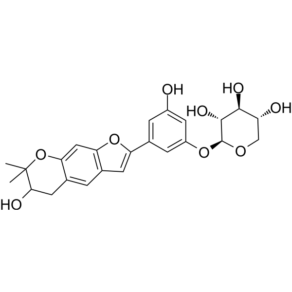
Mulberroside C
Based on 1 publication(s) in Google Scholar
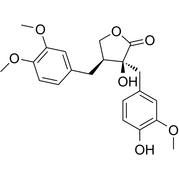
Mulberroside C is one of the main bioactive components in white mulberry (Morus alba L.). Mulberroside C exhibits antiplatelet, antiviral and neutrophil-regulating activities, and binds to EV-A71 VP1 with a Kd value of 1.289 nM. Mulberroside C reduces the phosphorylation level of ERK, promotes the phosphorylation of IP3RI at the Ser1756 site, and decreases calcium influx and calcium mobilization. Mulberroside C inhibits the expression of P-selectin (P-selectin). Mulberroside C upregulates the cyclic nucleotide signaling pathway in human platelets. Mulberroside C binds to IL-23R, upregulates the expression of G-CSF, GM-CSF and RASGRP1, and activates the RAS/ERK signaling pathway. Mulberroside C promotes neutrophil maturation, accelerates the recovery of leukopenia, and enhances the antibacterial activity of neutrophils. Mulberroside C inhibits the replication of hepatitis C virus in replicon cells. Mulberroside C prevents the uncoating and genome release of EV-A71, and inhibits the synthesis of viral proteins and RNA. Mulberroside C can be used in studies related to thrombosis-mediated cardiovascular diseases, chemotherapy- and radiotherapy-induced leukopenia, hepatitis C virus infection, and hand, foot and mouth disease.
For research use only. We do not sell to patients.
- Purity: 99.77%
- CAS No.: 102841-43-0
- Formula: C24H26O9
- Molecular Weight:458.46
-
Storage:
4°C, protect from light
* In solvent : -80°C, 6 months; -20°C, 1 month (protect from light)

To place orders, for customer services and technical support, please contact: MedChemExpress USA
Tel: 609-228-6898 E-mail: [email protected] [email protected]


-
 Biological Activity
-
Chemical Information
-
Solvent & Solubility
- Purity & Documentation
- References
-
 Help & FAQs





Publications Citing Use of MedChemExpress (MCE) Mulberroside C
MoreAll Calcium Channel Isoforms
MoreAll DNA/RNA Synthesis Isoforms
More

Biological Activity
|
EV-A71 VP1 1.289 nM (Kd) |
Mulberroside C (50-150 μM; 2 min preincubation, 5 min aggregation) dose-dependently inhibits washed human platelet aggregation induced by Collagen (HY-NP003), Thrombin (HY-114164) and U46619 (HY-108566), with an IC50 of 77.3 μM against collagen-induced aggregation, and shows no cytotoxicity at the tested concentrations[1].
Mulberroside C (50-150 μM; 3 min preincubation) reduces collagen-induced intracellular Ca2+ mobilization in washed human platelets in a dose-dependent manner[1].
Mulberroside C (50-150 μM; 2 min preincubation, 5 min Collagen stimulation) regulates platelet signaling pathways in washed human platelets by upregulating the phosphorylation levels of IP3RI and VASP, and downregulating the phosphorylation levels of ERK, cPLA2, p38 MAPK, PI3K, Akt and PLCγ2[1].
Mulberroside C (50-150 μM; 0-6 min) dose-dependently inhibits collagen-induced P-selectin expression (α-granule release), serotonin and ATP release (δ-granule secretion), and thromboxane A2 production in washed human platelets. It also suppresses collagen-induced fibrinogen binding to αIIb/β3 in washed human platelets, and elevates intracellular cAMP and cGMP levels in human platelets[1].
Mulberroside C (50-150 μM; 30 min preincubation, 15 min clot reaction) dose-dependently inhibits thrombin-induced fibrin clot retraction in human platelet-rich plasma[1].
Mulberroside C directly binds to human IL-23R with a binding score of 6.36 kcal/mol[2].
Mulberroside C (5-20 μM; 5 days) dose-dependently increases the proportion of CD11b-positive differentiated myeloid cells in NB4 and HL-60 cell lines, induces morphological differentiation of cells into mature neutrophils, and promotes the functional differentiation of NB4 cells into mature neutrophils[2].
Mulberroside C (5-20 μM; 5 days) dose-dependently enhances the bactericidal activity of differentiated NB4 cells against *Staphylococcus aureus*[2].
Mulberroside C (5-20 μM; 5 days) dose-dependently upregulates the expression of IL-23R, G-CSF, GM-CSF and RASGRP1 in NB4 cells, as well as the expression of IL-23R, RASGRP1, RAS, phosphorylated ERK, and myeloid transcription factors PU.1, c-Fos, CEBPA and RUNX1 in NB4 cells[2].
Mulberroside C (3.125-400 µM; 48 h) exhibits low cytotoxicity towards RD cells (CC50 = 94.82 µM) and Vero cells (CC50 = 257.1 µM)[3].
Mulberroside C (1.5625-50 µM; 48 h) potently inhibits the cytopathic effects induced by EV-A71 (BrCr strain) in RD cells (IC50 = 2.09 µM) and Vero cells (IC50 = 35.48 µM)[3].
Mulberroside C (12.5-50 µM; 4 days) reduces plaque formation of EV-A71 (BrCr strain) in Vero cells in a dose-dependent manner[3].
Mulberroside C (12.5-50 µM; 24-48 h) dose-dependently inhibits EV-A71 (BrCr strain) RNA synthesis in RD cells, and at 48 h post-infection, the 50 µM concentration reduces viral RNA levels by more than 10,000-fold[3].
Mulberroside C (12.5-50 µM; 24 h) dose-dependently inhibits the VP1 protein expression of EV-A71 (BrCr strain) in RD cells[3].
Mulberroside C (25 µM, administered at post-infection π -1 to 12 h, 0 to 12 h, 1 to 12 h, 3 to 12 h, 6 to 12 h, and 9 to 12 h) mainly inhibits the replication of EV-A71 (BrCr strain) at the early stage of RD cell infection, and significant antiviral activity is observed when it is added within 3 h post-infection[3].
Mulberroside C (50 µM; mixed with virus for 1 h, followed by 1 h of cell incubation) significantly inhibits the adsorption of EV-A71 (BrCr strain) to RD cells[3].
Mulberroside C (1.5625-50 µM; 48 h) inhibits the replication of EV-A71 (strain CC063) (IC50 = 7.237 µM) and CV-A16 (strain CC045) (IC50 = 15.24 µM) in RD cells[3].
Mulberroside C (46.88-1500 µM; 30 s association, 30 s dissociation) binds to purified EV-A71 VP1 protein in a concentration-dependent manner, with a dissociation constant (Kd) of 1.289 nM[3].
MedChemExpress (MCE) has not independently confirmed the accuracy of these methods. They are for reference only.
-
Cell Line:NB4, HL-60 human myeloid leukemia cell lines
-
Concentration:5, 10, 20 μM
-
Incubation Time:5 days
-
Result:Significantly increased the proportion of CD11b-positive cells in a dose-dependent manner: 10.0% at 5 μM, 16.1% at 10 μM, and 20.2% at 20 μM in NB4 cells, compared to 5.87% in the control group.\nDose-dependently reduced the nucleus-to-cytoplasm ratio and enhanced nuclear segmentation in cells: ~7% differentiated cells at 5 μM, ~8% at 10 μM, and ~12% at 20 μM, compared to ~2% in the control group.
Caused a significant, dose-dependent rise in the number of NBT-positive cells: ~8% at 5 μM, ~10% at 10 μM, and ~15% at 20 μM, compared to ~5% in the control group.
-
Cell Line:NB4 human myeloid leukemia cell line
-
Concentration:5, 10, 20 μM
-
Incubation Time:5 days
-
Result:Dose-dependently upregulated the expression of genes associated with myeloid differentiation and signaling: IL-23R (2.2-fold at 5 μM, 2.5-fold at 10 μM, 3.5-fold at 20 μM), G-CSF (1.6-fold at 5 μM, 2.2-fold at 10 μM, 3.2-fold at 20 μM), GM-CSF (1.7-fold at 5 μM, 2.4-fold at 10 μM, 3.8-fold at 20 μM), and RASGRP1 (1.3-fold at 5 μM, 1.4-fold at 10 μM, 1.8-fold at 20 μM), compared to control levels.
-
Cell Line:NB4 human myeloid leukemia cell line
-
Concentration:5, 10, 20 μM
-
Incubation Time:5 days
-
Result:Dose-dependently upregulated the expression of key proteins involved in myeloid differentiation and signaling: IL-23R (0.75-fold at 5 μM, 0.85-fold at 10 μM, 1.1-fold at 20 μM), RASGRP1 (1.2-fold at 5 μM, 1.4-fold at 10 μM, 1.4-fold at 20 μM), RAS (1.6-fold at 5 μM, 1.9-fold at 10 μM, 1.9-fold at 20 μM), phosphorylated ERK (1.3-fold at 5 μM, 1.6-fold at 10 μM, 1.6-fold at 20 μM), PU.1 (0.5-fold at 5 μM, 0.7-fold at 10 μM, 0.7-fold at 20 μM), c-Fos (0.9-fold at 5 μM, 1.2-fold at 10 μM, 1.3-fold at 20 μM), CEBPA (1.8-fold at 5 μM, 2.8-fold at 10 μM, 2.8-fold at 20 μM), and RUNX1 (1.3-fold at 5 μM, 1.5-fold at 10 μM, 1.5-fold at 20 μM), all normalized to GAPDH or total ERK levels.
-
Cell Line:EV-A71 (BrCr strain)-infected RD cells
-
Concentration:12.5, 25, 50 µM
-
Incubation Time:24 h, 48 h
-
Result:Dose-dependently reduced EV-A71 RNA levels at both time points.
Reduced viral RNA copy numbers by more than 10,000-fold compared to untreated controls at 48 h post-infection with 50 µM treatment.
-
Cell Line:EV-A71 (BrCr strain)-infected RD cells
-
Concentration:12.5, 25, 50 µM
-
Incubation Time:24 h
-
Result:Dose-dependently inhibited the expression of EV-A71 VP1 protein.
Significantly reduced the relative VP1 protein level compared to untreated infected cells, with the lowest level observed at 50 µM.
Mulberroside C (2-8 mg/kg, i.p.; once daily; for 11 consecutive days) dose-dependently restores white blood cell counts, promotes the proliferation and maturation of neutrophils, enhances bacterial clearance capacity, and exhibits favorable safety profiles in a mouse model of irradiation-induced leukopenia[2].
Mulberroside C (2-8 mg/kg, i.p.; once daily for 7 consecutive days) dose-dependently promotes the recovery of peripheral blood leukocyte levels in mice with chemotherapy-induced leukopenia[2].
Mulberroside C (25-50 mg/kg, i.p.; once daily; for 5 consecutive days) increases the survival rate of neonatal ICR mice to 30% at the dose of 50 mg/kg, reduces clinical scores, decreases viral loads in multiple tissues, and alleviates EV-A71-induced tissue damage; while the 25 mg/kg dose delays mouse death, as well as reduces viral loads and alleviates tissue damage[3].
MedChemExpress (MCE) has not independently confirmed the accuracy of these methods. They are for reference only.
-
Animal Model:Transgenic zebrafish (Tg(mpeg1: EGFP), Tg(lyz: DsRed2)) (2 days postfertilization larvae; irradiation-induced with 4 Gy X-rays at 2 dpf)[2]
-
Dosage:5 μM; 10 μM; 20 μM
-
Administration:immersion; continuous; from 3 dpf to 6 dpf
-
Result:Significantly increased counts of mpx:EGFP-positive neutrophils in the trunk compared to the irradiation model group, with a dose-dependent increase observed.
Significantly increased counts of lyz:DsRed2-positive neutrophils compared to the irradiation model group at 20 μM.
-
Animal Model:Transgenic zebrafish (Tg(mpeg1: EGFP), Tg(lyz: DsRed2)) (2 days postfertilization larvae; chemotherapy-induced with a single 200 mg/L dose of cyclophosphamide at 2 dpf)[2]
-
Dosage:5 μM; 10 μM; 20 μM
-
Administration:immersion; continuous; from 3 dpf to 6 dpf
-
Result:Significantly increased counts of mpx:EGFP-positive neutrophils in the trunk compared to the cyclophosphamide model group, with a dose-dependent increase observed.
Significantly increased counts of lyz:DsRed2-positive neutrophils compared to the cyclophosphamide model group at 20 μM.
-
Animal Model:Kunming (KM) mice (8 weeks old, ~25 g, equal male/female; irradiation-induced with a single 4 Gy total body X-ray dose)[2]
-
Dosage:2 mg/kg; 4 mg/kg; 8 mg/kg
-
Administration:i.p.; daily; 11 days
-
Result:Significantly increased peripheral white blood cell, neutrophil, monocyte, and lymphocyte counts from days 5 to 11 compared to the irradiation model group.
Significantly increased the percentage of CD11b+Ly6G+ neutrophils in peripheral blood, bone marrow, and spleen on day 9.
Significantly increased the percentage of CD34+CD117+ hematopoietic stem cells and CD117+Ly6G+ myeloid progenitor cells in bone marrow and spleen on day 9.
Significantly increased the percentage of CD3+ T cells and CD19+ B cells in peripheral blood, bone marrow, and spleen on day 9.
Induced a dose-dependent increase in Ki67+Ly6G+ proliferating immature neutrophils in bone marrow and spleen, with the 8 mg/kg group approaching normal levels.
Significantly increased bacterial clearance compared to the model group, with clearance superior to the G-CSF control group.
Caused no pathological changes in heart, liver, lungs, or kidneys at any dose.
-
Animal Model:Kunming (KM) mice (8 weeks old, ~25 g, equal male/female; chemotherapy-induced with 100 mg/kg cyclophosphamide i.p. daily for 3 days)[2]
-
Dosage:2 mg/kg; 4 mg/kg; 8 mg/kg
-
Administration:i.p.; daily; 7 days
-
Result:Significantly increased peripheral white blood cell, neutrophil, monocyte, and lymphocyte counts from days 7 to 10 compared to the cyclophosphamide model group.
Maintained significantly higher leukocyte counts than the model group even at the lowest count on day 5.
-
Animal Model:ICR mice (1-day-old, specific pathogen-free, intracerebral inoculation with EV-A71 CC063 strain)[5]
-
Dosage:25 mg/kg; 50 mg/kg
-
Administration:i.p.; daily; 5 consecutive days
-
Result:Improved survival rate to 30% (50 mg/kg group, vs. 0% in virus control group).
Delayed death of infected mice (25 mg/kg group).
Reduced clinical scores of infected mice compared to the virus control group (50 mg/kg group).
Significantly reduced viral loads in heart, brain, lung, intestine, and hind-limb muscle tissues compared to the virus control group (both 25 mg/kg and 50 mg/kg groups).
Prevented lung focal hemorrhages, preserved intestinal mucosa structure, and partially alleviated inflammatory cell infiltration in hind-limb muscle (50 mg/kg group).
Reduced tissue damage compared to virus control mice (25 mg/kg group).


Chemical Information
-
CAS No. 102841-43-0
-
Appearance Solid
-
Molecular Weight 458.46
-
Formula C24H26O9
-
Color White to off-white
-
SMILES
OC1=CC(O[C@@H]2OC[C@@H](O)[C@H](O)[C@H]2O)=CC(C3=CC4=C(C=C(OC(C)(C)C(O)C5)C5=C4)O3)=C1
-
Structure Classification
-
Initial Source
-
Shipping
Room temperature in continental US; may vary elsewhere.
-
Storage
4°C, protect from light
* In solvent : -80°C, 6 months; -20°C, 1 month (protect from light)


Publications (1)
-
Journal Impact Factor
-
Most Recent
-
Vet Microbiol
The Chinese medicine monomer Schisandrin C inhibits PRRSV infection by regulating the OGT-PI3K/AKT/mTOR signaling pathway. [Abstract]2026 May:316:110992. PMID: 41865607


Solvent & Solubility
DMSO : 62.5 mg/mL (136.33 mM; Need ultrasonic; Hygroscopic DMSO has a significant impact on the solubility of product, please use newly opened DMSO)
Please refer to the solubility information to select the appropriate solvent. Once prepared, please aliquot and store the solution to prevent product inactivation from repeated freeze-thaw cycles.
Storage method and period of stock solution: -80°C, 6 months; -20°C, 1 month (protect from light). When stored at -80°C, please use it within 6 months. When stored at -20°C, please use it within 1 month.
Please refer to the solubility information to select the appropriate solvent. Once prepared, please aliquot and store the solution to prevent product inactivation from repeated freeze-thaw cycles.
Storage method and period of stock solution: -80°C, 6 months; -20°C, 1 month (protect from light). When stored at -80°C, please use it within 6 months. When stored at -20°C, please use it within 1 month.
Concentration (start) × Volume (start) = Concentration (final) × Volume (final)
Select the appropriate dissolution method based on your experimental animal and administration route.
- For the following dissolution methods, please ensure to first prepare a clear stock solution using an In Vitro approach and then sequentially add co-solvents:
- To ensure reliable experimental results, the clarified stock solution can be appropriately stored based on storage conditions. As for the working solution for In Vivo experiments, it is recommended to prepare freshly and use it on the same day.
- The percentages shown for the solvents indicate their volumetric ratio in the final prepared solution. If precipitation or phase separation occurs during preparation, heat and/or sonication can be used to aid dissolution.
Add each solvent one by one: 10% DMSO 40% PEG300 5% Tween-80 45% Saline
Solubility: ≥ 2.08 mg/mL (4.54 mM); Clear solution
This protocol yields a clear solution of ≥ 2.08 mg/mL (saturation unknown).
Taking 1 mL working solution as an example, add 100 μL DMSO stock solution (20.8 mg/mL) to 400 μL PEG300, and mix evenly; then add 50 μL Tween-80 and mix evenly; then add 450 μL Saline to adjust the volume to 1 mL.
Preparation of Saline: Dissolve 0.9 g sodium chloride in ddH₂O and dilute to 100 mL to obtain a clear Saline solution.
Add each solvent one by one: 10% DMSO 90% (20% SBE-β-CD in Saline)
Solubility: ≥ 2.08 mg/mL (4.54 mM); Clear solution
This protocol yields a clear solution of ≥ 2.08 mg/mL (saturation unknown).
Taking 1 mL working solution as an example, add 100 μL DMSO stock solution (20.8 mg/mL) to 900 μL 20% SBE-β-CD in Saline, and mix evenly.
Preparation of 20% SBE-β-CD in Saline (4°C, storage for one week): 2 g SBE-β-CD powder is dissolved in 10 mL Saline, completely dissolve until clear.
Please enter the basic information of animal experiments:
-
-
-
-
Recommended: Prepare an additional quantity of animals to account for potential losses during experiments.
Please enter your animal formula composition:
-
%DMSO +
Recommended: Keep the proportion of DMSO in working solution below 2% if your animal is weak.
-
%+
-
+%Tween-80 + +
-
%Saline +
The co-solvents required include: DMSO, . All of co-solvents are available by MedChemExpress (MCE). , Tween 80. All of co-solvents are available by MedChemExpress (MCE).
Working solution concentration: 0.22 mg/mL
Method for preparing stock solution: mg drug dissolved in μL DMSO. Stock solution concentration: mg/mL. * In solvent : -80°C, 6 months; -20°C, 1 month (protect from light)
1. Take μL DMSO stock solution;
2. Add μL .
μL , mix evenly;
3. Then add μL Tween 80, mix evenly;
4. Then add μL
Please ensure that the stock solution in the first step is dissolved to a clear state, and add co-solvents in sequence. You can use ultrasonic heating (ultrasonic cleaner, recommended frequency 20-40 kHz), vortexing, etc. to assist dissolution.


Purity & Documentation
-
Data Sheet (296 KB)
-
SDS (251 KB)
- English - EN (251 KB)
- Français - FR (251 KB)
- Deutsch - DE (251 KB)
- Norwegian - NO (251 KB)
- Español - ES (251 KB)
- Swedish - SV (251 KB)
- Italian - IT (251 KB)
- Korean - KR (251 KB)
- Portuguese - PT (251 KB)
-
Handling Instructions (2659 KB)
References
[1]. Kwon HW, et al. In Vitro Antiplatelet Activity of Mulberroside C through the Up-Regulation of Cyclic Nucleotide Signaling Pathways and Down-Regulation of Phosphoproteins. Genes. 2021 Jun 30;12(7):1024. [Content Brief]
[2]. Zhang LW, et al. IL-23 Receptor Agonism by Mulberroside C Activates the RASGRP1/RAS/ERK Pathway Contributing to Leukopenia Treatments. Phytotherapy research : PTR. 2025 Aug;39(8):3578-3600. [Content Brief]
[3]. Cao Y, et al. Antiviral activity of Mulberroside C against enterovirus A71 in vitro and in vivo. European journal of pharmacology. 2021 Sep 05;906:174204. [Content Brief]


Complete Stock Solution Preparation Table
Please refer to the solubility information to select the appropriate solvent. Once prepared, please aliquot and store the solution to prevent product inactivation from repeated freeze-thaw cycles.
Storage method and period of stock solution: -80°C, 6 months; -20°C, 1 month (protect from light). When stored at -80°C, please use it within 6 months. When stored at -20°C, please use it within 1 month.
| Optional Solvent | Concentration Solvent Mass | 1 mg | 5 mg | 10 mg | 25 mg |
|---|---|---|---|---|---|
| DMSO | 1 mM | 2.1812 mL | 10.9061 mL | 21.8122 mL | 54.5304 mL |
| 5 mM | 0.4362 mL | 2.1812 mL | 4.3624 mL | 10.9061 mL | |
| 10 mM | 0.2181 mL | 1.0906 mL | 2.1812 mL | 5.4530 mL | |
| 15 mM | 0.1454 mL | 0.7271 mL | 1.4541 mL | 3.6354 mL | |
| 20 mM | 0.1091 mL | 0.5453 mL | 1.0906 mL | 2.7265 mL | |
| 25 mM | 0.0872 mL | 0.4362 mL | 0.8725 mL | 2.1812 mL | |
| 30 mM | 0.0727 mL | 0.3635 mL | 0.7271 mL | 1.8177 mL | |
| 40 mM | 0.0545 mL | 0.2727 mL | 0.5453 mL | 1.3633 mL | |
| 50 mM | 0.0436 mL | 0.2181 mL | 0.4362 mL | 1.0906 mL | |
| 60 mM | 0.0364 mL | 0.1818 mL | 0.3635 mL | 0.9088 mL | |
| 80 mM | 0.0273 mL | 0.1363 mL | 0.2727 mL | 0.6816 mL | |
| 100 mM | 0.0218 mL | 0.1091 mL | 0.2181 mL | 0.5453 mL |


Mulberroside C Related Classifications
HY-N0620 Related Classifications
 Powered by Bioz