



Adaptaquin
Based on 3 publication(s) in Google Scholar
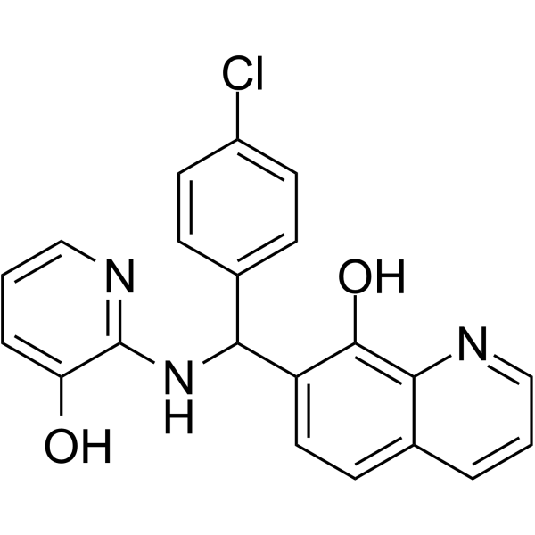
Adaptaquin is a BBB-penetrable HIF-PHDs inhibitor. Adaptaquin has anti-inflammatory and neuroprotective effects. Adaptaquin can effectively inhibit lipid peroxidation, maintain mitochondrial function, and reduce neuronal death. Adaptaquin can be used in the research of nervous system diseases such as Parkinson's disease.
For research use only. We do not sell to patients.
- Purity : 99.87%
- CAS No.: 385786-48-1
- Formula: C21H16ClN3O2
- Molecular Weight:377.82
-
Storage:Powder -20°C, 3 years , 4°C, 2 years ; In solvent -80°C, 6 months , -20°C, 1 month

To place orders, for customer services and technical support, please contact: MedChemExpress USA
Tel: 609-228-6898 E-mail: [email protected] [email protected]


-
 Biological Activity
-
Chemical Information
-
Solvent & Solubility
- Protocols
- Purity & Documentation
- References
-
 Help & FAQs


-
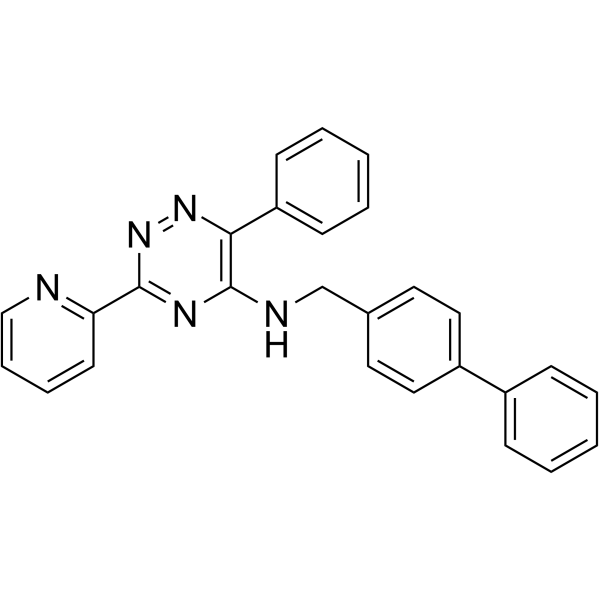
HY-12754
ML228HIF Activator -
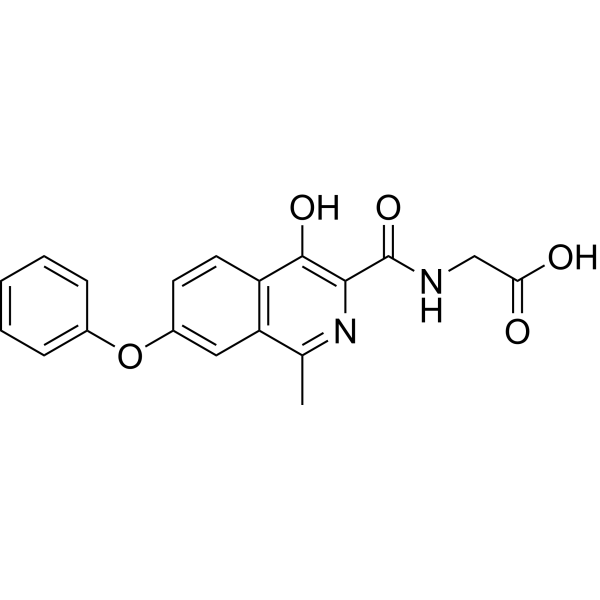
HY-13426
RoxadustatHIF-PH Inhibitor -
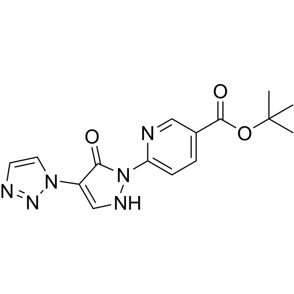
HY-120110
IOX4PHD2 Inhibitor -
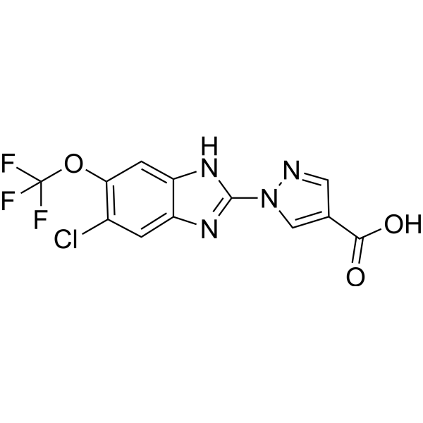
HY-12832
JNJ-42041935PHD Inhibitor -
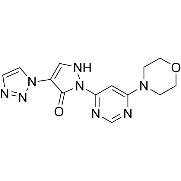
HY-12654
MolidustatHypoxia-Inducible Factor Prolyl Hydroxylase Inhibitor -
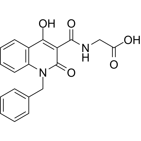
HY-15468
IOX2HIF/HIF Prolyl-Hydroxylase Inhibitor -
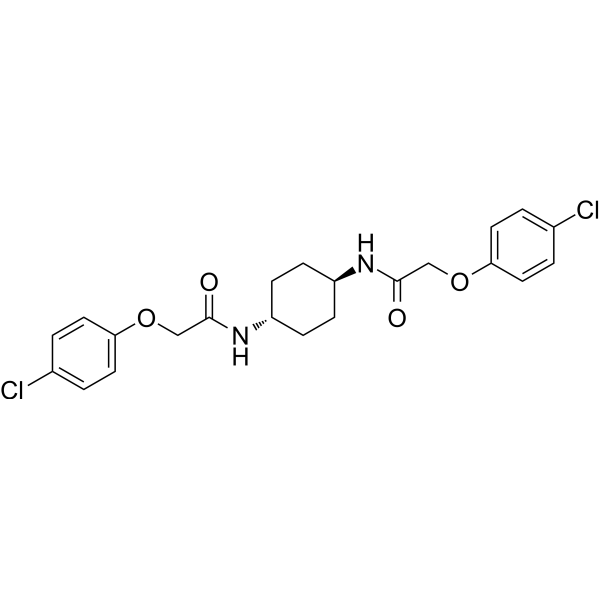
HY-12495
ISRIB (trans-isomer)PERK Inhiitor -
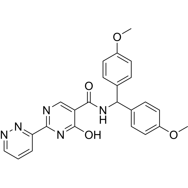
HY-101023
MK-8617HIF PHD1-3 Inhibitor -

HY-P99001
Human IgG1 kappa, Isotype ControlIgG1κ Isotype Control -
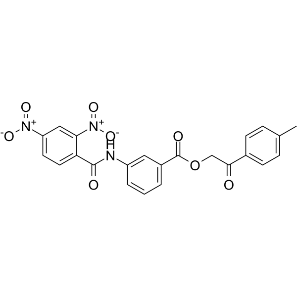
HY-15672
FM19G11HIF-1α Inhibitor -
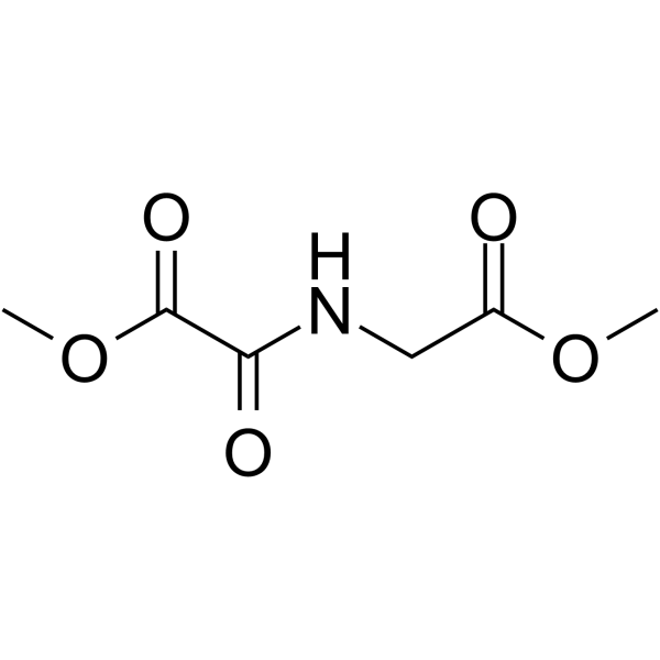
HY-15893
DMOGHIF-PH Inhibitor -
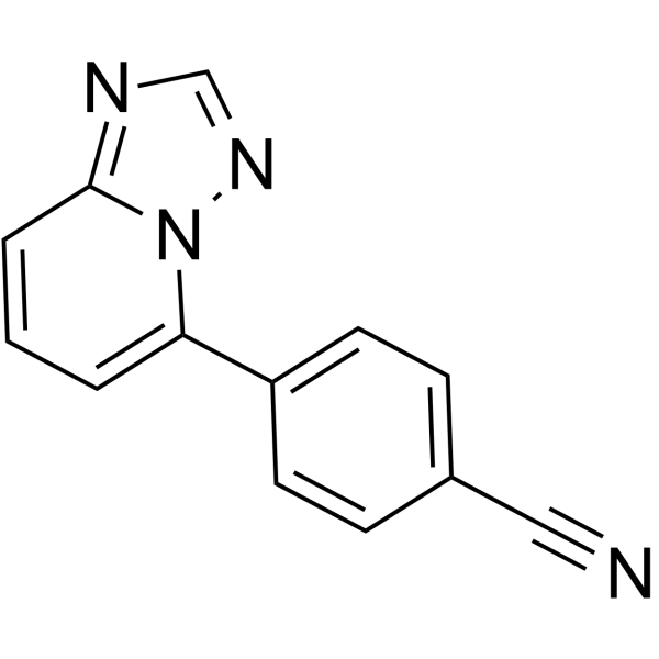
HY-136300
PHD-1-IN-1PHD-1 Inhibitor
Publications Citing Use of MedChemExpress (MCE) Adaptaquin
More

Biological Activity
Description
In Vitro
Adaptaquin (0.5-5 μM; 4-15 h) can increase the viability of glutamate-treated HT-22 cells and inhibit lipid peroxidation and ROS production[1].
Adaptaquin (0.1-5 μM; 24-48 h) can significantly protect PC12 cells and ventral midbrain dopaminergic neurons from cell death induced by Oxidopamine (HY-B1081) (6-OHDA) and MPP+ (HY-W008719) and maintain cell morphology[2].
Adaptaquin (0.5 μM; 8-16 h) can inhibit the expression of Trib3, ATF4, and CHOP induced by 6-OHDA/MPP+ and maintain the Parkin protein level in PC12 cells[2].
MedChemExpress (MCE) has not independently confirmed the accuracy of these methods. They are for reference only.
-
Cell Line:6-OHDA/ MPP+ treated PC12
-
Concentration:0.5 μM
-
Incubation Time:8 h/16 h
-
Result:Reduced the mRNA levels of Trib3, ATF4 and CHOP.
In Vivo
MedChemExpress (MCE) has not independently confirmed the accuracy of these methods. They are for reference only.
-
Animal Model:Adult male C57BL/6 mice (10-12 weeks) treated 6-OHDA hydrobromide (HY-B1081A)[2]
-
Dosage:30 mg/kg
-
Administration:Intraperitoneal injection; 7 days
-
Result:Enhanced survival of dopaminergic neurons and substantially protected their striatal projections.
Significantly enhanced retention of nigrostriatal function.


Chemical Information
-
CAS No. 385786-48-1
-
Appearance Solid
-
Molecular Weight 377.82
-
Formula C21H16ClN3O2
-
Color White to yellow
-
SMILES
OC1=C2N=CC=CC2=CC=C1C(C3=CC=C(Cl)C=C3)NC4=NC=CC=C4O
-
Shipping
Room temperature in continental US; may vary elsewhere.
-
Storage
Powder -20°C 3 years 4°C 2 years In solvent -80°C 6 months -20°C 1 month


Publications (3)
-
Journal Impact Factor
-
Most Recent
-
J Cell Sci
Biophysical and biochemical studies support PHD inhibitor development as a TPI deficiency therapy. [Abstract]2026 May 1;139(9):jcs264664. PMID: 41949155 -
-


Solvent & Solubility
In Vitro:
DMSO : 100 mg/mL (264.68 mM; Need ultrasonic; Hygroscopic DMSO has a significant impact on the solubility of product, please use newly opened DMSO)
Please refer to the solubility information to select the appropriate solvent. Once prepared, please aliquot and store the solution to prevent product inactivation from repeated freeze-thaw cycles.
Storage method and period of stock solution: -80°C, 6 months; -20°C, 1 month. When stored at -80°C, please use it within 6 months. When stored at -20°C, please use it within 1 month.
Please refer to the solubility information to select the appropriate solvent. Once prepared, please aliquot and store the solution to prevent product inactivation from repeated freeze-thaw cycles.
Storage method and period of stock solution: -80°C, 6 months; -20°C, 1 month. When stored at -80°C, please use it within 6 months. When stored at -20°C, please use it within 1 month.
Concentration (start) × Volume (start) = Concentration (final) × Volume (final)


Purity & Documentation
-
Data Sheet (280 KB)
-
SDS (393 KB)
- English - EN (393 KB)
- Français - FR (393 KB)
- Deutsch - DE (393 KB)
- Norwegian - NO (393 KB)
- Español - ES (393 KB)
- Swedish - SV (393 KB)
- Italian - IT (393 KB)
- Korean - KR (393 KB)
- Portuguese - PT (393 KB)
-
Handling Instructions (2659 KB)
References
[1]. Neitemeier S, et, al. Inhibition of HIF-prolyl-4-hydroxylases prevents mitochondrial impairment and cell death in a model of neuronal oxytosis. Cell Death Dis. 2016 May 5;7(5):e2214. [Content Brief]
[2]. Aimé P, et al. The drug adaptaquin blocks ATF4/CHOP-dependent pro-death Trib3 induction and protects in cellular and mouse models of Parkinson's disease. Neurobiol Dis. 2020 Mar;136:104725. [Content Brief]
[3]. Karuppagounder SS, et al. Therapeutic targeting of oxygen-sensing prolyl hydroxylases abrogates ATF4-dependent neuronal death and improves outcomes after brain hemorrhage in several rodent models. Sci Transl Med. 2016 Mar 2;8(328):328ra29. [Content Brief]


Complete Stock Solution Preparation Table
Please refer to the solubility information to select the appropriate solvent. Once prepared, please aliquot and store the solution to prevent product inactivation from repeated freeze-thaw cycles.
Storage method and period of stock solution: -80°C, 6 months; -20°C, 1 month. When stored at -80°C, please use it within 6 months. When stored at -20°C, please use it within 1 month.
| Optional Solvent | Concentration Solvent Mass | 1 mg | 5 mg | 10 mg | 25 mg |
|---|---|---|---|---|---|
| DMSO | 1 mM | 2.6468 mL | 13.2338 mL | 26.4676 mL | 66.1691 mL |
| 5 mM | 0.5294 mL | 2.6468 mL | 5.2935 mL | 13.2338 mL | |
| 10 mM | 0.2647 mL | 1.3234 mL | 2.6468 mL | 6.6169 mL | |
| 15 mM | 0.1765 mL | 0.8823 mL | 1.7645 mL | 4.4113 mL | |
| 20 mM | 0.1323 mL | 0.6617 mL | 1.3234 mL | 3.3085 mL | |
| 25 mM | 0.1059 mL | 0.5294 mL | 1.0587 mL | 2.6468 mL | |
| 30 mM | 0.0882 mL | 0.4411 mL | 0.8823 mL | 2.2056 mL | |
| 40 mM | 0.0662 mL | 0.3308 mL | 0.6617 mL | 1.6542 mL | |
| 50 mM | 0.0529 mL | 0.2647 mL | 0.5294 mL | 1.3234 mL | |
| 60 mM | 0.0441 mL | 0.2206 mL | 0.4411 mL | 1.1028 mL | |
| 80 mM | 0.0331 mL | 0.1654 mL | 0.3308 mL | 0.8271 mL | |
| 100 mM | 0.0265 mL | 0.1323 mL | 0.2647 mL | 0.6617 mL |


Adaptaquin Related Classifications
HY-101449 Related Classifications
 Powered by Bioz