



Oleclumab
Based on 1 publication(s) in Google Scholar
Oleclumab (MEDI9447) is a human IgG1λ monoclonal antibody targeting CD73 and inhibits the exonuclease activity of the extracellular enzyme CD73. Oleclumab can adjust the composition of bone marrow and lymphoid infiltrating leukocyte populations in the tumor microenvironment and has anti-tumor activity.
For research use only. We do not sell to patients.
- Purity : 99.90%
- CAS No.: 1803176-05-7
- Molecular Weight:143.14 kDa
-
Storage:
Please store the product under the recommended conditions in the Certificate of Analysis.


To place orders, for customer services and technical support, please contact: MedChemExpress USA
Tel: 609-228-6898 E-mail: [email protected] [email protected]


-
 Biological Activity
-
Chemical Information
- Protocols
- Purity & Documentation
- References
-
 Help & FAQs


-
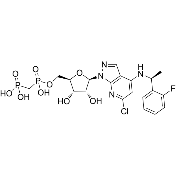
HY-125286
AB-680CD73 Inhibitor -

HY-138298A
Trastuzumab deruxtecanHER2 ADC -

HY-P99169
UliledlimabCD73 Inhibitor -
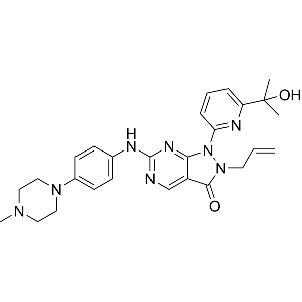
HY-10993
AdavosertibWee1 Inhibitor -
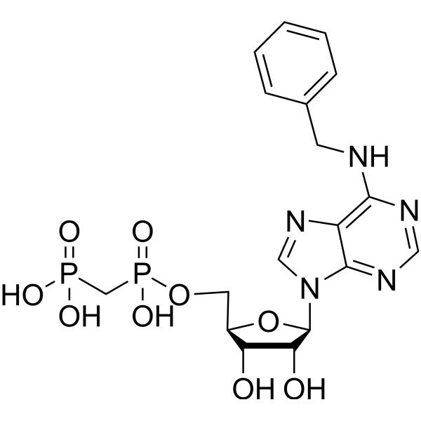
HY-100747
PSB-12379CD73 Inhibitor -
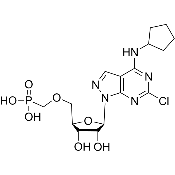
HY-131967
CD73-IN-4CD73 Inhibitor -
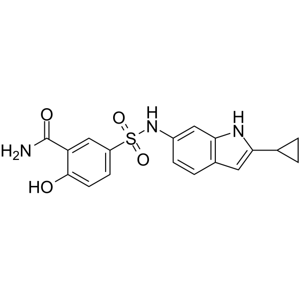
HY-103695
CD73-IN-1CD73 Inhibitor -
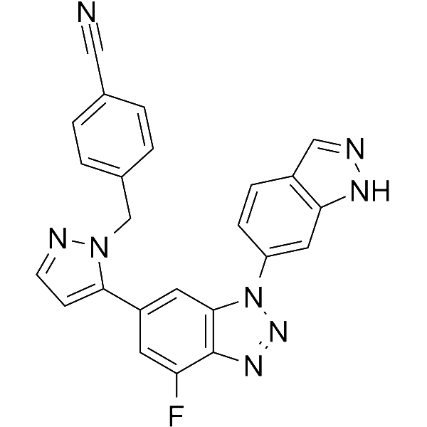
HY-145334
CD73-IN-5CD73 Inhibitor -

HY-103695R
CD73-IN-1 (Standard)Reference Standard -
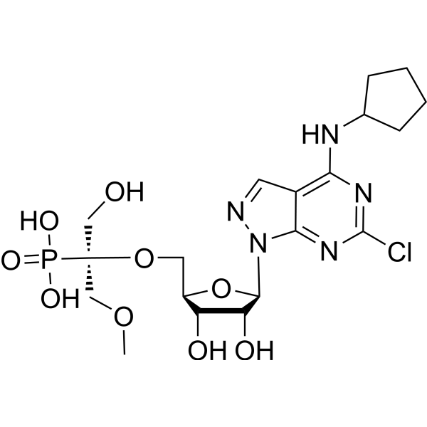
HY-136978
OP-5244CD73 Inhibitor -

HY-100747R
PSB-12379 (Standard)CD73 Inhibitor -
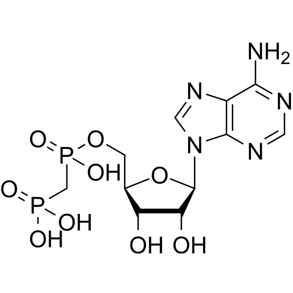
HY-112502
MethADPCD73 Inhibitor
Publications Citing Use of MedChemExpress (MCE) Oleclumab
More

Biological Activity
Description
Isotype
Human IgG1 lambda2
Recommend Isotype Controls
Species Reactivity
Human
IC50 & Target
NT5E/CD73
In Vivo
MedChemExpress (MCE) has not independently confirmed the accuracy of these methods. They are for reference only.
-
Animal Model:Murine, Balb/c, syngeneic CT26 colon carcinoma tumor model[2]
-
Dosage:10 mg/kg
-
Administration:ip; 2 time per week, for 2 weeks
-
Result:Inhibited tumor growth by 50% or greater from study day 7 to study day 16 when compared with isotype control antibody-treated groups.
Clinical Trial
| NCT Number | Sponsor | Condition | Start Date |
Phase

|
|---|---|---|---|---|
| NCT01329991 | Plexxikon| | 2011-05 | PHASE1 |
Gene ID
Accession
Conjugated
Unconjugated
Reconsititution
The product can be reconstituted/diluted with sterile PBS or saline.
Format
-
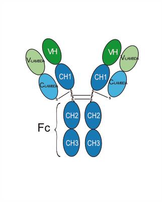
Human IgG1 lambda
Application
ELISA, FACS, Functional assay
Verified Bioactivity
-
 Immobilized CD73/5'-Nucleotidase Protein, Human (HEK293, His, HY-P70010) can bind Oleclumab. The EC50 for this effect is 3.27 ng/mL.
 -
 Flow Cytometry analysis of A431 cells labelling NT5E/CD73 (red) with Oleclumab (anti-NT5E/CD73) (HY-P99039). Goat Anti-Human IgG (Alexa Fluor 488) (HY-P83776) at a dilution of 1/1000 was used as the secondary antibody. Blue-Human IgG1 lambda2 (HY-P990096). Black-Unlabelled control, cells without incubation with primary antibody.


Chemical Information
-
CAS No. 1803176-05-7
-
Appearance Liquid
-
Molecular Weight 143.14 kDa
-
Color Colorless to light yellow
-
SMILES
[Oleclumab]
-
Synonyms
MEDI9447
-
Shipping
Shipping with dry ice.
-
Formulation
Please refer to the lot-specific COA for specific buffer information.
-
Storage
Please store the product under the recommended conditions in the Certificate of Analysis.


Publications (1)
-
Journal Impact Factor
-
Most Recent

Protocols
-
Multiplex immunofluorescence IHC
Multiplex immunofluorescence IHC detects multiple protein biomarkers in one tissue section by sequential antibody staining, HRP-mediated tyramide fluorophore deposition, heat-mediated antibody stripping, nuclear counterstaining, multispectral imaging, spectral unmixing, and digital cell phenotyping; TSA deposits fluorophore near the antigen so the fluorescence signal remains after primary and secondary antibodies are removed, enabling repeated staining cycles, including with antibodies from the same host species. Classic FFPE tumor immune-profiling applications use panels such as CD3, CD8, CD68/CD163, FOXP3, PD-1, PD-L1, pancytokeratin, Ki67, and DAPI to identify tumor cells, immune-cell subsets, checkpoint-marker expression, co-expression phenotypes, cell density, and spatial relationships in the tumor microenvironment.
-
Multiplex immunohistochemistry
Multiplex immunohistochemistry (mIHC), also known as tyramide dignal amplification (TSA), is an enzymatic detection method that uses horseradish peroxidase (HRP) to perform high-density in-situ labeling of target proteins or nucleic acids.
-
Research Protocol for Cancer Immunology
Cancer immunology studies how the immune system recognizes, suppresses, edits, or fails to eliminate malignant cells through tumor antigen release, antigen presentation, T-cell priming, immune trafficking, tumor-cell killing, and feedback inhibition in the tumor microenvironment. The cancer-immunity cycle links tumor antigenicity, dendritic-cell priming, CD8+ T-cell infiltration, cytotoxic function, and immune-checkpoint regulation to tumor rejection or immune escape. Immune-checkpoint pathways such as PD-1/PD-L1 and CTLA-4 suppress antitumor T-cell activity and can be therapeutically blocked, but many tumors remain resistant because of poor antigen presentation, weak T-cell infiltration, suppressive myeloid cells, regulatory T cells, and tumor-intrinsic immune-exclusion programs. Unresolved questions include which immune-cell states predict response, how tumor-intrinsic pathways exclude immune cells, how myeloid suppression limits checkpoint blockade, and which combination strategies


Purity & Documentation
-
Data Sheet (261 KB)
-
SDS (251 KB)
- English - EN (251 KB)
- Français - FR (251 KB)
- Deutsch - DE (251 KB)
- Norwegian - NO (251 KB)
- Español - ES (251 KB)
- Swedish - SV (251 KB)
- Italian - IT (251 KB)
- Korean - KR (251 KB)
- Portuguese - PT (251 KB)
-
Inhibitory Antibodies User Guide (603 KB)
References
[1]. Roy S Herbst, et al. COAST: An Open-Label, Phase II, Multidrug Platform Study of Durvalumab Alone or in Combination With Oleclumab or Monalizumab in Patients With Unresectable, Stage III Non-Small-Cell Lung Cancer. J Clin Oncol. 2022 Apr 22;JCO2200227. [Content Brief]
[2]. Hay CM, et al. Targeting CD73 in the tumor microenvironment with MEDI9447. Oncoimmunology. 2016 Jul 11;5(8):e1208875. [Content Brief]

Calculators
Concentration (start) × Volume (start) = Concentration (final) × Volume (final)


Oleclumab Related Classifications
HY-P99039 Related Classifications
 Powered by Bioz