
Phoenixin-14 TFA
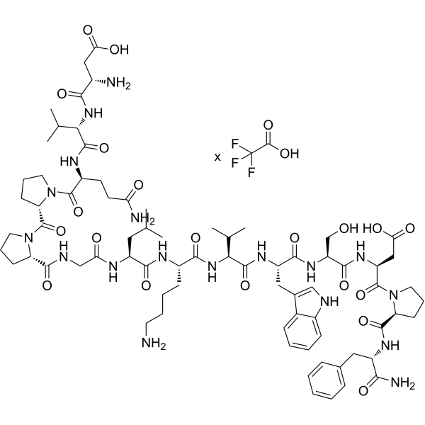
Phoenixin-14 (PNX-14) TFA is an endogenous neuropeptide with multiple biological activities, and serves as the endogenous ligand of GPR173. Phoenixin-14 TFA reduces ROS production by inhibiting the HMGB1/TLR4/MyD88/NF-κB signaling axis, thereby exerting antioxidant and mitochondrial protective effects. Phoenixin-14 TFA inhibits FOXO3 phosphorylation by upregulating SIRT3 expression, suppresses apoptosis, and improves myocardial systolic/diastolic function. Phoenixin-14 TFA resists ferroptosis by activating the ATF4/SLC7A11/GPX4 axis; it activates ERK1/2 phosphorylation via GPR173. Phoenixin-14 TFA can be used in researches on neuroprotection, diabetes, cardiomyopathy, reproductive protection and so on.
For research use only. We do not sell to patients.
- Formula: C75H110N18O20.xC2HF3O2
- Molecular Weight:1583.78 (free base)
-
Storage:
Please store the product under the recommended conditions in the Certificate of Analysis.
Biological Activity
Description
In Vitro
Phoenixin-14 (1-1000 nM; 24 h) TFA directly stimulates progesterone and estradiol secretion by porcine luteal cells on days 10-12 of the estrous cycle via upregulating the expression of steroidogenic enzymes; whereas combined treatment with luteinizing hormone (LH) (HY-P2293) attenuates progesterone secretion and the expression of steroidogenic enzymes[1].
Phoenixin-14 (1-1000 nM; 24 h) TFA regulates prostaglandin secretion in porcine luteal cells on days 10-12 of the estrous cycle by increasing PGE2 levels, decreasing PGF2α levels, and downregulating prostaglandin receptor expression[1].
Phoenixin-14 (1-1000 nM; 24 h) TFA upregulates the expression of GPR173 protein in porcine luteal cells on days 10-12 of the estrous cycle, and activates multiple kinase signaling pathways, including enhancing the phosphorylation of ERK1/2 and AKT, reducing the phosphorylation of PKA, and exerting time-dependent mixed effects on the phosphorylation of AMPKα and PKC[1].
The luteotropic effect of Phoenixin-14 (10 nM; 24 h) TFA on steroid and prostaglandin secretion in porcine luteal cells on days 10-12 of the estrous cycle is mediated via the GPR173 and ERK1/2 signaling pathways, independent of the PKA pathway[1].
Phoenixin-14 (50-100 nM; 24 h) TFA dose-dependently alleviates cisplatin-induced injury, oxidative stress and ferroptosis in KGN cells by restoring the ATF4/SLC7A11/GPX4 axis[2].
Phoenixin-14 (5-10 nM; 30 h) TFA dose-dependently inhibits the OGD/R-induced increases in mRNA and protein levels of TNF-α, IL-1β and IL-6 in BV2 microglia[4].
Phoenixin-14 (5-10 nM; 30 h) TFA dose-dependently reduces OGD/R-induced ROS production and restores depleted GSH levels in BV2 microglia[4].
Phoenixin-14 (5-10 nM; 30 h) TFA dose-dependently reverses the OGD/R-induced decrease in mitochondrial membrane potential in BV2 microglia, and the 10 nM dose almost completely abolishes this decrease[4].
Phoenixin-14 (5-10 nM; 30 h) TFA dose-dependently attenuates the decrease in viability of BV2 microglia induced by OGD/R, with the 10 nM dose almost maintaining the baseline cell viability[4].
Phoenixin-14 (5-10 nM; 30 h) TFA dose-dependently inhibits the OGD/R-induced increase in HMGB1 protein level in BV2 microglia[4].
Phoenixin-14 (5-10 nM; 30 h) TFA dose-dependently inhibits the OGD/R-induced increases in mRNA and protein levels of TLR4 and MyD88 in BV2 microglia[4].
Phoenixin-14 (5-10 nM; 30 h) TFA inhibits OGD/R-induced nuclear translocation of NF-κB p65 and activation of NF-κB luciferase activity in BV2 microglial cells in a dose-dependent manner[4].
Phoenixin-14 (10 nM; 30 h) TFA exerts a protective anti-inflammatory effect against OGD/R injury in BV2 microglia, and this effect depends on the GPR173 signaling pathway[4].
Phoenixin-14 (100 nM; 24 h) TFA reverses the decreases in viability, angiogenesis and migration capacity of human umbilical vein endothelial cells induced by conditioned media from iSMC and SMC, and also reverses the apoptosis induced by these media[5].
MedChemExpress (MCE) has not independently confirmed the accuracy of these methods. They are for reference only. Further protocols information, click here.
In Vivo
Phoenixin-14 (100 mg/kg; i.p.; daily; 2 months) TFA protects against streptozotocin-induced diabetic cardiomyopathy in C57BL/6 mice by improving cardiac function, reducing myocardial injury, ameliorating cardiac hypertrophy, mitigating oxidative stress and inflammation, and restoring SIRT3/FOXO3 signaling, with no observed effects on normal mice[3].
Phoenixin-14 (15 nmol; i.c.v.; single dose; 1 h prior to MCAO) TFA reduces ischemia/reperfusion-induced brain infarct volume to just under 20% and suppresses microglial activation in a rat MCAO ischemic stroke model[4].
MedChemExpress (MCE) has not independently confirmed the accuracy of these methods. They are for reference only.
Clinical Trial
| NCT Number | Sponsor | Condition | Start Date |
Phase
|
|---|---|---|---|---|
| NCT01329991 | Plexxikon| | 2011-05 | PHASE1 |
Chemical Information
-
Molecular Weight 1583.78 (free base)
-
Formula C75H110N18O20.xC2HF3O2
-
Synonyms
PNX-14 TFA
-
Sequence
Asp-Val-Gln-Pro-Pro-Gly-Leu-Lys-Val-Trp-Ser-Asp-Pro-Phe-NH2
-
Sequence Shortening
DVQPPGLKVWSDPF-NH2
-
Shipping
Room temperature in continental US; may vary elsewhere.
-
Storage
Please store the product under the recommended conditions in the Certificate of Analysis.
Protocols
-
Kinase activity and phosphorylation assays
Kinase activity assays measure the ability of kinases to transfer phosphate groups from ATP to specific substrates, while phosphorylation assays detect the presence and levels of phosphorylated proteins. Common methods include radiolabeled ATP incorporation (e. g. ,), ADP release detection via bioluminescence (e. g. ,[3]), enzyme-linked immunosorbent assays (ELISA) for phospho-specific epitopes (e. g. ,[6]), and microtiter-based formats for high-throughput screening (e. g. ,[8]). The ADP-Glo assay quantifies kinase activity by measuring ADP produced during phosphorylation using a luciferase-based system. Radiometric assays involve autoradiography or scintillation counting after incorporation of 32P-labeled ATP into substrate proteins. ELISA-based approaches rely on phospho-specific antibodies to detect activated kinases in cell lysates or purified samples.
-
Western Blot
Western blotting (WB) is a commonly used experimental method in molecular biology, biochemistry, and immunogenetics for identifying and quantifying target proteins. It combines gel electrophoresis with immunoassay, enabling researchers to analyze protein expression, post-translational modifications, and molecular weight.
-
RNA extraction experimental
By lysing cells, releasing RNA, and removing impurities such as proteins and DNA, high-purity RNA products are finally obtained. The commonly used traditional method is the guanidine isothiocyanate/phenol/chloroform method (Trizol), which is suitable for a variety of animal materials including animal tissues, microorganisms, cultured cells, etc., and most plant materials.
-
Apoptosis
Apoptosis, also called programmed cell death, is generally characterized by distinct morphological characteristics.
-
TUNEL staining for apoptotic DNA fragmentation
TUNEL staining detects DNA strand breaks by using terminal deoxynucleotidyl transferase to add labeled nucleotides to exposed 3′-OH DNA termini, generating either microscopic staining in fixed cells or tissue sections, or fluorescence/cytometric signal in cell suspensions. TUNEL positivity reflects DNA fragmentation but should not be interpreted alone as definitive apoptosis, because TUNEL can also label necrotic, autolytic, mechanically damaged, or DNA-repair-associated DNA breaks.
-
Annexin V plus membrane-impermeant dye apoptosis staining
Annexin V-based apoptosis assays rely on the detection of phosphatidylserine (PS) externalization from the inner leaflet of the plasma membrane to the outer leaflet, an early biochemical hallmark of apoptosis. Fluorescently labeled Annexin V binds PS in a calcium-dependent manner, enabling identification of early apoptotic cells by flow cytometry or fluorescence microscopy. When combined with a membrane-impermeant DNA-binding dye (e. g. , propidium iodide), this approach allows discrimination between viable (Annexin V−/dye−), early apoptotic (Annexin V+/dye−), and late apoptotic or necrotic (Annexin V+/dye+) cell populations by assessing membrane integrity and PS exposure.
-
ROS/oxidative-stress fluorescent staining
ROS/oxidative-stress fluorescent staining uses cell-permeant fluorogenic probes that become fluorescent after oxidation inside cells or tissues; commonly used examples include DCFH-DA/DCFDA for broad cellular oxidant detection, DHE for superoxide-related signal detection, MitoSOX for mitochondrial superoxide-related signal detection, and CellROX probes for oxidative-stress-associated fluorescence readouts. The assay detects probe oxidation rather than a single ROS species unless the probe and analysis method have been chemically validated for that species. DCFH-DA enters cells, is deacetylated by intracellular esterases to DCFH, and produces fluorescent DCF after oxidation, so the readout is used as an operational measure of total cellular oxidative stress rather than a species-specific ROS measurement. DHE and MitoSOX can report superoxide-related oxidation, but red fluorescence alone can include non-specific ethidium-like oxidation products; HPLC or optimized spectral approaches are
-
Apoptosis Solutions
Apoptosis is a regulated, generally non-lytic cell-death pathway that removes unwanted, damaged, infected, or abnormal cells through coordinated morphological changes, caspase activation, DNA fragmentation, and membrane remodeling. The intrinsic apoptosis pathway is controlled mainly by mitochondrial outer membrane permeabilization, BCL-2 family proteins, cytochrome c release, apoptosome formation, caspase-9 activation, and downstream executioner caspase-3/7 activation. The extrinsic apoptosis pathway is initiated by death receptors such as Fas, TNFR, and TRAIL receptors, which recruit adaptor proteins and activate caspase-8 before engaging executioner caspases or mitochondrial amplification through BID cleavage. Apoptosis is linked to many phenotypes, including cancer cell killing, tissue homeostasis, immune regulation, neurodegeneration, infection response, and treatment-induced cytotoxicity; unresolved questions include how apoptosis interacts with necroptosis, pyroptosis, ferroptos
-
Research Protocol for Inflammation-related Diseases
The NLRP3 inflammasome is a cytosolic innate immune signaling platform that integrates priming signals and danger-signal activation to promote caspase-1 activation, maturation of IL-1β and IL-18, and gasdermin D-mediated pyroptotic cell death. The core experimental logic is to determine whether inflammatory disease phenotypes are driven by increased NLRP3 expression, ASC-containing inflammasome assembly, caspase-1 cleavage, GSDMD cleavage, and extracellular release of IL-1β/IL-18 rather than by nonspecific cell injury alone. The pathway is strongly linked to inflammation-related disease phenotypes because monosodium urate crystals activate NALP3/NLRP3 inflammasome signaling in gout-like crystal inflammation, cholesterol crystals activate NLRP3 inflammasomes in atherogenesis models, and DSS-induced intestinal inflammation has been reported to involve NLRP3 inflammasome activity. However, experimental colitis studies also show context-dependent protective effects of NLRP3 inflammasome co
-
Protocol for Kinase activity and phosphorylation assays
Kinase activity assays measure transfer of phosphate from ATP to a protein or peptide substrate, generating phosphorylated substrate, ADP, or incorporated radiolabeled phosphate as the readout; phosphorylation assays measure site-specific phosphorylation in cells or tissues as a proxy for kinase-pathway activation, inhibition, or substrate regulation. Phosphorylation can be detected by phospho-specific Western blot, immunoprecipitation kinase assay, phospho-immunofluorescence, phospho-flow cytometry, luminescent ADP detection, radiolabeled ATP incorporation, or reporter-based pathway assays, and these readouts can be applied to cancer cells, primary neurons, mouse tumors, organoids, inflammatory macrophages, ferroptosis studies, and mitophagy studies when the kinase target is biologically relevant.
-
Ferroptosis Solutions
Ferroptosis is an iron-dependent, non-apoptotic form of regulated cell death characterized by lethal lipid peroxidation and sensitivity to suppression by iron chelators or lipophilic radical-trapping antioxidants. The core pathway links cystine uptake through system Xc−, glutathione availability, GPX4-dependent detoxification of phospholipid hydroperoxides, iron-dependent oxidative reactions, and polyunsaturated-phospholipid metabolism into a cell-death program that is biochemically and morphologically distinct from apoptosis, necrosis, and autophagy. The ferroptosis pathway is experimentally linked to phenotype through chemical and genetic perturbation. Erastin induces ferroptosis by inhibiting cystine uptake through system Xc− and weakening antioxidant defenses, while GPX4 inhibition or depletion causes lipid peroxide accumulation and ferroptotic cancer-cell death. ACSL4 and oxidizable arachidonoyl- or adrenoyl-containing phosphatidylethanolamines shape ferroptosis sensitivity by con
-
Research Protocol for Metabolic Diseases
AMP-activated protein kinase, AMPK, is a conserved cellular energy sensor that responds to reduced cellular energy status and coordinates metabolism by increasing ATP-generating catabolic pathways while suppressing ATP-consuming anabolic processes. In metabolic disease research, the AMPK pathway is experimentally relevant because it regulates hepatic lipid synthesis, fatty acid oxidation, glucose production, skeletal-muscle glucose disposal, mTORC1-linked biosynthesis, autophagy, mitochondrial homeostasis, and whole-body energy balance. The central pathway logic is that energy stress, metformin, exercise-like stimulation, or direct AMPK activators increase AMPKα Thr172 phosphorylation and downstream substrate phosphorylation, including ACC and RAPTOR. Phosphorylation of ACC suppresses lipogenesis and supports fatty acid oxidation, whereas phosphorylation of RAPTOR suppresses mTORC1 signaling and links cellular energy status to growth and protein synthesis control. The pathway is linked
Purity & Documentation
References
[1]. Mlyczyńska E, et al. Phoenixin-14 as a novel direct regulator of porcine luteal cell functions†. Biol Reprod. 2024;110(1):154-168. [Content Brief]
[2]. Wu R, et al. Phoenixin-14 Alleviates Premature Ovarian Failure by Inhibiting Ferroptosis Through SLC7A11/GPX4. Drug Dev Res. 2025;86(5):e70110. [Content Brief]
[3]. Yao B, et al. Phoenixin-14 protects cardiac damages in a streptozotocin-induced diabetes mice model through SIRT3. Arch Physiol Biochem. 2024;130(1):110-118. [Content Brief]
[4].
Ma H, et al. Phoenixin 14 inhibits ischemia/reperfusion-induced cytotoxicity in microglia. Arch Biochem Biophys. 2020 Aug 15;689:108411.
[Content Brief]
[5]. Ling C, et al. Phoenixin-14 alleviates inflammatory smooth muscle cell-induced endothelial cell dysfunction in vitro. Cytokine. 2022;157:155973. [Content Brief]
Calculators
Concentration (start) × Volume (start) = Concentration (final) × Volume (final)

