



(±)5,6-DHET lactone
5,6-DiHET lactone ((±)5,6-DiHETrE lactone) is a lactonized form of 5,6-EET and 5,6-DiHET. In solution, 5(6)-EET degrades into 5(6)-DiHET and 5(6)-δ-lactone, which can be converted to 5(6)-DiHET and quantified by GC-MS.1 5,6-DiHET potently induces vasodilation of isolated canine coronary arterioles, with 41 and 100% inhibition occurring at 0.01 and 100 pM, respectively. It also induces vasodilation in isolated human microvessels and increases intracellular calcium levels in a dose-dependent manner, an effect that can be blocked by the nitric oxide scavenger L-NAME.
For research use only. We do not sell to patients.
- CAS No.: 213126-92-2
- Formula: C20H32O3
- Molecular Weight:320.47
-
Storage:
Please store the product under the recommended conditions in the Certificate of Analysis.


To place orders, for customer services and technical support, please contact: MedChemExpress USA
Tel: 609-228-6898 E-mail: [email protected] [email protected]


-
 Biological Activity
-
Chemical Information
- Protocols
- Purity & Documentation
- References
-
 Help & FAQs


-
HY-118332
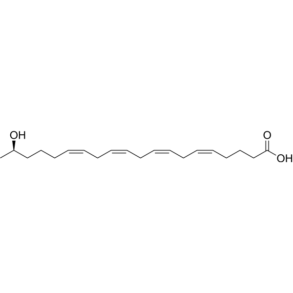
19(R)-HETEVasodilator -

HY-132184
5,6-Epoxyeicosatrienoic acidMetabolite -
HY-139114
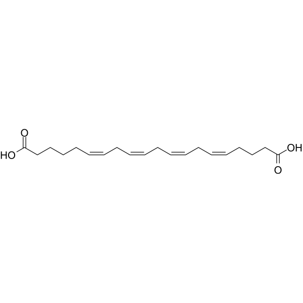
20-Carboxyarachidonic acidPPARalpha/PPARgamma Activator -

HY-P0206A
Bradykinin acetateBradykinin Recepto -
HY-113439
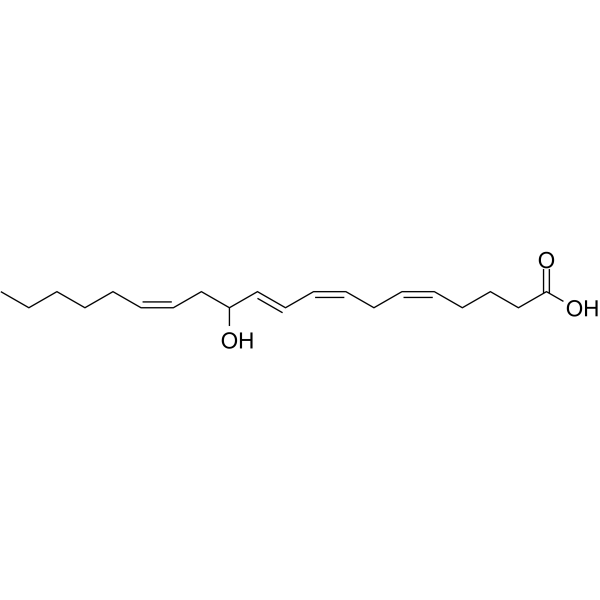
12-HETEVascular Function Regulator -
HY-12753A
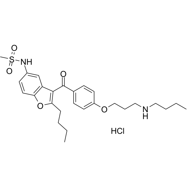
Debutyldronedarone hydrochlorideCytochrome P450 Enzyme/Thyroid Hormone Receptor Inhibitor -
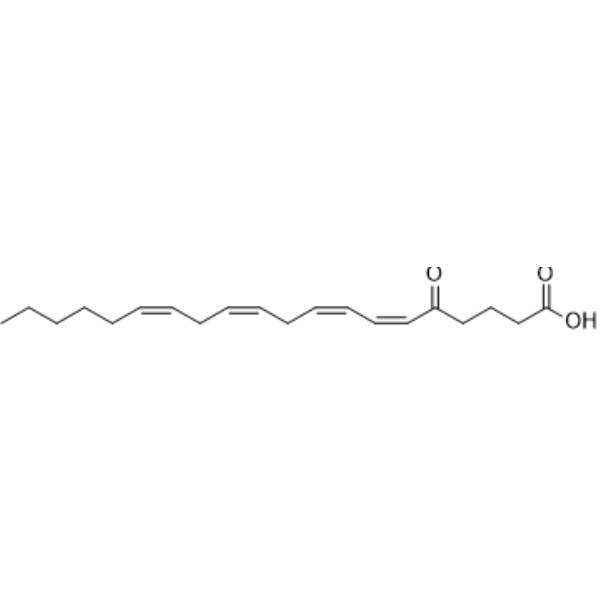
HY-112549
5-oxo-ETEApoptosis Inhibitor -
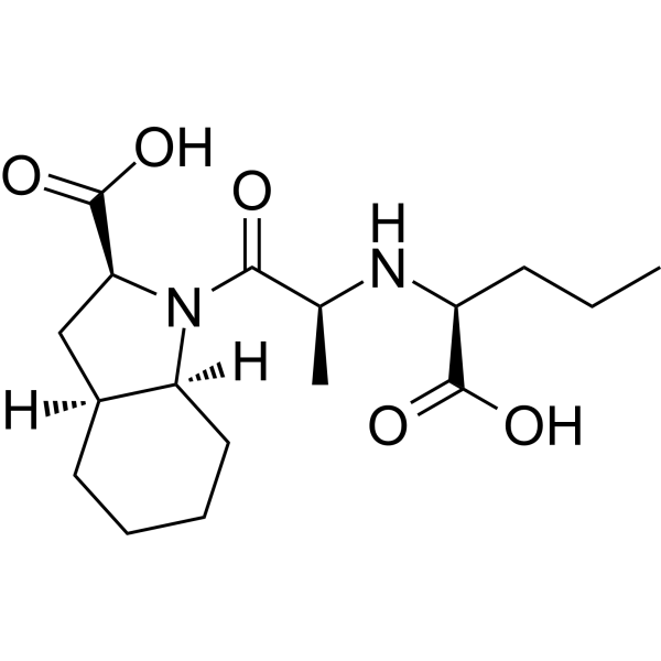
HY-B1433
PerindoprilatACE Inhibitor -
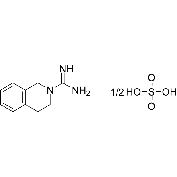
HY-B1624A
Debrisoquin hemisulfateTMPRSS2 Inhibitor -
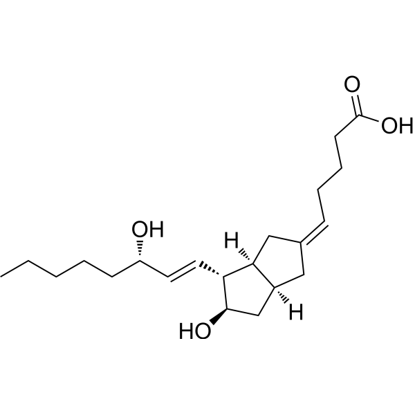
HY-124363
5-cis CarbaprostacyclinPGI2 Analog -
HY-130522
6β-Prostaglandin I1Prostacycline Analogue -
HY-135077
Prostaglandin A1 ethyl esterProstaglandin A1 Prodrug


Biological Activity
Description


Chemical Information
-
CAS No. 213126-92-2
-
Molecular Weight 320.47
-
Formula C20H32O3
-
SMILES
O=C1CCCC(C(O)C/C=C\C/C=C\C/C=C\CCCCC)O1
-
Synonyms
(±)5,6-DiHETrE lactone
-
Shipping
Room temperature in continental US; may vary elsewhere.
-
Storage
Please store the product under the recommended conditions in the Certificate of Analysis.

Protocols
-
Calcium Spark Assay
Calcium sparks are localized, transient increases in intracellular calcium concentration ([Ca2+]i) that occur in cardiac myocytes and represent elementary events underlying excitation-contraction coupling. These events are generated by the coordinated opening of clusters of ryanodine receptors (RyRs) on the sarcoplasmic reticulum membrane, leading to a brief release of Ca2+ into the cytosol. The detection and analysis of calcium sparks provide insights into the mechanisms of calcium handling and signaling in cardiac cells. Imaging techniques using fluorescent calcium indicators such as Fluo-3 are employed to visualize these subcellular calcium transients with high spatial and temporal resolution. The protocol is based on established methodologies described in primary literature for both experimental measurement and automated analysis of calcium sparks.
-
Ca2+ Staining Technique
Ca2+ staining is an experimental technique that utilizes specific fluorescent probes (such as Fluo-4 AM, Fura-2, etc.) to qualitatively or quantitatively detect dynamic changes in intracellular Ca2+ concentrations; this is achieved by monitoring the changes in fluorescent signals generated when these probes bind to free intracellular calcium ions. The underlying principle relies primarily on the presence of chelating groups within the probe's molecular structure that possess high affinity for calcium ions.


Purity & Documentation
References

Calculators
Concentration (start) × Volume (start) = Concentration (final) × Volume (final)


(±)5,6-DHET lactone Related Classifications
HY-130229 Related Classifications
 Powered by Bioz