



ReAsH-EDT2

Based on 1 Customer Validation
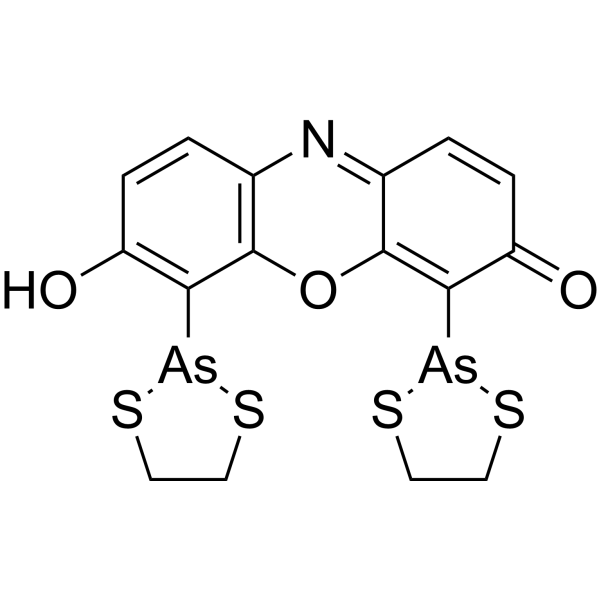
ReAsH-EDT2 is a red fluorescent dye that marks proteins. ReAsH-EDT2 is a membrane-permeable biarsenical compound that binds covalently to tetracysteine sequences which allows the protein to be imaged. ReAsH-EDT2 can be used for protein localization and trafficking. (λex=530 nm, λem=592 nm).
For research use only. We do not sell to patients.
- Purity : 95.0%
- CAS No.: 438226-89-2
- Formula: C16H13As2NO3S4
- Molecular Weight:545.38
-
Storage:
-20°C, sealed storage, away from moisture and light
* In solvent : -80°C, 6 months; -20°C, 1 month (sealed storage, away from moisture and light)

To place orders, for customer services and technical support, please contact: MedChemExpress USA
Tel: 609-228-6898 E-mail: [email protected] [email protected]


-
 Biological Activity
-
Chemical Information
- Protocols
- Purity & Documentation
- References
-
 Help & FAQs


-
HY-130412
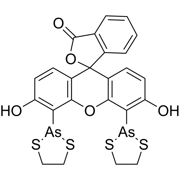
FlAsH-EDT2Protein Labeling Reagent -
HY-101894
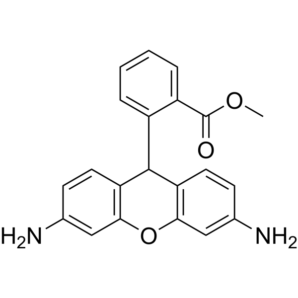
Dihydrorhodamine 123ROS Indicator -
HY-100041
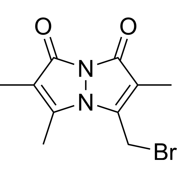
BromobimaneDye Reagent -
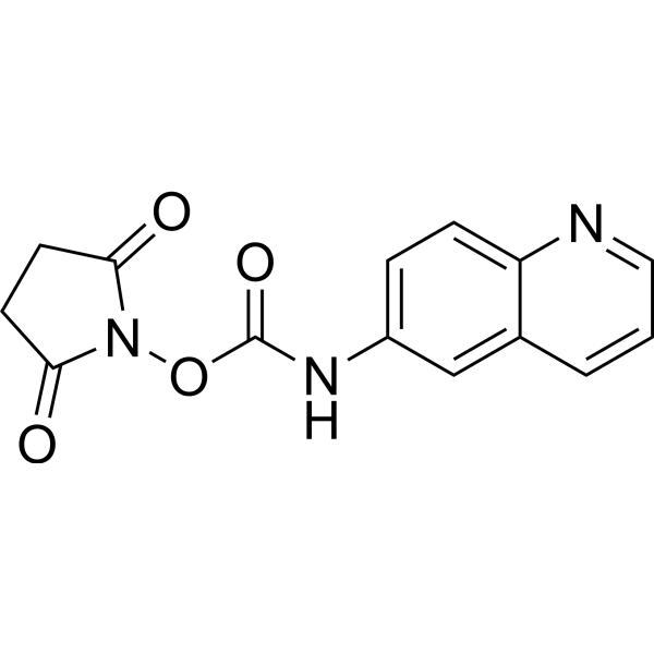
HY-117695
AQCAmino Acid Sequence Detection Reagent -
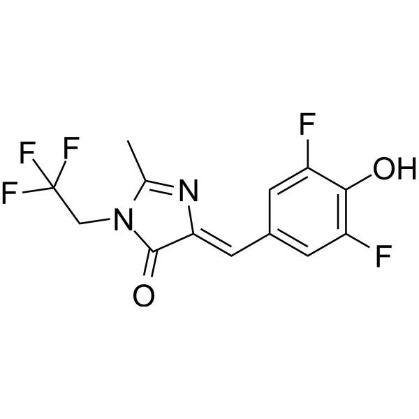
HY-110251
DFHBI-1TRNA Aptamers Probe -
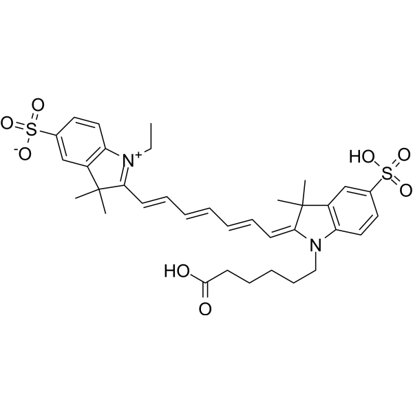
HY-D0825
CY7Fluorescent Dye -
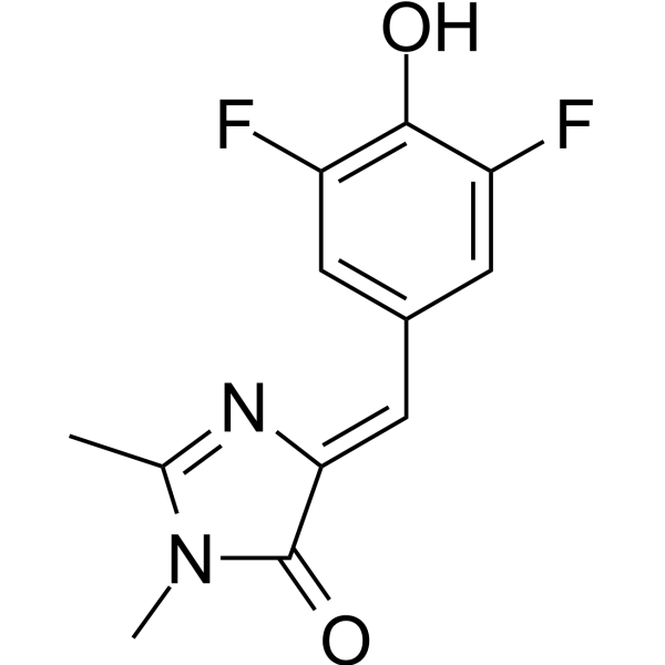
HY-110250
DFHBIRNA Fluorogen -
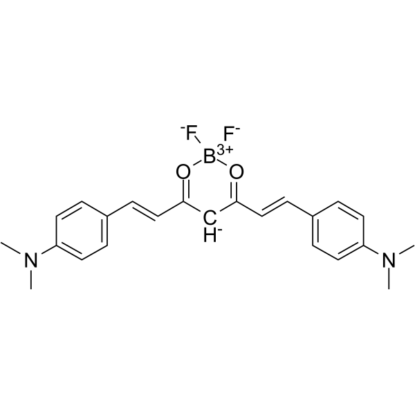
HY-103242
CRANAD-2Fluorescent Probe -
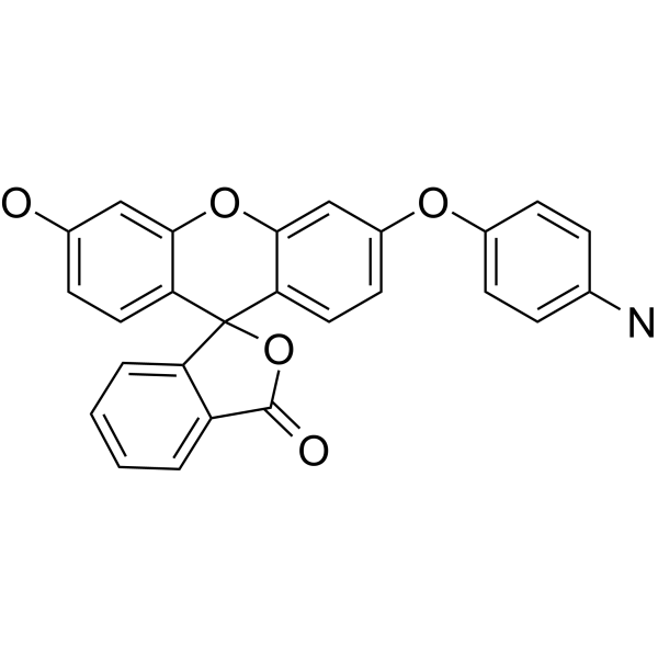
HY-119977
APFFluorescence Probe -

HY-DY1037
Dihydrorhodamine 123 (solution)ROS Indicator -
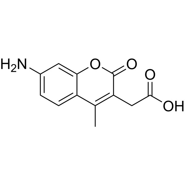
HY-D0025
7-Amino-4-methylcoumarin-3-acetic acidFluorescent Protein Labeling Agent -
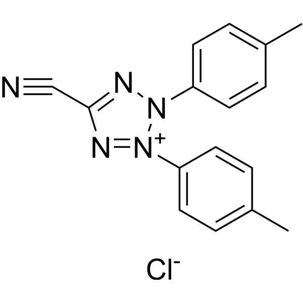
HY-D0016
5-Cyano-2,3-di-(p-tolyl)tetrazolium chlorideFluorescent Dye


Biological Activity
Description
In Vitro
ReAsH-EDT2 tags with GyrB.FGFR1KD.TC can appear with red fluorescence.
MedChemExpress (MCE) has not independently confirmed the accuracy of these methods. They are for reference only. Further protocols information, click here.
Emission (Em)
592
Excitation (Ex)
530


Chemical Information
-
CAS No. 438226-89-2
-
Appearance Solid
-
Molecular Weight 545.38
-
Formula C16H13As2NO3S4
-
Color Brown to reddish brown
-
SMILES
O=C1C=CC2=NC3=C(C([As]4SCCS4)=C(O)C=C3)OC2=C1[As]5SCCS5
-
Shipping
Room temperature in continental US; may vary elsewhere.
-
Storage
-20°C, sealed storage, away from moisture and light
* In solvent : -80°C, 6 months; -20°C, 1 month (sealed storage, away from moisture and light)

Protocols
-
Antibody-based immunofluorescence/immunocytochemistry staining
Antibody-based immunofluorescence/immunocytochemistry detects the cellular or subcellular localization of a target antigen by binding a primary antibody to the target and detecting that antibody directly with a fluorophore-conjugated primary antibody or indirectly with a fluorophore-conjugated secondary antibody. Indirect immunofluorescence can amplify signal because multiple secondary antibodies can bind one primary antibody. The assay readout is fluorescence intensity and localization measured by fluorescence or confocal microscopy, and the result reflects antigen distribution only when the antibody has been validated for the target, sample type, fixation condition, and imaging workflow. Antibody specificity must not be assumed from catalog information alone, and appropriate validation or control experiments are required for serious interpretation.
- Immunocytochemistry/Immunofluorescence


Purity & Documentation
-
Data Sheet (274 KB)
-
SDS (480 KB)
- English - EN (480 KB)
- Français - FR (480 KB)
- Deutsch - DE (480 KB)
- Norwegian - NO (480 KB)
- Español - ES (480 KB)
- Swedish - SV (480 KB)
- Italian - IT (480 KB)
- Korean - KR (480 KB)
- Portuguese - PT (480 KB)
-
Handling Instructions (2659 KB)
References
[1]. Adams SR, et, al. Preparation of the membrane-permeant biarsenicals FlAsH-EDT2 and ReAsH-EDT2 for fluorescent labeling of tetracysteine-tagged proteins. Nat Protoc. 2008;3(9):1527-34. [Content Brief]
[2]. Perdios L, et, al. Time-resolved FRET reports FGFR1 dimerization and formation of a complex with its effector PLCγ1. Adv Biol Regul. 2016 Jan;60:6-13. [Content Brief]

Calculators
Concentration (start) × Volume (start) = Concentration (final) × Volume (final)


ReAsH-EDT2 Related Classifications
HY-130533 Related Classifications
 Powered by Bioz