



HEPACAM2 Protein, Rat (HEK293, His)

Based on 1 Customer Validation
The HEPACAM2 gene encodes a protein associated with an immunoglobulin superfamily that plays a role in mitosis. Knocking down this gene can lead to prophase cell arrest, abnormal nuclear morphology and apoptosis. HEPACAM2 can be used as a biomarker for the prognosis of colorectal adenocarcinoma. HEPACAM2 Protein, Rat (HEK293, His) is the recombinant rat-derived HEPACAM2 protein, expressed by HEK293 , with C-His labeled tag.
- Species: Rat
- Source: HEK293
-
Storage:Stored at -20°C for 2 years from date of receipt. After reconstitution, it is stable at 4°C for 1 week or -20°C for longer (with carrier protein). It is recommended to freeze aliquots at -20°C or -80°C for extended storage.


To place orders, for customer services and technical support, please contact: MedChemExpress USA
Tel: 609-228-6898 E-mail: [email protected] [email protected]


-
 Biological Activity
-
Technical Parameters
-
Product Properties
- Documentation
- References
- Help & FAQs




Biological Activity
Description
The HEPACAM2 gene encodes a protein associated with an immunoglobulin superfamily that plays a role in mitosis. Knocking down this gene can lead to prophase cell arrest, abnormal nuclear morphology and apoptosis. HEPACAM2 can be used as a biomarker for the prognosis of colorectal adenocarcinoma. HEPACAM2 Protein, Rat (HEK293, His) is the recombinant rat-derived HEPACAM2 protein, expressed by HEK293 , with C-His labeled tag.
Background
HepaCAM, an N-linked glycoprotein encoding a member of the immunoglobulin superfamily, is a tumor suppressor gene that mediates multiple cellular biological functions. HepaCAM may be a novel therapeutic target for inhibiting bladder cancer proliferation through the AKT/FoxO pathway. The HEPACAM2 gene encodes a protein associated with an immunoglobulin superfamily that plays a role in mitosis. Knocking down this gene can lead to prophase cell arrest, abnormal nuclear morphology and apoptosis. The polymerization (ADP-ribosylation) of the encoding protein promotes its translocation to the centrosome, which may stimulate centrosome maturation. Deletion of chromosomes containing this gene may be associated with myeloid leukemia and myelodysplastic syndrome in human patients. HEPACAM2 can be used as a biomarker for the prognosis of colorectal adenocarcinoma[1][2][3].
MCE Validation Data
-
Purity - SDS-PAGE
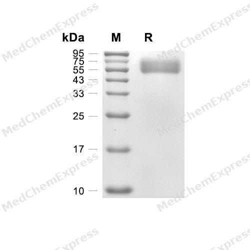
Purity - SDS-PAGE


Technical Parameters
-
Species Rat
-
Source HEK293
-
Tag C-His
-
Accession
B5DEN8 (L33-L350)
-
Molecular Construction
-
N-term
-
HEPACAM2 (L33-L350)
Accession # B5DEN8 -
His
-
C-term
-
-
Protein Length
Partial
-
Synonyms
HEPACAM2; FLJ38683; HEPACAM Family Member 2; MIKI; Mitotic Kinetics Regulator; Alternative Protein HEPACAM2
-
AA Sequence
LKVTVPSHTVHGIRGQALYLPVHYGFHTPASDIQIIWLFERSHTTPKYLLGSVNKSVVPDLEYQHKFTMMPPNASLLINPLQFTDEGNYIVKVNIQGNGTLSASQKIQVTVDDPVMKPMVQFHPASGAVEYVGNITLTCRVEGGTRLVYQWRKNGKPININSSHSFSPQNHTLRIVPATKEDIGNYTCLVSNPVSEMESDIIIPTIYYGPYGLQVNSDKGLKVGEVFTVDLGEAVLFDCSADSYPPNTYSWIRRSANTTRVIKHGPRLEVASDKVAQKTADYVCCAYNNITGRRDETHFTVIITSVGLEKLAQRGKSL
-
Molecular Weight
Approximately 60 kDa, based on SDS-PAGE under reducing conditions, due to the glycosylation.
-
Purity
≥ 95%, as determined by reducing SDS-PAGE.


Product Properties
Lyophilized powder.
Lyophilized from a 0.22 μm filtered solution of PBS, pH 7.4.
<1 EU/μg, determined by LAL method.
It is not recommended to reconstitute to a concentration less than 100 μg/mL in ddH2O. For long term storage it is recommended to add a carrier protein (0.1% BSA, 5% HSA, 10% FBS or 5% Trehalose).
Stored at -20°C for 2 years from date of receipt. After reconstitution, it is stable at 4°C for 1 week or -20°C for longer (with carrier protein). It is recommended to freeze aliquots at -20°C or -80°C for extended storage.
Room temperature in continental US; may vary elsewhere.


Documentation
-
Data Sheet (264 KB)
-
SDS (252 KB)
- English - EN (252 KB)
- Français - FR (252 KB)
- Deutsch - DE (252 KB)
- Norwegian - NO (252 KB)
- Español - ES (252 KB)
- Swedish - SV (252 KB)
- Italian - IT (252 KB)
- Korean - KR (252 KB)
- Portuguese - PT (252 KB)
-
Handling Instructions (2659 KB)


References
[1]. Klopfleisch R, et al. HEPACAM1 and 2 are differentially regulated in canine mammary adenomas and carcinomas and its lymph node metastases. BMC Vet Res. 2010 Mar 15;6:15. [Content Brief]
[2]. Tang M, et al. Overexpression of HepaCAM inhibits bladder cancer cell proliferation and viability through the AKT/FoxO pathway. J Cancer Res Clin Oncol. 2017 May;143(5):793-805. [Content Brief]

Calculators
Concentration (start) × Volume (start) = Concentration (final) × Volume (final)


 Powered by Bioz

