



PEPD Protein, Human

Based on 1 Customer Validation
PEPD Protein, a member of the peptidase family, is a dipeptidase which hydrolyze dipeptides with proline or hydroxyproline at the carboxy terminus. It plays an important role in collagen metabolism because the high level of iminoacids in collagen. PEPD suppression of p53 is essential for cell survival and tumor growth. PEPD Protein, Human is the recombinant human-derived PEPD protein, expressed by E. coli , with tag free.
- Species: Human
- Source: E. coli
-
Almacenamiento:Stored at -80°C for 1 year from date of receipt. It is stable at -20°C for 3 months after opening. It is recommended to freeze aliquots at -80°C for extended storage. Avoid repeated freeze-thaw cycles.


To place orders, for customer services and technical support, please contact: MedChemExpress USA
Tel: 609-228-6898 E-mail: [email protected] [email protected]


-
 Actividad biológica
-
Technical Parameters
-
Product Properties
- Documentación
- Help & FAQs




Actividad biológica
Descripciòn
PEPD Protein, a member of the peptidase family, is a dipeptidase which hydrolyze dipeptides with proline or hydroxyproline at the carboxy terminus. It plays an important role in collagen metabolism because the high level of iminoacids in collagen. PEPD suppression of p53 is essential for cell survival and tumor growth. PEPD Protein, Human is the recombinant human-derived PEPD protein, expressed by E. coli , with tag free.
Background
PEPD, a member of the peptidase family, is a dipeptidase which hydrolyze dipeptides with proline or hydroxyproline at the carboxy terminus. The preferred dipeptide substrate is Gly-Pro, but other Xaa-Pro dipeptides, such as Ala-Pro, Met-Pro, Phe-Pro, Val-Pro and Leu-Pro, can be cleaved by PEPD. It plays an important role in collagen metabolism because the high level of iminoacids in collagen. PEPD modulates expression of interferon α/β receptor IFNAR1. PEPD directly binds to p53 in the nucleus and cytoplasm and suppresses both transcription-dependent and transcription-independent activities of p53, which is essential for cell survival and tumor growth. In addition, PEPD stimulates proliferation and migration of fibroblasts via activation of the EGFR-downstream PI3K/Akt/mTOR signaling pathway, which also stimulates the expression of β1-integrin and IGF-1 receptors and proteins downstream to these receptors such as FAK, Grb2, and ERK1/2[1][2][3][4].
Verified Bioactivity
Measured by its ability to cleave a peptide substrate, Gly-Pro-AMC. Read at excitation and emission wavelengths of 380 nm and 460 nm (top read). The specific activity is 5013.428 pmol/min/µg. (Activation description: The proenzyme needs to be activated in activation buffer for an activated form.)
MCE Validation Data
-
Purity - SDS-PAGE
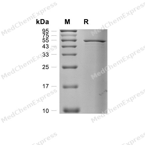
Purity - SDS-PAGE

Assay Procedure
Materials
Activation buffer: 0.05 M Tris, 0.01 M CaCl2, 0.15 M NaCl, 0.05% Brij-35, MnCl2, pH 7.5
Assay buffer: 0.05 M Tris, 0.1 M NaCl, 0.01% Brij-35, pH 8.5
PEPD Protein, Human (HY-P70992)
Substrate: Gly-Pro-AMC (HY-137834)
Standard: 7-amino, 4-Methyl Coumarin
Procedure
1. Dilute standard in assay buffer to concentrations of 0, 0.078125, 0.15625, 0.3125, 0.625, 1.25, 2.5, 5, 10, 20, 40, 80 μM.
2. Add 100 μL of each standard concentration to the wells.
3. Read the fluorescence in kinetic mode for 5 minutes with excitation and emission wavelengths of 380 nm and 460 nm (top read) .
4. Plot the standard concentrations on the y-axis and the measured RFU on the x-axis to create the standard curve and obtain the standard curve equation.
5. Dilute Human PEPD to 0.2 μg/mL in activation buffer.
6. Dilute the substrate to 50 μM in assay buffer.
7. Add 50 μL of 0.2 μg/mL Human PEPD to the wells, followed by 50 μL of 50 μM substrate to initiate the reaction.
For the control group, add 50 μL of assay buffer and 50 μL of 50 μM substrate.
8. Read the fluorescence in kinetic mode for 5 minutes with excitation and emission wavelengths of 380 nm and 460 nm (top read).
9. Calculate specific activity:
Specific Activity (pmol/min/μg) = | Adjusted Vmax* (RFU/min) x Conversion Factor ** (pmol/RFU) |
| amount of enzyme (μg) |
*Adjusted for Substrate Blank
**Derived using calibration standard
Per Well:
Human PEPD: 0.01 μg
Substrate: 25 μM


Technical Parameters
-
Species Human
-
Source E. coli
-
Tag Tag Free
-
Accession
AAH28295.1 (A2-K493)
-
Molecular Construction
-
N-term
-
PEPD (A2-K493)
Accession # AAH28295.1 -
C-term
-
-
Protein Length
Full Length of Mature Protein
-
Synonyms
PEPD; Prolidase; Peptidase D; X-Pro Dipeptidase; Xaa-Pro Dipeptidase; Testicular Tissue Protein Li 138; Imidodipeptidase; Aminoacyl-L-Proline Hydrolase; Proline Dipeptidase; PRD
-
AA Sequence
AAATGPSFWLGNETLKVPLALFALNRQRLCERLRKNPAVQAGSIVVLQGGEETQRYCTDTGVLFRQESFFHWAFGVTEPGCYGVIDVDTGKSTLFVPRLPASHATWMGKIHSKEHFKEKYAVDDVQYVDEIASVLTSQKPSVLLTLRGVNTDSGSVCREASFDGISKFEVNNTILHPEIVECRVFKTDMELEVLRYTNKISSEAHREVMKAVKVGMKEYELESLFEHYCYSRGGMRHSSYTCICGSGENSAVLHYGHAGAPNDRTIQNGDMCLFDMGGEYYCFASDITCSFPANGKFTADQKAVYEAVLRSSRAVMGAMKPGVWWPDMHRLADRIHLEELAHMGILSGSVDAMVQAHLGAVFMPHGLGHFLGIDVHDVGGYPEGVERIDEPGLRSLRTARHLQPGMVLTVEPGIYFIDHLLDEALADPARASFFNREVLQRFRGFGGVRIEEDVVVTDSGIELLTCVPRTVEEIEACMAGCDKAFTPFSGPK
-
Peso molecular
55-60 kDa
-
Pureza
≥ 90%, as determined by reducing SDS-PAGE.


Product Properties
Solution
1.Supplied as a 0.22 μm filtered solution of 25 mM Tris-HCl, 100 mM Glycine, 10% glycerol, pH 8.5.
2.Supplied as a 0.22 μm filtered solution of 50 mM Tris-HCl, 300 mM NaCl, pH 7.4, 10% glycerol.
Please refer to the lot-specific COA for specific buffer information.
Note: For SPR assay, please replace the buffer. Primary amine components (e.g., Tris, imidazole) can affect protein-coupled chips.
<1 EU/μg, determined by LAL method.
Stored at -80°C for 1 year from date of receipt. It is stable at -20°C for 3 months after opening. It is recommended to freeze aliquots at -80°C for extended storage. Avoid repeated freeze-thaw cycles.
Shipping with dry ice.


Documentación
-
Ficha de datos (238 KB)
-
SDS (252 KB)
- English - EN (252 KB)
- Français - FR (252 KB)
- Deutsch - DE (252 KB)
- Norwegian - NO (252 KB)
- Español - ES (252 KB)
- Swedish - SV (252 KB)
- Italian - IT (252 KB)
- Korean - KR (252 KB)
- Portuguese - PT (252 KB)
-
Instrucciones de manejo (2659 KB)

Calculators
Concentration (start) × Volume (start) = Concentration (final) × Volume (final)


 Powered by Bioz

