



RuBi-4AP
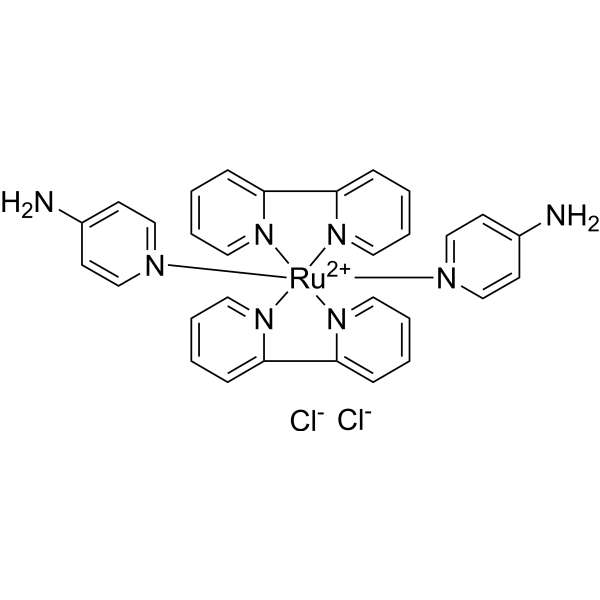
RuBi-4AP, a derivative of 4-aminopyridine (4AP; HY-B0604), is a caged Kv channel blocker. RuBi-4AP contains a photolabile protecting group, allowing its effect to be controlled precisely in both space and time with light. RuBi-4AP can be used for the research of neuronal excitability, synaptic transmission, and signal propagation.
Nur für Forschungszwecke. Wir verkaufen nicht an Patienten.
- CAS. Nr.: 851956-02-0
- Formel: C30H28Cl2N8Ru
- Molecular Weight:672.57
-
Speicherung:
Please store the product under the recommended conditions in the Certificate of Analysis.

To place orders, for customer services and technical support, please contact: MedChemExpress USA
Tel: 609-228-6898 E-mail: [email protected] [email protected]


-
 Biologische Aktivität
-
Chemical Information
- Protokoll
- Reinheit & Dokumentation
- Verweise
-
 Help & FAQs


-
HY-N6693
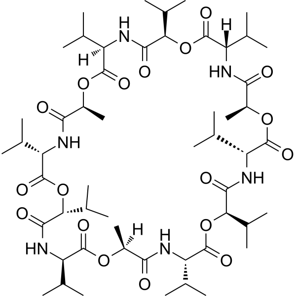
ValinomycinAntibiotic -
HY-B0112
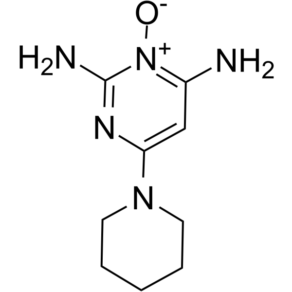
MinoxidilEndogenous Metabolite -
HY-B0232
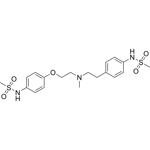
DofetilideIKr Blocker -
HY-B0401
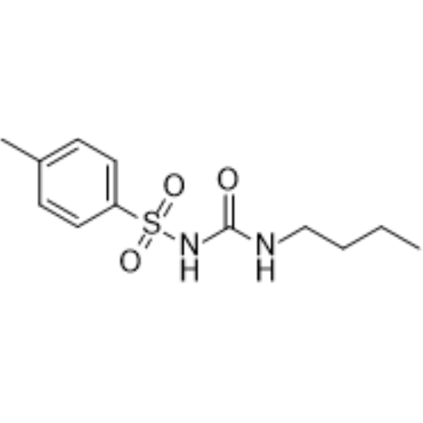
TolbutamideKATP Inhibitor -

HY-B0401R
Tolbutamide (Standard)KATP Inhibitor -
HY-15551
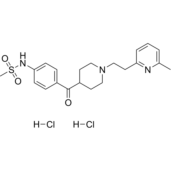
E-4031hERG Channels Blocker -
HY-P0256
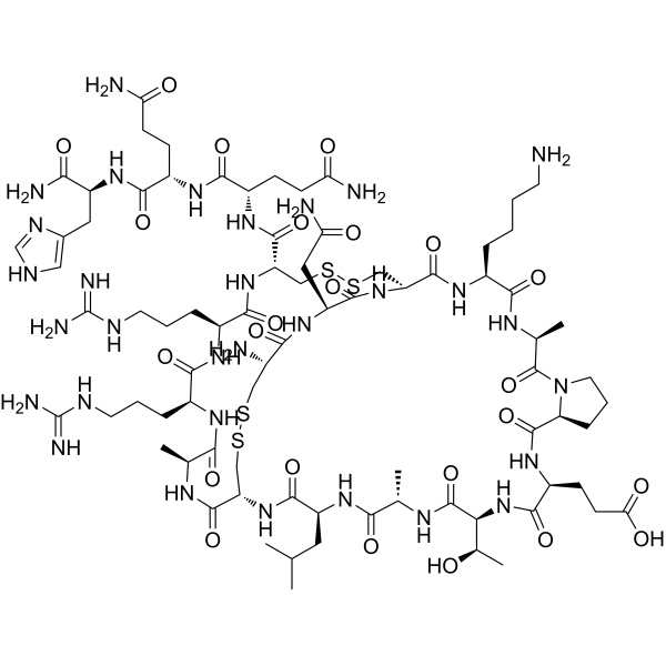
ApaminPotassium Channel Inhibitor -
HY-15209
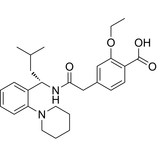
RepaglinideInsulin Secretagogue -
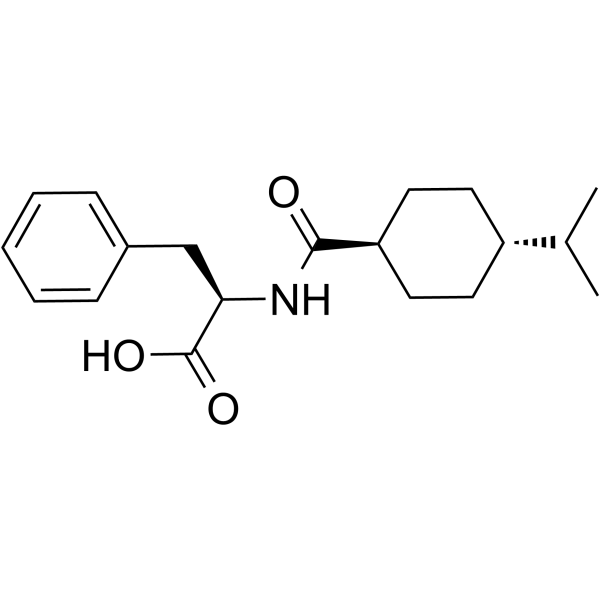
HY-B0422
NateglinideK+ Channel Inhibitor -
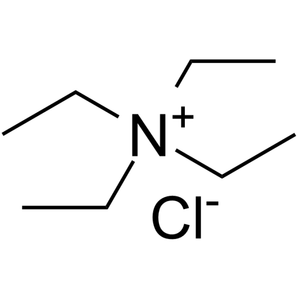
HY-B1793
Tetraethylammonium chloridePotassium Channel Inhibitor -
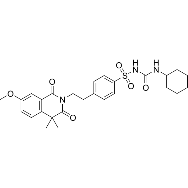
HY-B1114
GliquidoneAnti-Diabetic Drug -
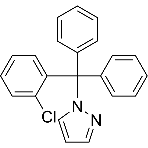
HY-13519
TRAM-34Potassium Channel Inhibitor


Biologische Aktivität
Beschreibung


Chemical Information
-
CAS. Nr. 851956-02-0
-
Molecular Weight 672.57
-
Formel C30H28Cl2N8Ru
-
SMILES
NC1=CC=[N]([Ru+2]23([N]4=CC=CC=C4C5=CC=CC=[N]35)([N]6=CC=C(N)C=C6)[N]7=CC=CC=C7C8=CC=CC=[N]28)C=C1.[Cl-].[Cl-]
-
Versand
Room temperature in continental US; may vary elsewhere.
-
Speicherung
Please store the product under the recommended conditions in the Certificate of Analysis.


Reinheit & Dokumentation
-
Data Sheet (268 KB)
-
SDS (252 KB)
- Français - FR (252 KB)
- Deutsch - DE (252 KB)
- Norwegian - NO (252 KB)
- Español - ES (252 KB)
- Swedish - SV (252 KB)
- Italian - IT (252 KB)
- Korean - KR (252 KB)
- Portuguese - PT (252 KB)
-
Handling Instructions (2659 KB)
Verweise
[1]. Zayat L, et al. A new strategy for neurochemical photodelivery: metal-ligand heterolytic cleavage. J Am Chem Soc. 2003 Jan 29;125(4):882-3. [Content Brief]
[2]. Rowan MJ, et al. Distinct Kv channel subtypes contribute to differences in spike signaling properties in the axon initial segment and presynaptic boutons of cerebellar interneurons. J Neurosci. 2014 May 7;34(19):6611-23. [Content Brief]

Calculators
Konzentration (Stammlösung) × Volumen (Stammlösung) = Konzentration (Ziellösung) × Volumen (Ziellösung)


RuBi-4AP Related Classifications
HY-108578 Related Classifications
 Powered by Bioz