



SAAP 148 TFA

Based on 1 Customer Validation
SAAP 148 TFA is a synthetic antimicrobial peptide (bacteria) that interacts with and disrupts the lipid bilayer of bacterial cytoplasmic membranes, thereby inducing changes in membrane permeability and bacterial death. SAAP 148 TFA kills drug-resistant, multidrug-resistant and persister bacterial strains, inhibits biofilm formation, eliminates established biofilms, and blocks bacterial colonization on implant surfaces. SAAP 148 TFA retains its activity after modification or immobilization, exhibits variable cytotoxicity in different human cell models, and shows reduced efficacy in protein-rich environments. SAAP 148 TFA can be used in infection-related research.
For research use only. We do not sell to patients.
- Purity : 99.55%
- Formula: C157H261N49O27.xC2HF3O2
- Molecular Weight:3267.06 (free base)
-
Storage:
Sealed storage, away from moisture.
Powder -80°C, 2 years , -20°C, 1 year* In solvent : -80°C, 6 months; -20°C, 1 month (sealed storage, away from moisture)


To place orders, for customer services and technical support, please contact: MedChemExpress USA
Tel: 609-228-6898 E-mail: [email protected] [email protected]


-
 Biological Activity
-
Chemical Information
-
Solvent & Solubility
- Protocols
- Purity & Documentation
- References
-
 Help & FAQs




Biological Activity
Description
In Vitro
SAAP 148 TFA kills ≥99.9% of planktonic Staphylococcus aureus JAR060131 with an LC99.9 of 1.6 μM in PBS and 12.8 μM in PBS with 50% human plasma[1].
SAAP 148 TFA kills ≥99.9% of planktonic Pseudomonas aeruginosa PAO1 with an LC99.9 of 1.6 μM in PBS and 12.8 μM in PBS with 50% human plasma[1].
SAAP 148 (1.6-12.8 μM; 24 hours) TFA dose-dependently prevents biofilm formation by Staphylococcus aureus JAR060131, Acinetobacter baumannii RUH875, and both ica-dependent and ica-independent Staphylococcus epidermidis O-47[1].
SAAP 148 (1.6-102.4 μM; 2 hours) TFA dose-dependently eradicates established biofilms of Staphylococcus aureus JAR060131, Acinetobacter baumannii RUH875, and both ica-dependent and ica-independent Staphylococcus epidermidis O-47[1].
SAAP 148 (1.6-3.2 μM; 2-4 hours) TFA completely eradicates Staphylococcus aureus JAR060131 persister cells[1].
SAAP 148 (0.4-3.2 μM; 0.5-120 minutes) TFA rapidly permeabilizes the membranes of Staphylococcus aureus JAR060131 and Acinetobacter baumannii RUH875, leading to complete bacterial killing[1].
SAAP 148 (0.125-4 μM) TFA strongly perturbs the hydrophobic core of bacterial cytoplasmic membrane-mimicking liposomes and induces complete fluorochrome leakage from POPG liposomes at concentrations ≥2 μM[1].
SAAP 148 TFA exhibits bactericidal activity against MRSA LUH14616, but its bactericidal efficacy is reduced by interaction with precipitated proteins in plasma or eschar extracts[3].
SAAP 148 TFA preincubation of SAAP-148 with Novomaix collagen-elastin 3D matrix for 24 h reduces bactericidal activity against MRSA LUH14616, while preincubation with 3% BSA causes an immediate, sustained reduction in efficacy[3].
SAAP 148 (0-100 nmol; 1 h) TFA induces dose-dependent cytotoxicity in 2D-cultured human fibroblasts and keratinocytes, with 0.23 nmol causing 50% LDH release after 1 h incubation[3].
MedChemExpress (MCE) has not independently confirmed the accuracy of these methods. They are for reference only. Further protocols information, click here.
In Vivo
SAAP 148 (0.125-2% (w/w); topical; single dose; 4 hours) TFA single 4-hour topical treatment effectively eradicates acute and established biofilm-associated MRSA and A. baumannii infections in murine abraded skin[1].
MedChemExpress (MCE) has not independently confirmed the accuracy of these methods. They are for reference only.


Chemical Information
-
Appearance Solid
-
Molecular Weight 3267.06 (free base)
-
Formula C157H261N49O27.xC2HF3O2
-
Color White to off-white
-
Sequence
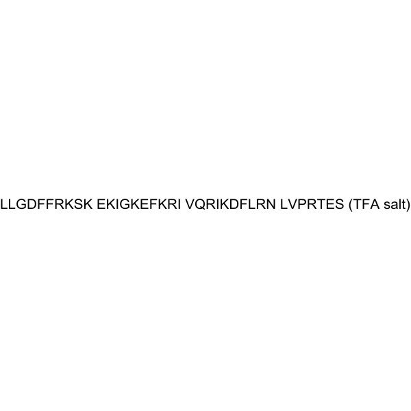
Ac-Leu-Lys-Arg-Val-Trp-Lys-Arg-Val-Phe-Lys-Leu-Leu-Lys-Arg-Tyr-Trp-Arg-Gln-Leu-Lys-Lys-Pro-Val-Arg-NH2
-
Sequence Shortening
Ac-LKRVWKRVFKLLKRYWRQLKKPVR-NH2
-
Shipping
Room temperature in continental US; may vary elsewhere.
-
Storage
Sealed storage, away from moisture
Powder -80°C 2 years -20°C 1 year * In solvent : -80°C, 6 months; -20°C, 1 month (sealed storage, away from moisture)


Solvent & Solubility
In Vitro:
H2O : 100 mg/mL (Need ultrasonic)

Protocols
-
Research Protocol for Infectious Diseases
Infectious-disease experiments test how pathogens interact with host barriers, innate immune receptors, inflammatory signaling, pathogen replication, and tissue injury; pattern-recognition receptors such as TLRs, RIG-I-like receptors, NOD-like receptors, and inflammasomes detect microbial molecules and activate NF-κB, interferon, and cytokine responses. The central hypothesis is that infection severity reflects the balance between pathogen burden and host response: protective inflammation restricts pathogen growth, whereas excessive or mislocalized inflammation contributes to tissue damage and disease phenotype. Unresolved questions include which host pathways are protective versus pathogenic, why some infection models fail to translate to human disease, and which combined readouts best predict clinically relevant infection outcomes.
-
Cell Cytotoxicity Assay
Cytotoxicity assays are usually based on the assessment of cell membrane damage, which can also be indirectly detected by measuring cell viability. Detection methods include MTT assay, CKK-8 assay, LDH assay and ATP assay, etc.
-
Mammalian live/dead viability and cytotoxicity staining
Live/dead viability and cytotoxicity staining assays are based on the simultaneous detection of intracellular esterase activity in metabolically active (viable) cells and membrane integrity loss in non-viable cells. In commonly used dual-staining approaches, membrane-permeant fluorogenic substrates are converted by intracellular esterases into fluorescent products in live cells, while impermeant DNA-binding dyes selectively enter cells with compromised plasma membranes and label nucleic acids in dead or dying cells, enabling discrimination between viable and non-viable populations by fluorescence microscopy or flow cytometry.
-
Transepithelial/transendothelial electrical resistance assay
TEER measures electrical resistance across epithelial or endothelial monolayers cultured on permeable supports, and the readout reflects ionic conductance through the cell barrier, especially the paracellular pathway regulated by junctional integrity. TEER can be measured without destroying the monolayer and is commonly used before or during transport, permeability, barrier-disruption, and barrier-maturation experiments. TEER values are influenced by biological maturation and technical conditions; reported factors include temperature, medium formulation, passage number, electrode geometry, membrane properties, and junctional length during early monolayer maturation. Therefore, TEER should be interpreted with blank-insert subtraction, area normalization, repeated readings, and, when possible, orthogonal barrier readouts such as FITC-dextran flux or tight-junction staining.
-
Bacterial live/dead nucleic-acid viability staining
The LIVE/DEAD bacterial viability staining method is based on differential permeability of nucleic-acid-binding fluorescent dyes, most commonly SYTO 9 and propidium iodide (PI), which enables discrimination of bacterial populations with intact versus compromised cytoplasmic membranes. SYTO 9 penetrates both intact and damaged bacterial membranes and binds nucleic acids to produce green fluorescence, whereas propidium iodide penetrates only cells with compromised membranes and fluoresces red while also reducing SYTO 9 signal through competitive binding and fluorescence interactions. The resulting fluorescence pattern is interpreted as a proxy for membrane integrity, which is widely used as an indicator of bacterial viability in microscopy, flow cytometry, and spectroscopic platforms. However, mechanistic studies show that SYTO 9 and PI interactions involve displacement and fluorescence resonance energy transfer effects, which can influence signal interpretation depending on dye ratios a
-
Fungal Biofilm Culture
Fungal biofilm culture is an in vitro method for growing surface-attached fungal communities, most commonly Candida albicans, on abiotic substrates such as polystyrene wells, silicone elastomer, or polymethylmethacrylate; the assay models adhesion, proliferation, filamentation, extracellular-matrix-associated maturation, and dispersion. Biofilm output can be read by optical density at 600 nm for adherent biomass, XTT reduction for metabolic activity, CFU recovery for viable attached or dispersed cells, and microscopy for architecture.
-
Cell Viability Determination by MTT Colorimetric Assay
The following protocol uses the MTT colorimetric assay as a classic literature-established method for assessing cell viability/metabolic activity in cultured mammalian cells. MTT[3-(4,5-dimethylthiazol-2-yl)-2,5-diphenyltetrazolium bromide] is reduced by metabolically active cells to a colored formazan product; the amount of formazan is quantified spectrophotometrically and provides an indirect measure of metabolically active viable cells. Importantly, MTT reduction reflects cellular oxidoreductase/metabolic activity rather than an absolute direct count of living cells, so changes in cellular metabolism can alter the signal independently of cell number.


Purity & Documentation
-
Data Sheet (281 KB)
-
SDS (252 KB)
- English - EN (252 KB)
- Français - FR (252 KB)
- Deutsch - DE (252 KB)
- Norwegian - NO (252 KB)
- Español - ES (252 KB)
- Swedish - SV (252 KB)
- Italian - IT (252 KB)
- Korean - KR (252 KB)
- Portuguese - PT (252 KB)
-
Handling Instructions (2659 KB)
References
[1]. de Breij A, et al. The antimicrobial peptide SAAP-148 combats drug-resistant bacteria and biofilms. Sci Transl Med. 2018;10(423):eaan4044. [Content Brief]
[2]. Atif M, et al. Antimicrobial Peptide SAAP-148-Functionalized Hydrogels from Photocrosslinkable Polymers with Broad Antibacterial Activity. Macromol Rapid Commun. 2024;45(24):e2400785. [Content Brief]

Calculators
Concentration (start) × Volume (start) = Concentration (final) × Volume (final)


SAAP 148 TFA Related Classifications
HY-P10233A Related Classifications
 Powered by Bioz
Keywords
- SAAP 148
- SAAP148
- SAAP-148
- Bacterial
- Pseudomonas aeruginosa PAO1
- ESBL-producing Escherichia coli
- multidrug-resistant Acinetobacter baumannii RUH875
- normal human dermal fibroblasts
- human keratinocytes
- colistin-resistant Escherichia coli LUH15117
- multidrug-resistant S. aureus LUH14616
- ica-independent Staphylococcus epidermidis O-47
- ica-dependent Staphylococcus epidermidis O-47
- Staphylococcus aureus JAR060131
- Inhibitor
- inhibitor
- inhibit