32 Results for "

intramolecular charge transfer

" in MedChemExpress (MCE) Product Catalog:
Products (32)

32 Results for "intramolecular charge transfer" in MCE Product Catalog:

Cat. No.: HY-152073
CAS No.: 2924598-24-1
Purity:  98.76%
Target:  

Fluorescent Dye

Research Areas:  

Others

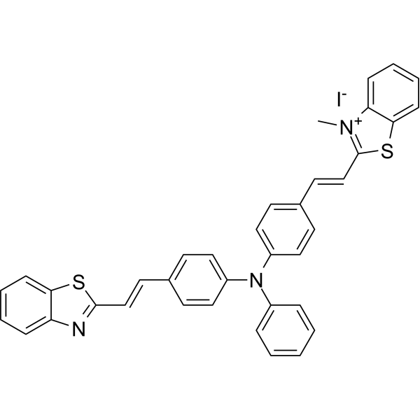
BETA-1 is the first twisted intramolecular charge transfer (TICT)-aggregation-induced emission (AIE) integration molecule. BETA-1 emits cyan fluorescence in lipid droplets (LDs) and red fluorescence in mitochondria. BETA-1 can be used for the simultaneous and dual-color imaging of LDs and mitochondria in vivo and in vitro .
loading...
    loading...
Cat. No.: HY-W024271
CAS No.: 63149-33-7
Synonyms: 9-Formyl-8-hydroxyjulolidine
8-Hydroxyjulolidine-9-carboxaldehyde (9-Formyl-8-hydroxyjulolidine) is a highly selective fluorescent probe for hydrogen sulfide (S 2−) and silicate (SiO3 2−). 8-Hydroxyjulolidine-9-carboxaldehyde operates via a mechanism of blocking intramolecular charge transfer (ICT), exhibiting significant fluorescence enhancement in the presence of weak acid ions. 8-Hydroxyjulolidine-9-carboxaldehyde is promising for research of fluorescence imaging of hydrogen sulfide within living cells .
loading...
    loading...
Cat. No.: HY-D2479
CAS No.: 2978635-30-0
DMANI is an intramolecular charge transfer (ICT)-based mitochondria-targeted ratiometric fluorescent probe. DMANI can monitor mitochondrial peroxynitrite (ONOO ) in living cells .
loading...
    loading...
Cat. No.: HY-D1470
CAS No.: 210832-86-3
Target:  

Fluorescent Dye

Research Areas:  

Others

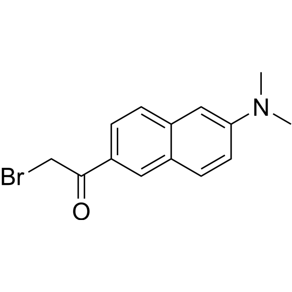
BADAN is a thiol-reactive, environment-sensitive fluorescent probe used for site-specific labeling of protein cysteine residues to detect protein topology, local polarity, protein-lipid interactions, and conformational changes. The bromoacetyl group of BADAN covalently binds to cysteine thiols, and its fluorescence originates from the intramolecular charge transfer (ICT) excited state. The emission peak position varies with the polarity of the surrounding medium: it blue-shifts to approximately 488 nm in nonpolar environments (such as the hydrophobic core of lipid bilayers) and red-shifts to approximately 496-507 nm in polar environments (such as aqueous phases), independent of enzymes, pH, or membrane potential. Ex = 380-402 nm; Em = 400-600 nm .
loading...
    loading...
Cat. No.: HY-D1259
CAS No.: 2414276-32-5
Synonyms: VDP-green (NEP)
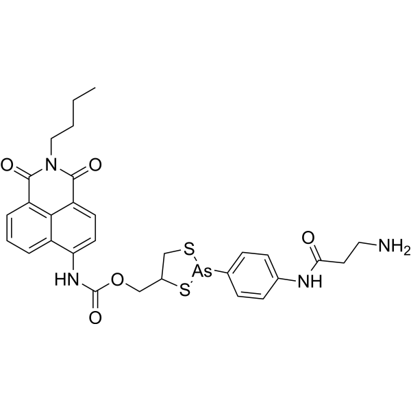
NEP (VDP-green (NEP)) is a turn-on fluorescent probe based on the intramolecular charge transfer (ICT) mechanism for sensing vicinal dithiol-containing proteins (VDPs). NEP exhibits high selectivity toward VDPs in live cells and in vivo and displays a strong green fluorescence signal (λexem=430/535 nm). NEP has the potential for parkinsonism .
loading...
    loading...
Cat. No.: HY-149203
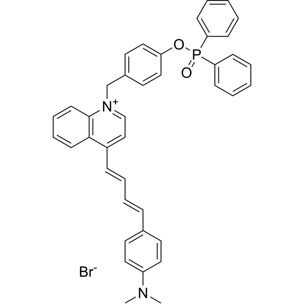
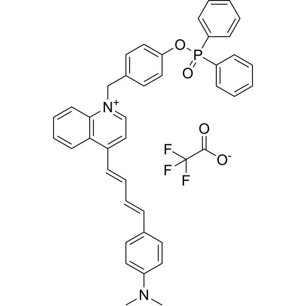
MQA-P is a multifunctional near-infrared (NIR) fluorescent probe for simultaneously detecting ONOO -, viscosity, and polarity within mitochondria. MQA-P exhibits a remarkable turn-on response to ONOO -em=645 nm) and is highly sensitive to viscosity/polarity in the NIR channel with λem>704 nm. MQA-P exhibits excited-state intramolecular charge transfer (ESICT) feature that is highly polarity-sensitive by engineering N,N-dimethylamino as the electron donor and a quinoline cationic unit as the electron acceptor. MQA-P is used for ferroptosis or cancer diagnosis in vitro and in vivo via dual-channel images .
loading...
    loading...
Cat. No.: HY-149203A
Purity:  97.36%
Target:  

Fluorescent Dye

Research Areas:  

Others

MQA-P is a multifunctional near-infrared (NIR) fluorescent probe that simultaneously detects ONOO -, viscosity, and polarity within mitochondria. MQA-P exhibits significant response to ONOO -, λem=645 nm; and NIR channel at λem>704 nm Medium is highly sensitive to viscosity/polarity. MQA-P possesses excited-state intramolecular charge transfer (ESICT) properties that are highly sensitive to polarity by designing the N,N-dimethylamino group as the electron donor and the quinoline cation unit as the electron acceptor. MQA-P is used for ferroptosis or cancer diagnosis in vitro and in vivo via dual-channel images .
loading...
    loading...
Cat. No.: HY-D3442
CAS No.: 3121565-89-4
ALDH2 probe 5 is a ALDH2 isoform-selective fluorescent probe with an emission wavelength centered at 494 nm. ALDH2 probe 5 is a turn-on fluorescent substrate for ALDH2; in its unreacted aldehyde form, twisted intramolecular charge transfer (TICT) occurs, resulting in fluorescence quenching. After ALDH2 probe 5 is catalytically oxidized to carboxylate by ALDH2, TICT is inhibited and fluorescence is significantly enhanced. ALDH2 probe 5 enables highly sensitive quantitative imaging of ALDH2 activity in recombinant enzymes, live cells, blood, tissue homogenates, and the brains of living mice. ALDH2 probe 5 can be used for studies related to neuroinflammation .
loading...
    loading...
Cat. No.: HY-D3071
CAS No.: 1510814-96-6
Target:  

Fluorescent Dye

Research Areas:  

Inflammation/Immunology

LW-1 is a fluorescent probe targeting polarity detection of lipid droplets (LD). LW-1 produces a strong solvatochromic effect via the intramolecular charge transfer (ICT) mechanism: as environmental polarity increases, the emission wavelength red-shifts and the fluorescence intensity decreases significantly (emission at 557 nm in toluene and 642 nm in acetonitrile, with an 85 nm red-shift); fluorescence enhances in low-polarity lipid droplets. LW-1 can be used for lipid droplet polarity detection, differentiation between cancer cells and normal cells, monitoring of lipid droplet polarity changes under different physiological conditions such as starvation, oleic acid treatment and oxidative stress, as well as ex vivo imaging of polarity changes in lung tissues of asthmatic mice. The detection wavelength for cell imaging with LW-1 is Ex/Em = 405/500-550 nm (green channel) .
loading...
    loading...
Cat. No.: HY-D3408
STB-2 is a fluorescent dye for near-infrared window II (NIR-II) fluorescence angiography. STB-2 achieves high-brightness NIR-II fluorescence by restricting the twisted intramolecular charge transfer state. STB-2, when modified with ApoE to form nanoparticles, can cross the blood-brain barrier and accumulate in gliomas (Ex/Em = 808/1020 nm). STB-2 is used in studies related to NIR-II fluorescence and single-/two-photon cerebrovascular imaging of gliomas .
loading...
    loading...
Cat. No.: HY-D3101
Target:  

Fluorescent Dye

Research Areas:  

Others

Nile-TPA is a fluorescent probe for polarity detection, which can be used for lipid droplet targeting, cell polarity analysis, in vivo polarity imaging, and blood polarity monitoring in circulating tumor mouse models. The detection mechanism of Nile-TPA relies on the dual intramolecular charge transfer (ICT) effect generated by the electron-donating triphenylamine and Nile Red moieties and the electron-withdrawing carbonyl group; this probe emits strong red fluorescence in a low-polarity environment, and as polarity increases, the fluorescence intensity decreases and a red shift occurs due to changes in the excited-state dipole moment. In spectral tests, its excitation wavelength is 540 nm, and the emission wavelength varies in the range of 550-800 nm depending on solvent polarity; for cell, zebrafish and mouse imaging, Ex/Em = 561/570-620 nm, and for in vivo mouse imaging, Ex/Em = 560/620 nm .
loading...
    loading...
Cat. No.: HY-D3125
Target:  

Fluorescent Dye

Research Areas:  

Others

NtHzBtd is a fluorescent probe for detecting Fe 3+. NtHzBtd is applicable to the selective fluorescent detection of Fe 3+ ions and live cell imaging studies. NtHzBtd can selectively coordinate with Fe 3+ to form a 1:1 complex, triggering chelation enhanced quenching (CHEQ) and intramolecular charge transfer (ICT) processes, which result in fluorescence turn-off, thereby enabling sensitive detection of Fe 3+ and live cell fluorescence imaging. After binding to Fe 3+, NtHzBtd reduces fluorescence intensity, exhibits a rapid response property, with a limit of detection of 0.036 μM and a response time of approximately 55 s. The detection wavelengths are Ex/Em = 334/401 nm (solution system) .
loading...
    loading...
Cat. No.: HY-D3104
CAS No.: 2767443-23-0
Target:  

Fluorescent Dye

Research Areas:  

Others

ERNT is a fluorescent probe for monitoring endoplasmic reticulum (ER) polarity and detecting endoplasmic reticulum stress (ERS) during the progression of dynamic liver injury. ERNT functions based on a donor-π-acceptor (D-π-A) structure with an intramolecular charge transfer (ICT) effect. ERNT has multiple excitation/emission pairs: for in vitro cell imaging, the green channel has Ex/Em = 488/500-550 nm, and the red channel has Ex/Em = 488/570-620 nm; for in vivo liver imaging, Ex/Em = 520/620 nm; for solvent optical tests, Ex = 470 nm, with the maximum emission peak at 548 nm in low-polarity toluene and 668 nm in high-polarity DMSO. ERNT can evaluate the efficacy of hepatoprotective interventions by detecting changes in ER polarity, and also exhibits excellent photostability and low cytotoxicity .
loading...
    loading...
Cat. No.: HY-D3117
CAS No.: 2146114-18-1
Target:  

Fluorescent Dye

Research Areas:  

Others

MBCB is a two-photon Fluorescent probe for dual-detection of mitochondrial SO₂ derivatives and viscosity. For SO₂ derivatives detection, the probe utilizes a Michael addition mechanism: nucleophilic addition of SO₂ derivatives to the C=C bond between the carbazole skeleton and 3-methylbenzothiazolium moiety destroys the strong intramolecular charge transfer (ICT) system between these groups, while enhancing the weak ICT system between the benzothiazole group and carbazole framework; this causes the red emission at 600 nm to decrease and the blue emission at 434 nm to increase, creating a ratiometric response based on the I₄₃₄ₙₘ/I₆₀₀ₙₘ intensity ratio. For viscosity detection, in low-viscosity environments, steric hindrance creates a twisted ICT (TICT) system with weak fluorescence, while in high-viscosity environments, intramolecular rotation is blocked, the TICT state is disrupted, and the strong ICT system is recovered, leading to a strong red emission at 567 nm with negligible change to the short-wavelength emission at 415 nm, creating a ratiometric response based on the I₅₆₇ₙₘ/I₄₁₅ₙₘ intensity ratio that has a logarithmic linear relationship with viscosity. The probe has excitation/emission wavelengths of Ex/Em = 351/434, 600 nm for SO₂ derivatives detection and Ex/Em = 351/567 nm for viscosity detection, with two-photon excitation at 740 nm for bioimaging; it also exhibits good mitochondrial targeting ability with a Pearson's colocalization coefficient of 0.93 when paired with Mito-Tracker Green. The probe shows high sensitivity and selectivity for SO₂ derivatives, has low cell cytotoxicity, and can be applied to detect exogenous/endogenous HSO₃⁻ in living cells and in vivo, as well as visualize mitochondrial viscosity changes induced by nystatin[1].
loading...
    loading...
Cat. No.: HY-D3105
Target:  

Fluorescent Dye

Research Areas:  

Others

DCA is a Fluorescent probe for visualization of phase behavior in ER membranes. DCA is an ER-targeting, polarity-responsive NIR ratiometric probe, with its p-toluenesulfonamide group responsible for ER localization; its sensitivity to polarity relies on its donor-π-acceptor (D-π-A) structure, where aniline acts as the donor and dicyanomethylene acts as the acceptor, driving an intramolecular charge transfer (ICT) process upon excitation. In environments with low polarity, such as the closely packed, low water content ERₒ phase of ER membranes, DCA emits at a shorter wavelength, while in high polarity environments like the loosely packed, higher water content ERd phase, ICT leads to a red-shifted emission, allowing discrimination of the two phases via dual NIR emission colors and ratiometric imaging. Ex/Em = 488/570–620 nm and 488/665–735 nm; additional excitation/emission pairs include Ex/Em = 488/631 nm in low polarity 1,4-dioxane and Ex/Em = 488/677 nm in 1,4-dioxane with 30% water, the higher polarity condition. It shows a large Stokes shift of ~170 nm, and pH, viscosity, and biologically relevant species including Cys, GSH, H₂O₂, and metal ions do not exert marked interference on its fluorescence spectra[1].
loading...
    loading...
Cat. No.: HY-D3084
Target:  

Fluorescent Dye

Research Areas:  

Cancer

C-TPA is a fluorescent probe used for hypochlorite detection and bioimaging of lipid droplet polarity in cancer cells. For hypochlorite detection, when its derivative C-TPA-S is exposed to hypochlorite, an oxidative desulfurization reaction converts the thiocarbonyl group of C-TPA-S to the carbonyl group of C-TPA, thus restoring bright fluorescence from the almost non-fluorescent C-TPA-S. For bioimaging of lipid droplet polarity, the intramolecular charge transfer process between its Triphenylamine (HY-W011998) donor group and Coumarin (HY-N0709) acceptor group endows C-TPA with solvatochromism-it exhibits stronger fluorescence in less polar environments (such as lipid droplets in cancer cells) and weaker fluorescence in more polar environments (such as lipid droplets in normal cells), enabling the differentiation of cancer cells and cancer tissues from normal cells and normal tissues. The excitation wavelength of C-TPA is 405 nm, and the emission wavelengths used for cell and tissue imaging cover 425-475 nm and 500-550 nm; in solvents, its emission peak shifts from 494 nm in non-polar toluene to 528 nm in polar water .
loading...
    loading...
Cat. No.: HY-D3224
CAS No.: 2413777-91-8
HCP is a two-photon fluorescent probe capable of crossing the blood-brain barrier, which is specifically designed for real-time detection of hypochlorous acid (HClO/ClO -) produced by myeloperoxidase (MPO). Chlorination of the quinoline skeleton of HCP generates HCP-Cl, which eliminates intramolecular charge transfer (ICT) quenching and thereby produces fluorescence. HCP is suitable for in vivo dynamic imaging of HClO in the brains of epileptic mice and high-throughput screening of antiepileptic drugs (HCP-Cl: Ex/Em = 800 nm/495 nm) .
loading...
    loading...
Cat. No.: HY-D3099
CAS No.: 3078752-74-3
Target:  

Fluorescent Dye

Research Areas:  

Others

CSMPP is a ratiometric lysosomal pH fluorescent probe with aggregation-induced emission (AIE) properties, which is used for real-time quantitative detection of lysosomal pH changes in living cells and in vivo. CSMPP has a cyanodistyrene backbone, and contains N-methylpiperazine and pyridine groups (pH-responsive); protonation of the pyridine group significantly enhances intramolecular charge transfer (ICT), leading to green fluorescence emission (Ex/Em = 405/503 nm) under neutral/basic conditions and red fluorescence emission (Ex/Em = 405/615 nm) under acidic conditions. CSMPP exhibits high selectivity, reversibility, good biocompatibility and excellent photostability .
loading...
    loading...
Cat. No.: HY-D3364
Research Areas:  

Others

RL480 is a green-emitting RNA fluorescent dye used for selective RNA labeling and imaging. RL480 contains an acylimidazole reactive group that undergoes selective acylation with the 2′-OH group of RNA ribose, and restricts intramolecular rotation through covalent binding to relieve the TICT quenching effect, thereby generating a fluorescence enhancement signal. RL480 enables selective fluorescent labeling of RNA over DNA, and is applicable to RNA gel detection, real-time RNA synthesis monitoring and live-cell RNA imaging. The detection wavelengths are Ex/Em = 480 nm/514 nm .
loading...
    loading...
Cat. No.: HY-D3365
Target:  

Fluorescent Dye

Research Areas:  

Others

RL560 is a red-emitting RNA fluorescent dye used for RNA-selective labeling and live-cell RNA imaging. RL560 achieves RNA-selective fluorescence turn-on via covalent acylation between its acylimidazole reactive group and the 2′-OH of RNA ribose, as well as fluorescence signal enhancement through restriction of TICT molecular motion. RL560 is applicable for RNA imaging and multicolor RNA detection, with detection wavelengths of Ex/Em = 560/639 nm .
loading...
    loading...

