23 Results for "

intramolecular charge-transfer

" in MedChemExpress (MCE) Product Catalog:
Products (23)

23 Results for "intramolecular charge-transfer" in MCE Product Catalog:

Cat. No.: HY-152073
CAS No.: 2924598-24-1
Purity:  98.76%
Target:  

Fluorescent Dye

Research Areas:  

Others

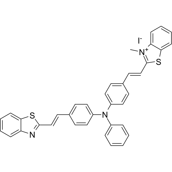
BETA-1 is the first twisted intramolecular charge transfer (TICT)-aggregation-induced emission (AIE) integration molecule. BETA-1 emits cyan fluorescence in lipid droplets (LDs) and red fluorescence in mitochondria. BETA-1 can be used for the simultaneous and dual-color imaging of LDs and mitochondria in vivo and in vitro .
loading...
    loading...
Cat. No.: HY-W024271
CAS No.: 63149-33-7
Synonyms: 9-Formyl-8-hydroxyjulolidine
8-Hydroxyjulolidine-9-carboxaldehyde (9-Formyl-8-hydroxyjulolidine) is a highly selective fluorescent probe for hydrogen sulfide (S 2−) and silicate (SiO3 2−). 8-Hydroxyjulolidine-9-carboxaldehyde operates via a mechanism of blocking intramolecular charge transfer (ICT), exhibiting significant fluorescence enhancement in the presence of weak acid ions. 8-Hydroxyjulolidine-9-carboxaldehyde is promising for research of fluorescence imaging of hydrogen sulfide within living cells .
loading...
    loading...
Cat. No.: HY-D2479
CAS No.: 2978635-30-0
DMANI is an intramolecular charge transfer (ICT)-based mitochondria-targeted ratiometric fluorescent probe. DMANI can monitor mitochondrial peroxynitrite (ONOO ) in living cells .
loading...
    loading...
Cat. No.: HY-D1470
CAS No.: 210832-86-3
Target:  

Fluorescent Dye

Research Areas:  

Others

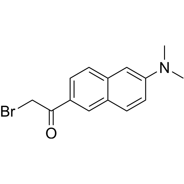
BADAN is a thiol-reactive, environment-sensitive fluorescent probe used for site-specific labeling of protein cysteine residues to detect protein topology, local polarity, protein-lipid interactions, and conformational changes. The bromoacetyl group of BADAN covalently binds to cysteine thiols, and its fluorescence originates from the intramolecular charge transfer (ICT) excited state. The emission peak position varies with the polarity of the surrounding medium: it blue-shifts to approximately 488 nm in nonpolar environments (such as the hydrophobic core of lipid bilayers) and red-shifts to approximately 496-507 nm in polar environments (such as aqueous phases), independent of enzymes, pH, or membrane potential. Ex = 380-402 nm; Em = 400-600 nm .
loading...
    loading...
Cat. No.: HY-D1259
CAS No.: 2414276-32-5
Synonyms: VDP-green (NEP)
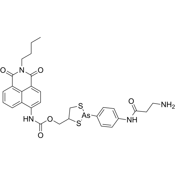
NEP (VDP-green (NEP)) is a turn-on fluorescent probe based on the intramolecular charge transfer (ICT) mechanism for sensing vicinal dithiol-containing proteins (VDPs). NEP exhibits high selectivity toward VDPs in live cells and in vivo and displays a strong green fluorescence signal (λexem=430/535 nm). NEP has the potential for parkinsonism .
loading...
    loading...
Cat. No.: HY-149203
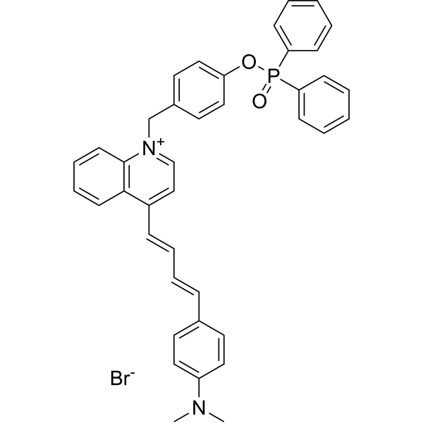
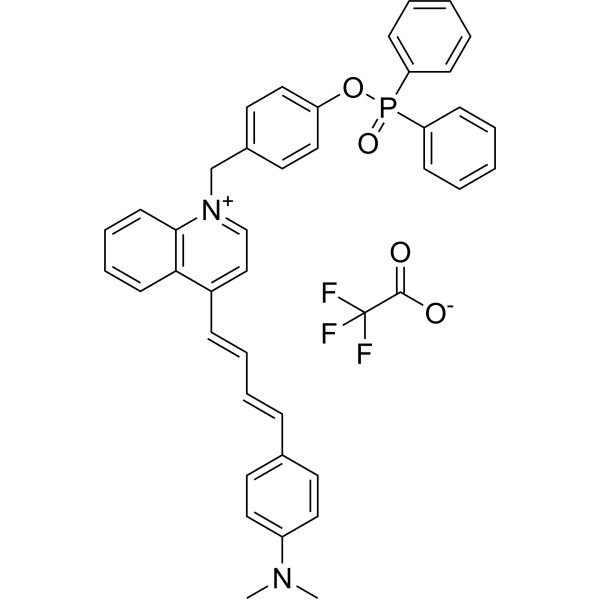
MQA-P is a multifunctional near-infrared (NIR) fluorescent probe for simultaneously detecting ONOO -, viscosity, and polarity within mitochondria. MQA-P exhibits a remarkable turn-on response to ONOO -em=645 nm) and is highly sensitive to viscosity/polarity in the NIR channel with λem>704 nm. MQA-P exhibits excited-state intramolecular charge transfer (ESICT) feature that is highly polarity-sensitive by engineering N,N-dimethylamino as the electron donor and a quinoline cationic unit as the electron acceptor. MQA-P is used for ferroptosis or cancer diagnosis in vitro and in vivo via dual-channel images .
loading...
    loading...
Cat. No.: HY-149203A
Purity:  97.36%
Target:  

Fluorescent Dye

Research Areas:  

Others

MQA-P is a multifunctional near-infrared (NIR) fluorescent probe that simultaneously detects ONOO -, viscosity, and polarity within mitochondria. MQA-P exhibits significant response to ONOO -, λem=645 nm; and NIR channel at λem>704 nm Medium is highly sensitive to viscosity/polarity. MQA-P possesses excited-state intramolecular charge transfer (ESICT) properties that are highly sensitive to polarity by designing the N,N-dimethylamino group as the electron donor and the quinoline cation unit as the electron acceptor. MQA-P is used for ferroptosis or cancer diagnosis in vitro and in vivo via dual-channel images .
loading...
    loading...
Cat. No.: HY-D3086
CAS No.: 1668565-72-7
Target:  

Fluorescent Dye

Research Areas:  

Cancer

Mito-polarity is a mitochondria-targeted polarity probe. Mito-polarity can be used to detect mitochondrial polarity. The detection mechanism of Mito-polarity is based on an intramolecular charge transfer (ICT) system designed with a donor-π-bridge-acceptor (D-π-A) structure. Among them, the 467 nm green emission generated by the Coumarin (HY-N0709) structure is extremely sensitive to polarity changes, while the 642 nm red emission produced by the extended π-conjugation and ICT effect between the Coumarin and benzothiazine structures shows only a weak response, thus forming a ratiometric fluorescence response that is linearly correlated with solvent polarity. When Mito-polarity is excited at 405 nm in methanol, its excitation/emission wavelengths are Ex/Em = 405/467 nm and Ex/Em = 405/642 nm, with maximum absorption wavelengths of 426 nm and 561 nm .
loading...
    loading...
Cat. No.: HY-D3224
CAS No.: 2413777-91-8
HCP is a two-photon fluorescent probe capable of crossing the blood-brain barrier, which is specifically designed for real-time detection of hypochlorous acid (HClO/ClO -) produced by myeloperoxidase (MPO). Chlorination of the quinoline skeleton of HCP generates HCP-Cl, which eliminates intramolecular charge transfer (ICT) quenching and thereby produces fluorescence. HCP is suitable for in vivo dynamic imaging of HClO in the brains of epileptic mice and high-throughput screening of antiepileptic drugs (HCP-Cl: Ex/Em = 800 nm/495 nm) .
loading...
    loading...
Cat. No.: HY-D3408
STB-2 is a fluorescent dye for near-infrared window II (NIR-II) fluorescence angiography. STB-2 achieves high-brightness NIR-II fluorescence by restricting the twisted intramolecular charge transfer state. STB-2, when modified with ApoE to form nanoparticles, can cross the blood-brain barrier and accumulate in gliomas (Ex/Em = 808/1020 nm). STB-2 is used in studies related to NIR-II fluorescence and single-/two-photon cerebrovascular imaging of gliomas .
loading...
    loading...
Cat. No.: HY-D3099
CAS No.: 3078752-74-3
Target:  

Fluorescent Dye

Research Areas:  

Others

CSMPP is a ratiometric lysosomal pH fluorescent probe with aggregation-induced emission (AIE) properties, which is used for real-time quantitative detection of lysosomal pH changes in living cells and in vivo. CSMPP has a cyanodistyrene backbone, and contains N-methylpiperazine and pyridine groups (pH-responsive); protonation of the pyridine group significantly enhances intramolecular charge transfer (ICT), leading to green fluorescence emission (Ex/Em = 405/503 nm) under neutral/basic conditions and red fluorescence emission (Ex/Em = 405/615 nm) under acidic conditions. CSMPP exhibits high selectivity, reversibility, good biocompatibility and excellent photostability .
loading...
    loading...
Cat. No.: HY-D3442
CAS No.: 3121565-89-4
ALDH2 probe 5 is a ALDH2 isoform-selective fluorescent probe with an emission wavelength centered at 494 nm. ALDH2 probe 5 is a turn-on fluorescent substrate for ALDH2; in its unreacted aldehyde form, twisted intramolecular charge transfer (TICT) occurs, resulting in fluorescence quenching. After ALDH2 probe 5 is catalytically oxidized to carboxylate by ALDH2, TICT is inhibited and fluorescence is significantly enhanced. ALDH2 probe 5 enables highly sensitive quantitative imaging of ALDH2 activity in recombinant enzymes, live cells, blood, tissue homogenates, and the brains of living mice. ALDH2 probe 5 can be used for studies related to neuroinflammation .
loading...
    loading...
Cat. No.: HY-D3118
CAS No.: 2989973-69-3
Target:  

Fluorescent Dye

Research Areas:  

Others

Biotin-V is a fluorescent probe targeting the biotin receptor (BiR), which can be used to detect changes in intracellular viscosity in cancer cells and tumor tissues. Biotin-V consists of a viscosity-responsive TICT (twisted internal charge transfer) fluorophore and a biotin-targeting moiety. In a high-viscosity environment, intramolecular rotation is restricted, which reduces non-radiative transitions and enhances fluorescence intensity and fluorescence lifetime. Biotin-V enters BiR-positive cancer cells via biotin receptor-mediated uptake, and mainly localizes to mitochondria, enabling dual-modal viscosity imaging (fluorescence intensity imaging and fluorescence lifetime imaging) (Ex/Em = 540/608 nm; cell imaging Ex/Em = 561/570-620 nm) .
loading...
    loading...
Cat. No.: HY-D3125
Target:  

Fluorescent Dye

Research Areas:  

Others

NtHzBtd is a fluorescent probe for detecting Fe 3+. NtHzBtd is applicable to the selective fluorescent detection of Fe 3+ ions and live cell imaging studies. NtHzBtd can selectively coordinate with Fe 3+ to form a 1:1 complex, triggering chelation enhanced quenching (CHEQ) and intramolecular charge transfer (ICT) processes, which result in fluorescence turn-off, thereby enabling sensitive detection of Fe 3+ and live cell fluorescence imaging. After binding to Fe 3+, NtHzBtd reduces fluorescence intensity, exhibits a rapid response property, with a limit of detection of 0.036 μM and a response time of approximately 55 s. The detection wavelengths are Ex/Em = 334/401 nm (solution system) .
loading...
    loading...
Cat. No.: HY-D3108
CAS No.: 2569032-08-0
Target:  

Fluorescent Dye

Research Areas:  

Others

BV-1 is a fluorescent probe for viscosity detection and imaging in living cells. BV-1 can be used to detect changes in intracellular viscosity and mitochondrial viscosity in living cells. BV-1 responds to environmental viscosity via a molecular rotor mechanism. In a low-viscosity environment, the centrally located unsaturated structure induces a twisted intramolecular charge transfer (TICT) state and promotes non-radiative transitions, resulting in fluorescence quenching; when viscosity increases, molecular rotation is restricted, the TICT state is attenuated, and fluorescence is enhanced with a red shift. BV-1 can be used for real-time monitoring of changes in cytoplasmic viscosity and the viscosity increase during cell apoptosis. Ex/Em = 480/570 nm .
loading...
    loading...
Cat. No.: HY-D3071
CAS No.: 1510814-96-6
Target:  

Fluorescent Dye

Research Areas:  

Inflammation/Immunology

LW-1 is a fluorescent probe targeting polarity detection of lipid droplets (LD). LW-1 produces a strong solvatochromic effect via the intramolecular charge transfer (ICT) mechanism: as environmental polarity increases, the emission wavelength red-shifts and the fluorescence intensity decreases significantly (emission at 557 nm in toluene and 642 nm in acetonitrile, with an 85 nm red-shift); fluorescence enhances in low-polarity lipid droplets. LW-1 can be used for lipid droplet polarity detection, differentiation between cancer cells and normal cells, monitoring of lipid droplet polarity changes under different physiological conditions such as starvation, oleic acid treatment and oxidative stress, as well as ex vivo imaging of polarity changes in lung tissues of asthmatic mice. The detection wavelength for cell imaging with LW-1 is Ex/Em = 405/500-550 nm (green channel) .
loading...
    loading...
Cat. No.: HY-D3101
Target:  

Fluorescent Dye

Research Areas:  

Others

Nile-TPA is a fluorescent probe for polarity detection, which can be used for lipid droplet targeting, cell polarity analysis, in vivo polarity imaging, and blood polarity monitoring in circulating tumor mouse models. The detection mechanism of Nile-TPA relies on the dual intramolecular charge transfer (ICT) effect generated by the electron-donating triphenylamine and Nile Red moieties and the electron-withdrawing carbonyl group; this probe emits strong red fluorescence in a low-polarity environment, and as polarity increases, the fluorescence intensity decreases and a red shift occurs due to changes in the excited-state dipole moment. In spectral tests, its excitation wavelength is 540 nm, and the emission wavelength varies in the range of 550-800 nm depending on solvent polarity; for cell, zebrafish and mouse imaging, Ex/Em = 561/570-620 nm, and for in vivo mouse imaging, Ex/Em = 560/620 nm .
loading...
    loading...
Cat. No.: HY-D3104
CAS No.: 2767443-23-0
Target:  

Fluorescent Dye

Research Areas:  

Others

ERNT is a fluorescent probe for monitoring endoplasmic reticulum (ER) polarity and detecting endoplasmic reticulum stress (ERS) during the progression of dynamic liver injury. ERNT functions based on a donor-π-acceptor (D-π-A) structure with an intramolecular charge transfer (ICT) effect. ERNT has multiple excitation/emission pairs: for in vitro cell imaging, the green channel has Ex/Em = 488/500-550 nm, and the red channel has Ex/Em = 488/570-620 nm; for in vivo liver imaging, Ex/Em = 520/620 nm; for solvent optical tests, Ex = 470 nm, with the maximum emission peak at 548 nm in low-polarity toluene and 668 nm in high-polarity DMSO. ERNT can evaluate the efficacy of hepatoprotective interventions by detecting changes in ER polarity, and also exhibits excellent photostability and low cytotoxicity .
loading...
    loading...
Cat. No.: HY-D3391
RMR-Tre is a fluorescent probe targeting the mycobacterial acyltransferase Ag85. Under the catalysis of Ag85, RMR-Tre undergoes 6-position mycoloylation and anchors to the mycobacterial membrane, while achieving fluorescence activation by inhibiting the intramolecular twisted charge transfer state transition. RMR-Tre can distinguish live mycobacteria from dead ones through metabolism-driven labeling, enabling rapid, wash-free, low-background detection of viable bacteria. RMR-Tre reports the drug resistance of Mycobacterium tuberculosis via the trehalose catalytic shift activity readout associated with TreS. In addition, RMR-Tre can be combined with flow cytometry or high-content imaging techniques to visualize and quantitatively analyze the metabolic heterogeneity of Mycobacterium tuberculosis related to persistence and drug resistance. RMR-Tre is widely used in tuberculosis-related research .
loading...
    loading...
Cat. No.: HY-D3088
Research Areas:  

Others

CQPP is a Fluorescent probe designed for monitoring polarity changes in the cellular microenvironment, including polarity tracking of lipid droplets/nuclei during ferroptosis. CQPP exhibits ratiometric fluorescence emission and fluorescence lifetime variations in response to polarity changes; a nonpolar environment stimulates fluorescence from the locally excited (LE) state, while a polar environment drives solvation relaxation to the intramolecular charge transfer (ICT) state, which attenuates the LE emission at ~470 nm and simultaneously enhances the ICT emission at ~670 nm. CQPP binds to intranuclear DNA through electrostatic interactions and hydrogen bonds in the DNA minor groove, which enhances its emission at ~670 nm and prolongs its fluorescence lifetime. CQPP can simultaneously target lipid droplets (green LE fluorescence) and the nucleus (red ICT fluorescence). The detection wavelengths of CQPP are Ex/Em = 405/470 nm and Ex/Em = 405/670 nm, and it also supports fluorescence lifetime imaging via 810 nm two-photon excitation .
loading...
    loading...

