



Pipecolic acid
Based on 2 publication(s) in Google Scholar
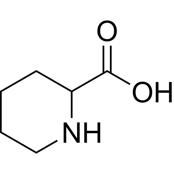
Pipecolic acid is an orally bioavailable, blood-brain barrier-permeable metabolite of lysine with antioxidant, inhibitor, and inducer activity. Pipecolic acid modulates the YAP-GPX4 signaling pathway, reduces retinal vascular tube formation, and mitigates ferroptosis. Pipecolic acid potentiates voltage-sensitive Ca2+ channel currents and induces neuronal apoptosis. Pipecolic acid can be used for the research of diabetic retinopathy.
For research use only. We do not sell to patients.
- Purity : 99.92%
- CAS No.: 535-75-1
- Formula: C6H11NO2
- Molecular Weight:129.16
-
Storage:Powder -20°C, 3 years , 4°C, 2 years ; In solvent -80°C, 6 months , -20°C, 1 month

To place orders, for customer services and technical support, please contact: MedChemExpress USA
Tel: 609-228-6898 E-mail: [email protected] [email protected]


-
 Biological Activity
-
Chemical Information
-
Solvent & Solubility
- Protocols
- Purity & Documentation
- References
-
 Help & FAQs


-
HY-N0067
γ-Aminobutyric acidNeurotransmitter -
HY-B0399
L-CarnitineCo-factor for β-oxidation -
HY-B0511
BiotinVitamin B7 -
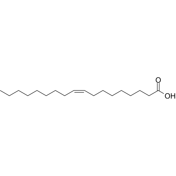
HY-N1446
Oleic acidMonounsaturated Fatty Acid -
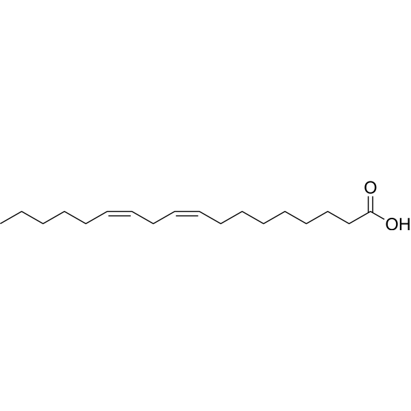
HY-N0729
Linoleic acidHuman Endogenous Metabolite -

HY-10071
Y-27632ROCK Inhibitor -

HY-15371
ForskolinAdenylate Cyclase Activator -
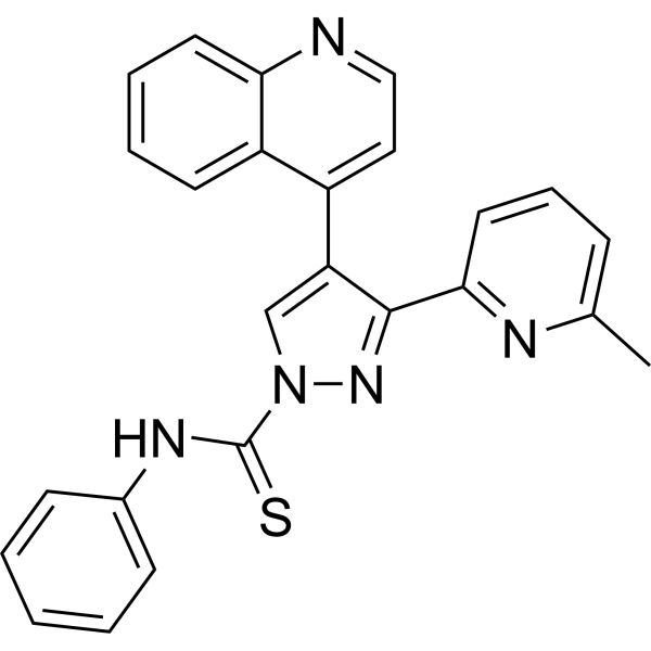
HY-10432
A 83-01ALK4/5/7 Inhibitor -
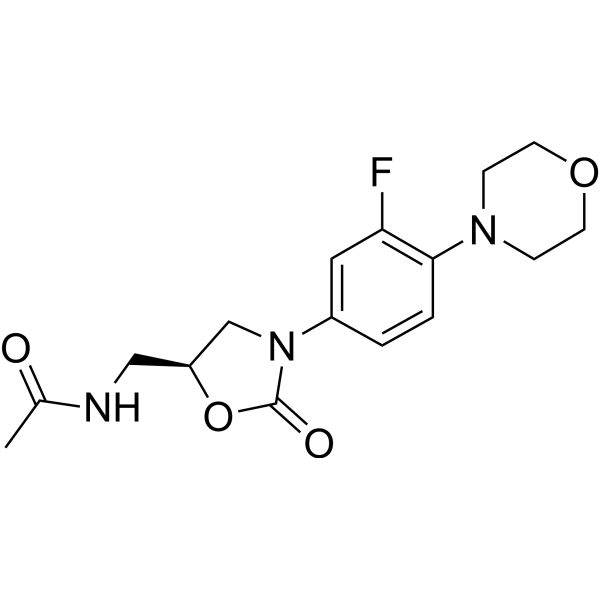
HY-10394
LinezolidAntibiotic -
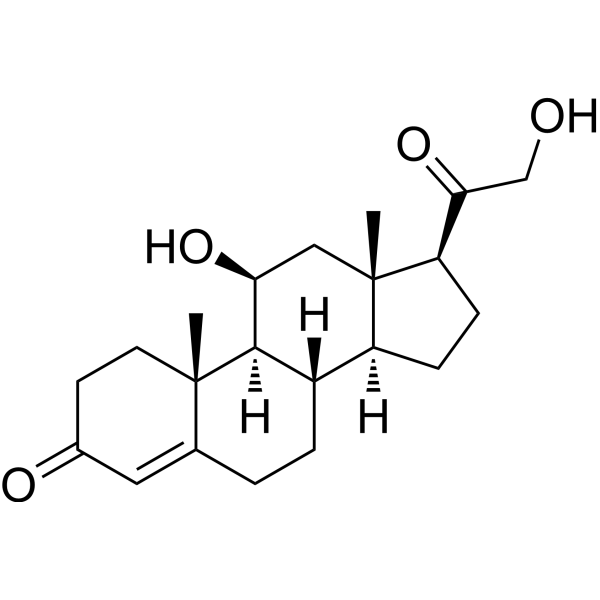
HY-B1618
CorticosteroneEndogenous Glucocorticoids -
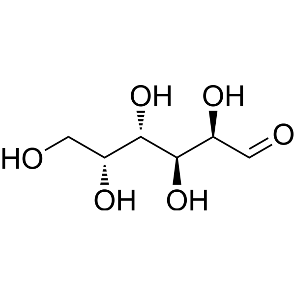
HY-N0210
D-GalactoseEndogenous Metabolite -

HY-135849
Catalase, Aspergillus nigerAntioxidant Enzyme
Publications Citing Use of MedChemExpress (MCE) Pipecolic acid
MoreAll Endogenous Metabolite Isoforms
MoreAll YAP Isoforms
More

Biological Activity
Description
IC50 & Target
|
Microbial Metabolite |
Human Endogenous Metabolite |
GPX4 |
In Vitro
Pipecolic acid mitigates high-glucose-induced oxidative stress, cell growth inhibition, and apoptosis, and downregulates YAP and GPX4 expression in human retinal capillary endothelial cells[1].
DL-pipecolic acid (1, 10, 100 μM; 20 min) potentiates population spike amplitude in the dentate gyrus of rat hippocampal slices at 10 and 100 μM, while D- and L-pipecolic acid show no significant inhibitory effect[2].
DL-pipecolic acid (10, 100 μM) does not significantly affect voltage-sensitive Na+ channel and voltage-sensitive K+ channel currents in Neuro-2A cells at 10 or 100 μM[2]. DL-pipecolic acid
DL-pipecolic acid (10, 100 μM) potentiates voltage-sensitive Ca2+ channel currents in cultured rat cortical neurons at 10 and 100 μM[2].
DL-pipecolic acid (1, 10, 100 μM; 72 h) reduces viability of Neuro-2A cells in a dose-dependent manner, with the most potent effect among D-, L-, and DL-enantiomers[2].
DL-pipecolic acid (100 μM; 72 h) induces cell death and apoptosis in Neuro-2A cells, as indicated by propidium iodide and Hoechst 33342 staining[2].
MedChemExpress (MCE) has not independently confirmed the accuracy of these methods. They are for reference only. Further protocols information, click here.
-
Cell Line:Neuro-2A cells (mouse neuroblastoma cell line)
-
Concentration:1, 10, 100 μM
-
Incubation Time:72 h
-
Result:Reduced cell viability in a dose-dependent manner to 68 ± 4% (1 μM), 60 ± 6% (10 μM), and 48 ± 4% of control levels (100 μM); exhibited the most potent effect among D-, L-, and DL-enantiomers.
In Vivo
Pipecolic acid (250 mg/kg; i.g.; daily; 14 days) improves body weight gain and reduces retinal pathological changes in YAP knockout mice with streptozotocin-induced diabetic retinopathy, though its efficacy is diminished relative to its effects in wild-type mice[1].
MedChemExpress (MCE) has not independently confirmed the accuracy of these methods. They are for reference only.
-
Animal Model:C57BL/6 (male, 8 weeks old, ~25 g, streptozotocin-induced type 1 diabetes)[1]
-
Dosage:250 mg/kg
-
Administration:i.g.; daily; 7 days
-
Result:Achieved significantly higher body weight gain (11.04 ± 1.85 g) compared to untreated diabetic mice; reduced serum glucose levels; significantly decreased the ratio of retinal positive areas to whole positive areas; reversed diabetic-induced oxidative stress changes by reducing retinal malondialdehyde, reactive oxygen species, and iron content, while increasing retinal glutathione peroxidase, catalase, and superoxide dismutase levels; significantly increased retinal YAP mRNA expression compared to untreated diabetic mice; reduced the ratio of endothelial cells to pericytes, the number of acellular capillaries, and the level of mitophagy autosomes/nucleus in retinal tissue compared to untreated diabetic mice.
-
Animal Model:YAP knockout (male, streptozotocin-induced type 1 diabetes)[1]
-
Dosage:250 mg/kg
-
Administration:i.g.; daily; 14 days
-
Result:Achieved significantly higher body weight gain compared to untreated YAP knockout diabetic mice; did not significantly affect blood glucose levels; reduced the ratio of retinal positive areas to whole positive areas, the ratio of endothelial cells to pericytes, the number of acellular capillaries, and the ratio of epithelial cells to retinal pericytes compared to untreated YAP knockout diabetic mice; significantly reduced retinal GPX4 and VEGF protein expression compared to untreated YAP knockout diabetic mice.


Chemical Information
-
CAS No. 535-75-1
-
Appearance Solid
-
Molecular Weight 129.16
-
Formula C6H11NO2
-
Color White to off-white
-
SMILES
O=C(O)C1CCCCN1
-
Shipping
Room temperature in continental US; may vary elsewhere.
-
Storage
Powder -20°C 3 years 4°C 2 years In solvent -80°C 6 months -20°C 1 month


Publications (2)
-
Journal Impact Factor
-
Most Recent
-
Sci Adv
Disrupted de novo pyrimidine biosynthesis impairs adult hippocampal neurogenesis and cognition in pyridoxine-dependent epilepsy. [Abstract]2024 Apr 5;10(14):eadl2764. PMID: 38579001 -
Mol Ther
Dysregulation of astrocyte-derived matrix gla protein impairs dendritic spine development in pyridoxine-dependent epilepsy. [Abstract]2025 Apr 2;33(4):1785-1802. PMID: 39980193


Solvent & Solubility
In Vitro:
H2O : 50 mg/mL (387.12 mM; Need ultrasonic)
Please refer to the solubility information to select the appropriate solvent. Once prepared, please aliquot and store the solution to prevent product inactivation from repeated freeze-thaw cycles.
Storage method and period of stock solution: -80°C, 6 months; -20°C, 1 month. When stored at -80°C, please use it within 6 months. When stored at -20°C, please use it within 1 month.
* Note: If you choose water as the stock solution, please dilute it to the working solution, then filter and sterilize it with a 0.22 μm filter before use.
Please refer to the solubility information to select the appropriate solvent. Once prepared, please aliquot and store the solution to prevent product inactivation from repeated freeze-thaw cycles.
Storage method and period of stock solution: -80°C, 6 months; -20°C, 1 month. When stored at -80°C, please use it within 6 months. When stored at -20°C, please use it within 1 month.
* Note: If you choose water as the stock solution, please dilute it to the working solution, then filter and sterilize it with a 0.22 μm filter before use.
Concentration (start) × Volume (start) = Concentration (final) × Volume (final)

Protocols
-
Apoptosis
Apoptosis, also called programmed cell death, is generally characterized by distinct morphological characteristics.
-
TUNEL staining for apoptotic DNA fragmentation
TUNEL staining detects DNA strand breaks by using terminal deoxynucleotidyl transferase to add labeled nucleotides to exposed 3′-OH DNA termini, generating either microscopic staining in fixed cells or tissue sections, or fluorescence/cytometric signal in cell suspensions. TUNEL positivity reflects DNA fragmentation but should not be interpreted alone as definitive apoptosis, because TUNEL can also label necrotic, autolytic, mechanically damaged, or DNA-repair-associated DNA breaks.
-
Annexin V plus membrane-impermeant dye apoptosis staining
Annexin V-based apoptosis assays rely on the detection of phosphatidylserine (PS) externalization from the inner leaflet of the plasma membrane to the outer leaflet, an early biochemical hallmark of apoptosis. Fluorescently labeled Annexin V binds PS in a calcium-dependent manner, enabling identification of early apoptotic cells by flow cytometry or fluorescence microscopy. When combined with a membrane-impermeant DNA-binding dye (e. g. , propidium iodide), this approach allows discrimination between viable (Annexin V−/dye−), early apoptotic (Annexin V+/dye−), and late apoptotic or necrotic (Annexin V+/dye+) cell populations by assessing membrane integrity and PS exposure.
-
Endothelial Tube Formation Assay
Endothelial tube formation assay evaluates the ability of endothelial cells to attach, migrate, align, and organize into capillary-like networks when cultured on gelled basement membrane extract or Matrigel; the readout is the morphology and quantity of tube-like networks, which reflects an in vitro endothelial morphogenesis step related to angiogenesis. Basement membrane extract/Matrigel provides laminin-rich extracellular matrix cues that support endothelial differentiation into capillary-like structures, but it can contain biologically active growth factors, so growth-factor-reduced matrix is preferred when testing defined angiogenic stimulators or inhibitors.
-
Apoptosis Solutions
Apoptosis is a regulated, generally non-lytic cell-death pathway that removes unwanted, damaged, infected, or abnormal cells through coordinated morphological changes, caspase activation, DNA fragmentation, and membrane remodeling. The intrinsic apoptosis pathway is controlled mainly by mitochondrial outer membrane permeabilization, BCL-2 family proteins, cytochrome c release, apoptosome formation, caspase-9 activation, and downstream executioner caspase-3/7 activation. The extrinsic apoptosis pathway is initiated by death receptors such as Fas, TNFR, and TRAIL receptors, which recruit adaptor proteins and activate caspase-8 before engaging executioner caspases or mitochondrial amplification through BID cleavage. Apoptosis is linked to many phenotypes, including cancer cell killing, tissue homeostasis, immune regulation, neurodegeneration, infection response, and treatment-induced cytotoxicity; unresolved questions include how apoptosis interacts with necroptosis, pyroptosis, ferroptos
-
Ferroptosis Solutions
Ferroptosis is an iron-dependent, non-apoptotic form of regulated cell death characterized by lethal lipid peroxidation and sensitivity to suppression by iron chelators or lipophilic radical-trapping antioxidants. The core pathway links cystine uptake through system Xc−, glutathione availability, GPX4-dependent detoxification of phospholipid hydroperoxides, iron-dependent oxidative reactions, and polyunsaturated-phospholipid metabolism into a cell-death program that is biochemically and morphologically distinct from apoptosis, necrosis, and autophagy. The ferroptosis pathway is experimentally linked to phenotype through chemical and genetic perturbation. Erastin induces ferroptosis by inhibiting cystine uptake through system Xc− and weakening antioxidant defenses, while GPX4 inhibition or depletion causes lipid peroxide accumulation and ferroptotic cancer-cell death. ACSL4 and oxidizable arachidonoyl- or adrenoyl-containing phosphatidylethanolamines shape ferroptosis sensitivity by con
-
Ca2+ Staining Technique
Ca2+ staining is an experimental technique that utilizes specific fluorescent probes (such as Fluo-4 AM, Fura-2, etc.) to qualitatively or quantitatively detect dynamic changes in intracellular Ca2+ concentrations; this is achieved by monitoring the changes in fluorescent signals generated when these probes bind to free intracellular calcium ions. The underlying principle relies primarily on the presence of chelating groups within the probe's molecular structure that possess high affinity for calcium ions.


Purity & Documentation
-
Data Sheet (278 KB)
-
SDS (393 KB)
- English - EN (393 KB)
- Français - FR (393 KB)
- Deutsch - DE (393 KB)
- Norwegian - NO (393 KB)
- Español - ES (393 KB)
- Swedish - SV (393 KB)
- Italian - IT (393 KB)
- Korean - KR (393 KB)
- Portuguese - PT (393 KB)
-
Handling Instructions (2659 KB)
References


Complete Stock Solution Preparation Table
Please refer to the solubility information to select the appropriate solvent. Once prepared, please aliquot and store the solution to prevent product inactivation from repeated freeze-thaw cycles.
Storage method and period of stock solution: -80°C, 6 months; -20°C, 1 month. When stored at -80°C, please use it within 6 months. When stored at -20°C, please use it within 1 month.
| Optional Solvent | Concentration Solvent Mass | 1 mg | 5 mg | 10 mg | 25 mg |
|---|---|---|---|---|---|
| H2O | 1 mM | 7.7423 mL | 38.7117 mL | 77.4233 mL | 193.5584 mL |
| 5 mM | 1.5485 mL | 7.7423 mL | 15.4847 mL | 38.7117 mL | |
| 10 mM | 0.7742 mL | 3.8712 mL | 7.7423 mL | 19.3558 mL | |
| 15 mM | 0.5162 mL | 2.5808 mL | 5.1616 mL | 12.9039 mL | |
| 20 mM | 0.3871 mL | 1.9356 mL | 3.8712 mL | 9.6779 mL | |
| 25 mM | 0.3097 mL | 1.5485 mL | 3.0969 mL | 7.7423 mL | |
| 30 mM | 0.2581 mL | 1.2904 mL | 2.5808 mL | 6.4519 mL | |
| 40 mM | 0.1936 mL | 0.9678 mL | 1.9356 mL | 4.8390 mL | |
| 50 mM | 0.1548 mL | 0.7742 mL | 1.5485 mL | 3.8712 mL | |
| 60 mM | 0.1290 mL | 0.6452 mL | 1.2904 mL | 3.2260 mL | |
| 80 mM | 0.0968 mL | 0.4839 mL | 0.9678 mL | 2.4195 mL | |
| 100 mM | 0.0774 mL | 0.3871 mL | 0.7742 mL | 1.9356 mL |
* Note: If you choose water as the stock solution, please dilute it to the working solution, then filter and sterilize it with a 0.22 μm filter before use.


Pipecolic acid Related Classifications
HY-Y0669 Related Classifications
 Powered by Bioz
Keywords
- Pipecolic acid
- 535-75-1
- Endogenous Metabolite
- YAP
- Glutathione Peroxidase
- Ferroptosis
- Zellweger syndrome
- ferroptosis
- human retinal capillary endothelial cells
- rat cortical neurons
- glutathione peroxidase 4
- Yes-associated protein
- diabetic retinopathy
- YAP-GPX4 signaling pathway
- Neuro-2A cell
- chronic liver diseases
- Inhibitor
- inhibitor
- inhibit