



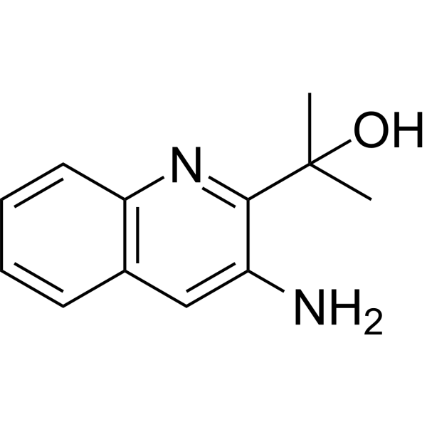
Acloproxalap

Based on 1 Customer Validation
Acloproxalap (ADX-629) is an orally active reactive aldehyde species (RASP) inhibitor. Acloproxalap binds covalently to free aldehydes, sequesters reactive aldehyde species, malondialdehyde, acetaldehyde and preformed malondialdehyde-acetaldehyde adducts, and reduces elevated RASP levels. Acloproxalap decreases the formation of aldehyde adducts, reduces liver and serum triglyceride levels, hepatic fat accumulation, circulating anti-malondialdehyde-acetaldehyde adduct antibody levels, and inhibits the release of IL-6 and MCP-1. Acloproxalap improves cell viability of liver slices exposed to ethanol. Acloproxalap blocks ethanol-induced damage in animal models of alcoholic liver disease. Acloproxalap is applicable to research related to alcoholic liver disease, alcoholic fatty liver disease, non-alcoholic steatohepatitis, chronic cough, rheumatoid arthritis, ulcerative colitis and mild atopic asthma.
For research use only. We do not sell to patients.
- Purity : 99.92%
- CAS No.: 1824609-67-7
- Formula: C12H14N2O
- Molecular Weight:202.25
-
Storage:Powder -20°C, 3 years , 4°C, 2 years ; In solvent -80°C, 6 months , -20°C, 1 month

To place orders, for customer services and technical support, please contact: MedChemExpress USA
Tel: 609-228-6898 E-mail: [email protected] [email protected]


-
 Biological Activity
-
Chemical Information
-
Solvent & Solubility
- Protocols
- Purity & Documentation
- References
-
 Help & FAQs




Biological Activity
Description
IC50 & Target
[4]|
IL-6 |
In Vitro
ADX-629 (0.1-100 μM; 24-72 h) reduces acetaldehyde levels in precision-cut liver slices from C57/BL6 mice exposed to 25 mM ethanol. Among the tested conditions, 0.1 μM (24 h) induces the greatest reduction within 24 hours, and the reduction effect of 1 μM (72 h) is statistically significant at the 72-hour time point[2].
ADX-629 (0.1-100 μM; 24-72 h) reduces ethanol-induced elevation of triglycerides in precision liver slices from C57/BL6 mice. Among the tested conditions, 0.1 μM (24 h) produces the greatest reduction within 24 hours, while 1 μM (24, 48, and 72 h) significantly decreases triglyceride levels to the level of the control group at all three time points[2].
ADX-629 increases the cell viability of precision liver slices from C57/BL6 mice exposed to 25 mM ethanol, and this effect is detected by measuring lactate dehydrogenase and adenosine triphosphate levels[2].
Acloproxalap (0-200 μM; 30-minute pre-treatment) potently reduces FIB-MAA-induced IL-6 and MCP-1 release in PMA (HY-18739)-differentiated human monocytic U-937 macrophages; it exerts significant inhibitory effects at a concentration of 1 μM, reduces cytokine release to background levels at concentrations ≥10 μM, and shows no cytotoxicity at all tested doses[4].
MedChemExpress (MCE) has not independently confirmed the accuracy of these methods. They are for reference only. Further protocols information, click here.
-
Cell Line:PMA-differentiated U-937 human monocytic macrophages
-
Concentration:0, 1, 10, 50, 100, 200 μM (30-minute pre-treatment)
-
Incubation Time:30 min (pre-treatment); 24 h (FIB-MAA incubation)
-
Result:Significantly reduced IL-6 and MCP-1 release at 1 μM.
Reduced cytokine levels to background (unmodified fibrinogen) levels at concentrations ≥10 μM.
Showed no toxic effects, with >99% cell viability confirmed via LDH assay.
In Vivo
MedChemExpress (MCE) has not independently confirmed the accuracy of these methods. They are for reference only.
-
Animal Model:C57/BL6 (10- to 12-week-old female, ≥20 g, chronic/binge alcoholic liver disease model)[1]
-
Dosage:400 mg/kg
-
Administration:p.o.; single dose
-
Result:Increased serum acetaldehyde to 37.5 µM from 8.3 µM in ethanol-only mice.
Decreased liver acetaldehyde to 146.2 µM/mg from 284.3 µM/mg in ethanol-only mice.
Reduced serum triglyceride levels to 112.2 mg/dL from 166.2 mg/dL in ethanol-only mice.
Reduced liver triglyceride levels to 157.4 mg/dL/mg of protein from 299.0 mg/dL/mg of protein in ethanol-only mice.
Reduced Oil Red O staining integrated density significantly.
Reduced Bodipy staining integrated density significantly.
Reduced liver malondialdehyde-acetaldehyde (MAA) antigen levels to 3.2 from 5.5 in ethanol-only mice.
Reduced serum anti-MAA antibody levels to 74.3 relative units from 232.6 relative units in ethanol-only mice.
Reduced serum alanine aminotransferase (ALT) levels to 26.3 IU/L from 43.8 IU/L in ethanol-only mice.
Reduced liver interferon-γ (IFN-γ) levels to 3.0 pg/mL/mg from 6.6 pg/mL/mg in ethanol-only mice.
Reduced liver monocyte chemoattractant protein-1 (MCP-1) levels to 15.62 pg/mL/mg from 47.5 pg/mL/mg in ethanol-only mice.
Clinical Trial
| NCT Number | Sponsor | Condition | Start Date |
Phase

|
|---|---|---|---|---|
| NCT01329991 | Plexxikon| | 2011-05 | PHASE1 |


Chemical Information
-
CAS No. 1824609-67-7
-
Appearance Solid
-
Molecular Weight 202.25
-
Formula C12H14N2O
-
Color Light yellow to yellow
-
SMILES
OC(C)(C)C1=NC2=C(C=C1N)C=CC=C2
-
Synonyms
ADX-629
-
Shipping
Room temperature in continental US; may vary elsewhere.
-
Storage
Powder -20°C 3 years 4°C 2 years In solvent -80°C 6 months -20°C 1 month


Solvent & Solubility
In Vitro:
DMSO : 100 mg/mL (494.44 mM; Need ultrasonic; Hygroscopic DMSO has a significant impact on the solubility of product, please use newly opened DMSO)
Please refer to the solubility information to select the appropriate solvent. Once prepared, please aliquot and store the solution to prevent product inactivation from repeated freeze-thaw cycles.
Storage method and period of stock solution: -80°C, 6 months; -20°C, 1 month. When stored at -80°C, please use it within 6 months. When stored at -20°C, please use it within 1 month.
Please refer to the solubility information to select the appropriate solvent. Once prepared, please aliquot and store the solution to prevent product inactivation from repeated freeze-thaw cycles.
Storage method and period of stock solution: -80°C, 6 months; -20°C, 1 month. When stored at -80°C, please use it within 6 months. When stored at -20°C, please use it within 1 month.
Concentration (start) × Volume (start) = Concentration (final) × Volume (final)
In Vivo:
Select the appropriate dissolution method based on your experimental animal and administration route.
- For the following dissolution methods, please ensure to first prepare a clear stock solution using an In Vitro approach and then sequentially add co-solvents:
- To ensure reliable experimental results, the clarified stock solution can be appropriately stored based on storage conditions. As for the working solution for In Vivo experiments, it is recommended to prepare freshly and use it on the same day.
- The percentages shown for the solvents indicate their volumetric ratio in the final prepared solution. If precipitation or phase separation occurs during preparation, heat and/or sonication can be used to aid dissolution.
Add each solvent one by one: 10% DMSO 40% PEG300 5% Tween-80 45% Saline
Solubility: ≥ 2.5 mg/mL (12.36 mM); Clear solution
This protocol yields a clear solution of ≥ 2.5 mg/mL (saturation unknown).
Taking 1 mL working solution as an example, add 100 μL DMSO stock solution (25.0 mg/mL) to 400 μL PEG300, and mix evenly; then add 50 μL Tween-80 and mix evenly; then add 450 μL Saline to adjust the volume to 1 mL.
Preparation of Saline: Dissolve 0.9 g sodium chloride in ddH₂O and dilute to 100 mL to obtain a clear Saline solution.
Add each solvent one by one: 10% DMSO 90% (20% SBE-β-CD in Saline)
Solubility: ≥ 2.5 mg/mL (12.36 mM); Clear solution
This protocol yields a clear solution of ≥ 2.5 mg/mL (saturation unknown).
Taking 1 mL working solution as an example, add 100 μL DMSO stock solution (25.0 mg/mL) to 900 μL 20% SBE-β-CD in Saline, and mix evenly.
Preparation of 20% SBE-β-CD in Saline (4°C, storage for one week): 2 g SBE-β-CD powder is dissolved in 10 mL Saline, completely dissolve until clear.
In Vivo Dissolution Calculator
Please enter the basic information of animal experiments:
-
-
-
-
Recommended: Prepare an additional quantity of animals to account for potential losses during experiments.
Please enter your animal formula composition:
-
%DMSO +
Recommended: Keep the proportion of DMSO in working solution below 2% if your animal is weak.
-
%+
-
+%Tween-80 + +
-
%Saline +
The co-solvents required include: DMSO, . All of co-solvents are available by MedChemExpress (MCE). , Tween 80. All of co-solvents are available by MedChemExpress (MCE).
Working solution concentration: 0.22 mg/mL
Method for preparing stock solution: mg drug dissolved in μL DMSO. Stock solution concentration: mg/mL.
1. Take μL DMSO stock solution;
2. Add μL .
μL , mix evenly;
3. Then add μL Tween 80, mix evenly;
4. Then add μL
Please ensure that the stock solution in the first step is dissolved to a clear state, and add co-solvents in sequence. You can use ultrasonic heating (ultrasonic cleaner, recommended frequency 20-40 kHz), vortexing, etc. to assist dissolution.

Protocols
-
Collagen-Induced Arthritis
Collagen-induced arthritis (CIA) is an autoimmune murine model of rheumatoid arthritis in which immunization with type II collagen (CII) emulsified in an adjuvant induces a T cell- and autoantibody-driven inflammatory arthritis characterized by synovial hyperplasia, immune cell infiltration, and joint destruction. The model typically relies on genetically susceptible mouse strains (e. g. , DBA/1) and reproduces key features of human rheumatoid arthritis, including anti-collagen immune responses and progressive joint inflammation. Disease onset generally occurs within ~3-4 weeks after immunization, depending on antigen/adjuvant combinations and protocol variation. The immunopathology is driven by adaptive immune activation against CII, leading to systemic and local joint inflammation mediated by pro-inflammatory cytokines and effector immune cells, making CIA a standard preclinical platform for evaluating immunomodulatory and anti-arthritic interventions.
-
Cell Cytotoxicity Assay
Cytotoxicity assays are usually based on the assessment of cell membrane damage, which can also be indirectly detected by measuring cell viability. Detection methods include MTT assay, CKK-8 assay, LDH assay and ATP assay, etc.
-
Mammalian live/dead viability and cytotoxicity staining
Live/dead viability and cytotoxicity staining assays are based on the simultaneous detection of intracellular esterase activity in metabolically active (viable) cells and membrane integrity loss in non-viable cells. In commonly used dual-staining approaches, membrane-permeant fluorogenic substrates are converted by intracellular esterases into fluorescent products in live cells, while impermeant DNA-binding dyes selectively enter cells with compromised plasma membranes and label nucleic acids in dead or dying cells, enabling discrimination between viable and non-viable populations by fluorescence microscopy or flow cytometry.
-
DSS-Induced Colitis
Dextran sulfate sodium (DSS)-induced colitis is generated by administering DSS in mouse drinking water, producing epithelial injury, barrier disruption, weight loss, diarrhea, fecal blood, colon shortening, histologic mucosal damage, and inflammatory mediator changes; the model is mainly used to study acute or chronic intestinal inflammation resembling selected features of ulcerative colitis. DSS injury is interpreted through clinical and tissue readouts rather than a single molecular endpoint: daily body weight, stool consistency, and bleeding are combined into a disease activity index, while colon length, histology, cytokines, myeloperoxidase activity, intestinal permeability, and tight-junction markers provide complementary measures of inflammation and barrier damage.
-
TNBS-Induced Colitis
TNBS-induced colitis is produced by intrarectal delivery of 2,4,6-trinitrobenzene sulfonic acid in ethanol, where ethanol disrupts the mucosal barrier and TNBS haptenates colonic proteins, generating immune-mediated colonic inflammation with weight loss, diarrhea, ulceration, transmural injury, inflammatory-cell infiltration, and cytokine responses. The model is used as an experimental intestinal inflammation model with Crohn’s disease–like features, especially when Th1-type responses, IL-12–dependent inflammation, chronic relapsing inflammation, or fibrosis-related endpoints are studied.
-
CCK-8/WST-8 Cell Proliferation Assay
The CCK-8/WST-8 assay is based on the reduction of the water-soluble tetrazolium salt WST-8 to a water-soluble formazan product by cellular dehydrogenases in metabolically active cells, where the generated formazan amount is proportional to the number of living cells and is quantified by measuring absorbance in the visible range, providing a colorimetric readout for cell viability and proliferation assessment. This class of tetrazolium-based assays improves upon earlier MTT-based systems by producing a water-soluble formazan, eliminating the need for organic solubilization steps and enabling direct spectrophotometric measurement in culture medium.
-
MTT Cell Proliferation Assay
The MTT assay is a colorimetric endpoint assay for estimating viable cell number, cell growth, cytotoxicity, or cell activation in cultured mammalian cells. Living cells reduce the yellow tetrazolium salt MTT into purple/blue formazan, while dead cells do not generate the same signal; the resulting color can be quantified with a multiwell spectrophotometer. MTT reduction is commonly interpreted as a readout of metabolic activity that often correlates with viable cell number, but it should not be treated as a direct cell-counting method unless the assay is optimized for the cell type and experimental condition. Studies show that MTT reduction can involve mitochondrial and non-mitochondrial reducing systems, and formazan may accumulate in intracellular lipid droplets rather than simply marking mitochondria.
-
Research Protocol for Metabolic Diseases
AMP-activated protein kinase, AMPK, is a conserved cellular energy sensor that responds to reduced cellular energy status and coordinates metabolism by increasing ATP-generating catabolic pathways while suppressing ATP-consuming anabolic processes. In metabolic disease research, the AMPK pathway is experimentally relevant because it regulates hepatic lipid synthesis, fatty acid oxidation, glucose production, skeletal-muscle glucose disposal, mTORC1-linked biosynthesis, autophagy, mitochondrial homeostasis, and whole-body energy balance. The central pathway logic is that energy stress, metformin, exercise-like stimulation, or direct AMPK activators increase AMPKα Thr172 phosphorylation and downstream substrate phosphorylation, including ACC and RAPTOR. Phosphorylation of ACC suppresses lipogenesis and supports fatty acid oxidation, whereas phosphorylation of RAPTOR suppresses mTORC1 signaling and links cellular energy status to growth and protein synthesis control. The pathway is linked
-
Cell Viability Determination by MTT Colorimetric Assay
The following protocol uses the MTT colorimetric assay as a classic literature-established method for assessing cell viability/metabolic activity in cultured mammalian cells. MTT[3-(4,5-dimethylthiazol-2-yl)-2,5-diphenyltetrazolium bromide] is reduced by metabolically active cells to a colored formazan product; the amount of formazan is quantified spectrophotometrically and provides an indirect measure of metabolically active viable cells. Importantly, MTT reduction reflects cellular oxidoreductase/metabolic activity rather than an absolute direct count of living cells, so changes in cellular metabolism can alter the signal independently of cell number.


Purity & Documentation
-
Data Sheet (281 KB)
-
SDS (393 KB)
- English - EN (393 KB)
- Français - FR (393 KB)
- Deutsch - DE (393 KB)
- Norwegian - NO (393 KB)
- Español - ES (393 KB)
- Swedish - SV (393 KB)
- Italian - IT (393 KB)
- Korean - KR (393 KB)
- Portuguese - PT (393 KB)
-
Handling Instructions (2659 KB)
References
[1]. Duryee MJ, Aripova N, Hunter CD, Ruskamp RJ, et al. A novel reactive aldehyde species inhibitor prevents the deleterious effects of ethanol in an animal model of alcoholic liver disease. Int Immunopharmacol. 2022 Dec;113(Pt A):109400. [Content Brief]
[3]. Guilleminault L, et al. Drugs Targeting Cough Receptors: New Therapeutic Options in Refractory or Unexplained Chronic Cough. Drugs. 2024 Jul;84(7):763-777. [Content Brief]


Complete Stock Solution Preparation Table
Please refer to the solubility information to select the appropriate solvent. Once prepared, please aliquot and store the solution to prevent product inactivation from repeated freeze-thaw cycles.
Storage method and period of stock solution: -80°C, 6 months; -20°C, 1 month. When stored at -80°C, please use it within 6 months. When stored at -20°C, please use it within 1 month.
| Optional Solvent | Concentration Solvent Mass | 1 mg | 5 mg | 10 mg | 25 mg |
|---|---|---|---|---|---|
| DMSO | 1 mM | 4.9444 mL | 24.7219 mL | 49.4438 mL | 123.6094 mL |
| 5 mM | 0.9889 mL | 4.9444 mL | 9.8888 mL | 24.7219 mL | |
| 10 mM | 0.4944 mL | 2.4722 mL | 4.9444 mL | 12.3609 mL | |
| 15 mM | 0.3296 mL | 1.6481 mL | 3.2963 mL | 8.2406 mL | |
| 20 mM | 0.2472 mL | 1.2361 mL | 2.4722 mL | 6.1805 mL | |
| 25 mM | 0.1978 mL | 0.9889 mL | 1.9778 mL | 4.9444 mL | |
| 30 mM | 0.1648 mL | 0.8241 mL | 1.6481 mL | 4.1203 mL | |
| 40 mM | 0.1236 mL | 0.6180 mL | 1.2361 mL | 3.0902 mL | |
| 50 mM | 0.0989 mL | 0.4944 mL | 0.9889 mL | 2.4722 mL | |
| 60 mM | 0.0824 mL | 0.4120 mL | 0.8241 mL | 2.0602 mL | |
| 80 mM | 0.0618 mL | 0.3090 mL | 0.6180 mL | 1.5451 mL | |
| 100 mM | 0.0494 mL | 0.2472 mL | 0.4944 mL | 1.2361 mL |


Acloproxalap Related Classifications
HY-147240 Related Classifications
 Powered by Bioz
Keywords
- Acloproxalap
- 1824609-67-7
- ADX-629
- ADX629
- ADX 629
- Interleukin Related
- malondialdehyde-acetaldehyde adducts
- malondialdehyde
- alcoholic liver disease
- U-937 human monocytic macrophages
- reactive aldehyde species
- C57/BL6 mouse precision cut liver slices
- macrophages
- acetaldehyde
- liver slices
- non-alcoholic steatohepatitis
- Inhibitor
- inhibitor
- inhibit