



11-Ketodihydrotestosterone
Based on 1 publication(s) in Google Scholar
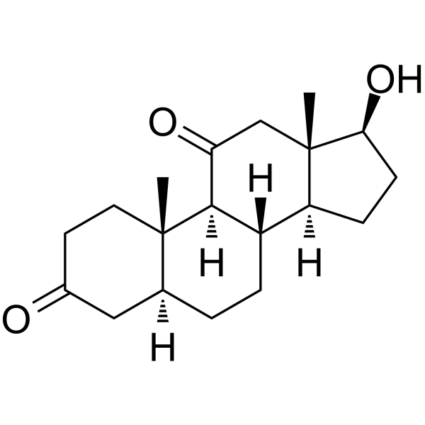
11-Ketodihydrotestosterone (11-KDHT; 5α-Dihydro-11-keto testosterone) is an endogenous steroid and a metabolite of 11β-Hydroxyandrostenedione. 11-Ketodihydrotestosterone is an active androgen and is also a potent androgen receptor (AR) agonist with a Ki of 20.4 nM and an EC50 of 1.35 nM for human AR. 11-Ketodihydrotestosterone drives gene regulation, protein expression and cell growth in androgen-dependent prostate cancer cells.
For research use only. We do not sell to patients.
- Purity : 98.0%
- CAS No.: 32694-37-4
- Formula: C19H28O3
- Molecular Weight:304.42
-
Storage:Powder -20°C, 3 years ; In solvent -80°C, 6 months , -20°C, 1 month

To place orders, for customer services and technical support, please contact: MedChemExpress USA
Tel: 609-228-6898 E-mail: [email protected] [email protected]


-
 Biological Activity
-
Chemical Information
-
Solvent & Solubility
- Protocols
- Purity & Documentation
- References
-
 Help & FAQs


-
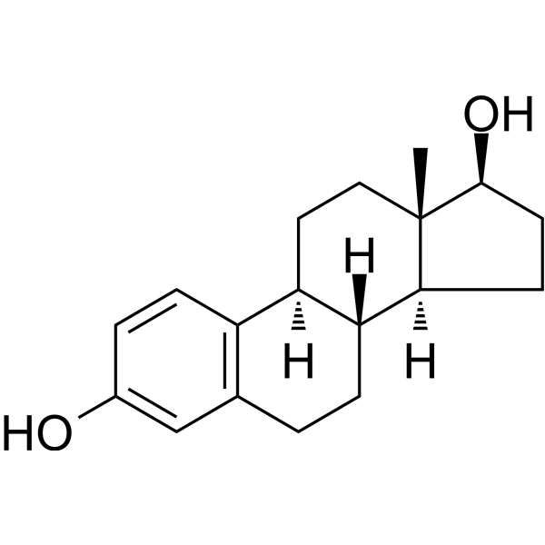
HY-B0141
EstradiolHuman Endogenous Metabolite -
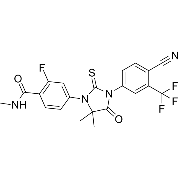
HY-70002
EnzalutamideAndrogen Receptor Antagonist -

HY-14648
DexamethasoneGR Agonist -
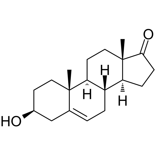
HY-14650
DHEASteroid Hormone -
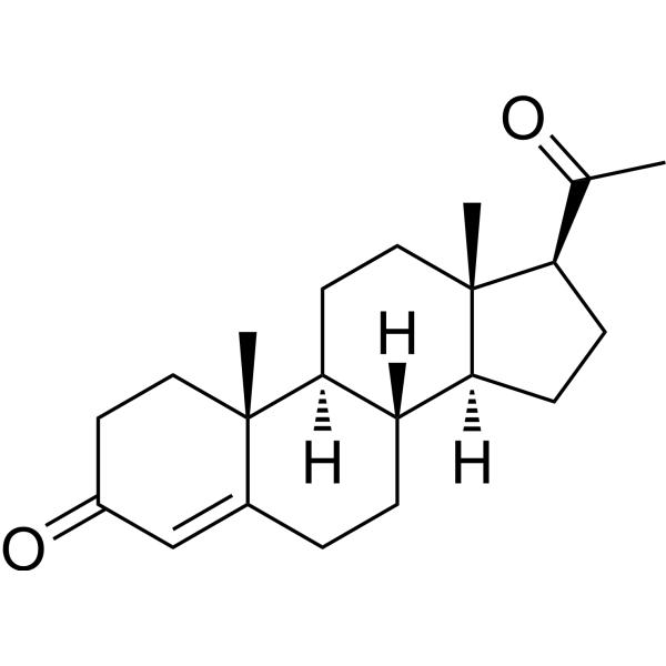
HY-N0437
ProgesteroneEndogenous Metabolite -
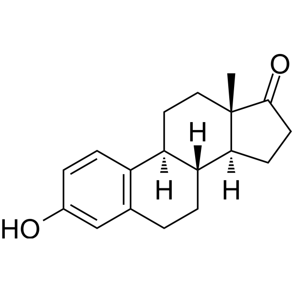
HY-B0234
EstroneEndogenous Metabolite -

HY-114118
SemaglutideGLP-1R Agonist -
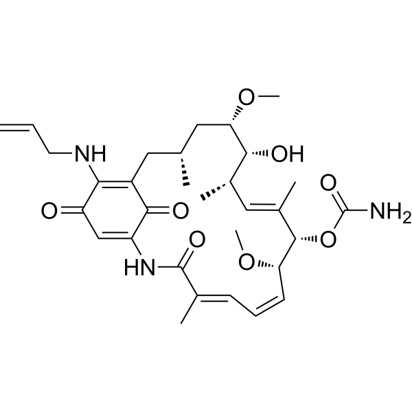
HY-10211
TanespimycinHSP90 Inhibitor -
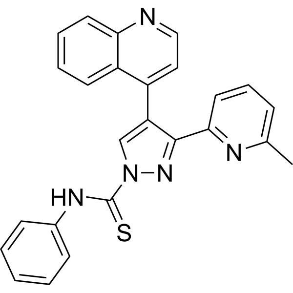
HY-10432
A 83-01ALK4/5/7 Inhibitor -
HY-12248
TelaglenastatGLS1 Inhibitor -
HY-16985
DarolutamideAndrogen Receptor Antagonist -
HY-17565
Bleomycin sulfateDNA Synthesis Inhibitor
Publications Citing Use of MedChemExpress (MCE) 11-Ketodihydrotestosterone
More

Biological Activity
Description
IC50 & Target
Ki: 20.4 nM (Human androgen receptor)[1]
EC50: 1.35 nM (Human androgen receptor)[1]
In Vitro
11-Ketodihydrotestosterone (11-KDHT; 1-10 nM; 24 hours; LNCaP and VCaP cells) treatment induces significant cell proliferation[1].
11-Ketodihydrotestosterone (11-KDHT; 0.1-10 nM; 7-10 days; LNCaP and VCaP cells) treatment results in the significant upregulation of KLK3, TMPRSS2 and FKBP5 in both LNCaP and VCaP cells, with the exception of KLK3 at 1 nM in LNCaP cells[1].
In PNT2 cells, only 20% of 11β-hydroxyandrostenedione (11OHA4) and 11β-hydroxytestosterone (11OHT) are metabolised with the former yielding 11keto-androstenedione (11KA4), 11-Ketodihydrotestosterone (11-KDHT) and 11β-hydroxy-5α-androstanedione (11OH-5αDIONE) and the latter yielding 11OHA4, 11KT and 11-Ketodihydrotestosterone with downstream products <0.03 μM[2].
In prostate cancer tissue, C11-oxy C19 metabolites at significantly higher levels than the C19 steroids are detected, with unconjugated 11-Ketodihydrotestosterone, 11KT and 11OHA4 levels ranging between 13 and 37.5 ng/g. Analyses of total steroid levels in plasma show significant levels of 11OHA4 (≈230-440 nM), 11KT (≈250-390 nM) and 11-Ketodihydrotestosterone (≈19 nM)[2].
MedChemExpress (MCE) has not independently confirmed the accuracy of these methods. They are for reference only. Further protocols information, click here.
-
Cell Line:LNCaP and VCaP cells
-
Concentration:0.1 nM, 1 nM or 10 nM
-
Incubation Time:7 days (LNCaP cells) or 10 days (VCaP cells)
-
Result:Induced significant cell proliferation.
-
Cell Line:LNCaP and VCaP cells
-
Concentration:1 nM, 10 nM
-
Incubation Time:24 hours
-
Result:Resulted in the significant upregulation of KLK3, TMPRSS2 and FKBP5 in both LNCaP (Fig 3) and VCaP (Fig 4) cells.


Chemical Information
-
CAS No. 32694-37-4
-
Appearance Solid
-
Molecular Weight 304.42
-
Formula C19H28O3
-
Color White to off-white
-
SMILES
C[C@@]1(C2)[C@@H](O)CC[C@@]1([H])[C@]3([H])CC[C@@]4([H])CC(CC[C@]4(C)[C@@]3([H])C2=O)=O
-
Synonyms
11-KDHT; 5α-Dihydro-11-keto testosterone
-
Shipping
Room temperature in continental US; may vary elsewhere.
-
Storage
Powder -20°C 3 years In solvent -80°C 6 months -20°C 1 month


Publications (1)
-
Journal Impact Factor
-
Most Recent
-
Sci Rep
Network pharmacology and experimental validation to elucidate the pharmacological mechanisms of Guben Xiezhuo decoction against renal fibrosis. [Abstract]2025 Aug 20;15(1):30479. PMID: 40830226


Solvent & Solubility
In Vitro:
DMSO : 100 mg/mL (328.49 mM; Need ultrasonic; Hygroscopic DMSO has a significant impact on the solubility of product, please use newly opened DMSO)
Please refer to the solubility information to select the appropriate solvent. Once prepared, please aliquot and store the solution to prevent product inactivation from repeated freeze-thaw cycles.
Storage method and period of stock solution: -80°C, 6 months; -20°C, 1 month. When stored at -80°C, please use it within 6 months. When stored at -20°C, please use it within 1 month.
Please refer to the solubility information to select the appropriate solvent. Once prepared, please aliquot and store the solution to prevent product inactivation from repeated freeze-thaw cycles.
Storage method and period of stock solution: -80°C, 6 months; -20°C, 1 month. When stored at -80°C, please use it within 6 months. When stored at -20°C, please use it within 1 month.
Concentration (start) × Volume (start) = Concentration (final) × Volume (final)
In Vivo:
Select the appropriate dissolution method based on your experimental animal and administration route.
- For the following dissolution methods, please ensure to first prepare a clear stock solution using an In Vitro approach and then sequentially add co-solvents:
- To ensure reliable experimental results, the clarified stock solution can be appropriately stored based on storage conditions. As for the working solution for In Vivo experiments, it is recommended to prepare freshly and use it on the same day.
- The percentages shown for the solvents indicate their volumetric ratio in the final prepared solution. If precipitation or phase separation occurs during preparation, heat and/or sonication can be used to aid dissolution.
Add each solvent one by one: 10% DMSO 40% PEG300 5% Tween-80 45% Saline
Solubility: ≥ 2.5 mg/mL (8.21 mM); Clear solution
This protocol yields a clear solution of ≥ 2.5 mg/mL (saturation unknown).
Taking 1 mL working solution as an example, add 100 μL DMSO stock solution (25.0 mg/mL) to 400 μL PEG300, and mix evenly; then add 50 μL Tween-80 and mix evenly; then add 450 μL Saline to adjust the volume to 1 mL.
Preparation of Saline: Dissolve 0.9 g sodium chloride in ddH₂O and dilute to 100 mL to obtain a clear Saline solution.
Add each solvent one by one: 10% DMSO 90% (20% SBE-β-CD in Saline)
Solubility: 2.5 mg/mL (8.21 mM); Suspended solution; Need ultrasonic
This protocol yields a suspended solution of 2.5 mg/mL. Suspended solution can be used for oral and intraperitoneal injection.
Taking 1 mL working solution as an example, add 100 μL DMSO stock solution (25.0 mg/mL) to 900 μL 20% SBE-β-CD in Saline, and mix evenly.
Preparation of 20% SBE-β-CD in Saline (4°C, storage for one week): 2 g SBE-β-CD powder is dissolved in 10 mL Saline, completely dissolve until clear.
In Vivo Dissolution Calculator
Please enter the basic information of animal experiments:
-
-
-
-
Recommended: Prepare an additional quantity of animals to account for potential losses during experiments.
Please enter your animal formula composition:
-
%DMSO +
Recommended: Keep the proportion of DMSO in working solution below 2% if your animal is weak.
-
%+
-
+%Tween-80 + +
-
%Saline +
The co-solvents required include: DMSO, . All of co-solvents are available by MedChemExpress (MCE). , Tween 80. All of co-solvents are available by MedChemExpress (MCE).
Working solution concentration: 0.22 mg/mL
Method for preparing stock solution: mg drug dissolved in μL DMSO. Stock solution concentration: mg/mL.
1. Take μL DMSO stock solution;
2. Add μL .
μL , mix evenly;
3. Then add μL Tween 80, mix evenly;
4. Then add μL
Please ensure that the stock solution in the first step is dissolved to a clear state, and add co-solvents in sequence. You can use ultrasonic heating (ultrasonic cleaner, recommended frequency 20-40 kHz), vortexing, etc. to assist dissolution.

Protocols
-
RNA extraction experimental
By lysing cells, releasing RNA, and removing impurities such as proteins and DNA, high-purity RNA products are finally obtained. The commonly used traditional method is the guanidine isothiocyanate/phenol/chloroform method (Trizol), which is suitable for a variety of animal materials including animal tissues, microorganisms, cultured cells, etc., and most plant materials.
-
Western Blot
Western blotting (WB) is a commonly used experimental method in molecular biology, biochemistry, and immunogenetics for identifying and quantifying target proteins. It combines gel electrophoresis with immunoassay, enabling researchers to analyze protein expression, post-translational modifications, and molecular weight.
-
Protocol for Cell Counting and Cell Density Analysis
Cell counting and cell-density analysis estimate the number of cells in a known volume or field area. Manual hemocytometer counting uses a chamber of defined geometry to convert counted cells into cells/mL, while automated counters and image-analysis workflows detect cell objects from optical, brightfield, fluorescence, impedance, or digital-image features. Trypan blue viability counting is based on dye exclusion: viable cells with intact membranes exclude dye, while non-viable cells with compromised membranes stain blue. The readout is total cell density, viable-cell density, dead-cell density, and percent viability. Cell density can also be estimated from microscopy images by counting objects per image area, from flow cytometry using calibrated volume or reference particles, or from in situ microscopy in bioreactors after calibration against reference methods such as hemocytometer or flow cytometry.
-
Research Protocol for Endocrine Diseases
Endocrine diseases often arise from disrupted hormone production, hormone signaling, or target-tissue responsiveness; for diabetes-focused endocrine disease models, insulin signaling regulates glucose uptake, hepatic glucose output, lipid metabolism, and β-cell compensation. Type 2 diabetes develops through interacting defects in insulin resistance, β-cell dysfunction, adipose inflammation, hepatic glucose overproduction, altered incretin signaling, and ectopic lipid metabolism. A major unresolved question is whether endocrine dysfunction is driven primarily by target-tissue insulin resistance, intrinsic β-cell failure, immune/inflammatory stress, or combined multi-organ failure that differs by disease stage.
-
Somatic Cell Culture
A method of simulating the in vivo environment in vitro to maintain the cell growth, differentation and main functions.
-
CCK-8/WST-8 Cell Proliferation Assay
The CCK-8/WST-8 assay is based on the reduction of the water-soluble tetrazolium salt WST-8 to a water-soluble formazan product by cellular dehydrogenases in metabolically active cells, where the generated formazan amount is proportional to the number of living cells and is quantified by measuring absorbance in the visible range, providing a colorimetric readout for cell viability and proliferation assessment. This class of tetrazolium-based assays improves upon earlier MTT-based systems by producing a water-soluble formazan, eliminating the need for organic solubilization steps and enabling direct spectrophotometric measurement in culture medium.
-
Cell Counting-Based Growth Curve Assay
Cell counting-based growth curve assays quantify cell proliferation by directly measuring changes in viable cell number over time using manual or automated counting methods such as hemocytometer-based counting or instrument-assisted cell enumeration, enabling construction of growth curves that reflect population expansion dynamics in response to culture conditions. A widely used approach is trypan blue exclusion with hemocytometer counting, where membrane-compromised (non-viable) cells take up the dye, allowing discrimination between viable and non-viable cells while simultaneously enabling total cell number quantification. Repeated sampling across time points allows estimation of proliferation rate, growth phases, and comparative growth kinetics between experimental conditions.
-
CRISPRi/CRISPRa gene-regulation editing
CRISPRi and CRISPRa use catalytically inactive Cas9, typically SpCas9 D10A/H840A, as an RNA-guided DNA-binding platform that targets genomic loci through sgRNA complementarity and an adjacent PAM without generating Cas9 nuclease-mediated DNA cleavage. CRISPRi represses transcription by recruiting dCas9 alone or dCas9 fused to repressor domains such as KRAB to promoters or transcription start site regions, while CRISPRa activates transcription by recruiting activation domains such as VP64, VPR, or SAM components to promoter-proximal regions. The primary readout is target-gene expression change, commonly measured by RT-qPCR, RNA-seq, reporter fluorescence, or protein-level assays, and the readout reflects transcriptional repression or activation at the targeted endogenous locus.
-
MTT Cell Proliferation Assay
The MTT assay is a colorimetric endpoint assay for estimating viable cell number, cell growth, cytotoxicity, or cell activation in cultured mammalian cells. Living cells reduce the yellow tetrazolium salt MTT into purple/blue formazan, while dead cells do not generate the same signal; the resulting color can be quantified with a multiwell spectrophotometer. MTT reduction is commonly interpreted as a readout of metabolic activity that often correlates with viable cell number, but it should not be treated as a direct cell-counting method unless the assay is optimized for the cell type and experimental condition. Studies show that MTT reduction can involve mitochondrial and non-mitochondrial reducing systems, and formazan may accumulate in intracellular lipid droplets rather than simply marking mitochondria.
-
Protocol For Protein Expression And Purification
Recombinant protein expression in Escherichia coli followed by purification of a His-tagged soluble protein by immobilized metal affinity chromatography (IMAC), with optional MBP fusion and TEV tag removal when the construct includes these elements. The biological readout is production of the encoded target protein, detected as an inducible band at the expected molecular mass by SDS-PAGE and quantified by total protein assay or chromatographic absorbance; the purification readout is enrichment of the target protein in elution fractions after selective binding of polyhistidine residues to immobilized Ni2+/metal-chelate resin and elution by imidazole-containing buffer. Expression is driven by an inducible bacterial expression system, commonly T7/lac-based, in which IPTG or lactose/auto-induction activates transcription and translation of the cloned gene; lower induction temperature, lower inducer concentration, induction timing, and solubility-enhancing fusion tags can influence the frac
-
Cell Viability Determination by MTT Colorimetric Assay
The following protocol uses the MTT colorimetric assay as a classic literature-established method for assessing cell viability/metabolic activity in cultured mammalian cells. MTT[3-(4,5-dimethylthiazol-2-yl)-2,5-diphenyltetrazolium bromide] is reduced by metabolically active cells to a colored formazan product; the amount of formazan is quantified spectrophotometrically and provides an indirect measure of metabolically active viable cells. Importantly, MTT reduction reflects cellular oxidoreductase/metabolic activity rather than an absolute direct count of living cells, so changes in cellular metabolism can alter the signal independently of cell number.


Purity & Documentation
-
Data Sheet (282 KB)
-
SDS (252 KB)
- English - EN (252 KB)
- Français - FR (252 KB)
- Deutsch - DE (252 KB)
- Norwegian - NO (252 KB)
- Español - ES (252 KB)
- Swedish - SV (252 KB)
- Italian - IT (252 KB)
- Korean - KR (252 KB)
- Portuguese - PT (252 KB)
-
Handling Instructions (2659 KB)
References
[1]. Pretorius E, et al. 1-Ketotestosterone and 11-Ketodihydrotestosterone in Castration Resistant Prostate Cancer: Potent Androgens Which Can No Longer Be Ignored. PLoS One. 2016 Jul 21;11(7):e0159867. [Content Brief]
[2]. du Toit T, et al. Profiling adrenal 11β-hydroxyandrostenedione metabolites in prostate cancer cells, tissue and plasma: UPC2-MS/MS quantification of 11β-hydroxytestosterone, 11keto-testosterone and 11keto-dihydrotestosterone. J Steroid Biochem Mol Biol. 2017 Feb;166:54-67. [Content Brief]


Complete Stock Solution Preparation Table
Please refer to the solubility information to select the appropriate solvent. Once prepared, please aliquot and store the solution to prevent product inactivation from repeated freeze-thaw cycles.
Storage method and period of stock solution: -80°C, 6 months; -20°C, 1 month. When stored at -80°C, please use it within 6 months. When stored at -20°C, please use it within 1 month.
| Optional Solvent | Concentration Solvent Mass | 1 mg | 5 mg | 10 mg | 25 mg |
|---|---|---|---|---|---|
| DMSO | 1 mM | 3.2849 mL | 16.4247 mL | 32.8494 mL | 82.1234 mL |
| 5 mM | 0.6570 mL | 3.2849 mL | 6.5699 mL | 16.4247 mL | |
| 10 mM | 0.3285 mL | 1.6425 mL | 3.2849 mL | 8.2123 mL | |
| 15 mM | 0.2190 mL | 1.0950 mL | 2.1900 mL | 5.4749 mL | |
| 20 mM | 0.1642 mL | 0.8212 mL | 1.6425 mL | 4.1062 mL | |
| 25 mM | 0.1314 mL | 0.6570 mL | 1.3140 mL | 3.2849 mL | |
| 30 mM | 0.1095 mL | 0.5475 mL | 1.0950 mL | 2.7374 mL | |
| 40 mM | 0.0821 mL | 0.4106 mL | 0.8212 mL | 2.0531 mL | |
| 50 mM | 0.0657 mL | 0.3285 mL | 0.6570 mL | 1.6425 mL | |
| 60 mM | 0.0547 mL | 0.2737 mL | 0.5475 mL | 1.3687 mL | |
| 80 mM | 0.0411 mL | 0.2053 mL | 0.4106 mL | 1.0265 mL | |
| 100 mM | 0.0328 mL | 0.1642 mL | 0.3285 mL | 0.8212 mL |


11-Ketodihydrotestosterone Related Classifications
HY-135794 Related Classifications
 Powered by Bioz