9(10)-Nitrooleate
Based on 1 Customer Validation
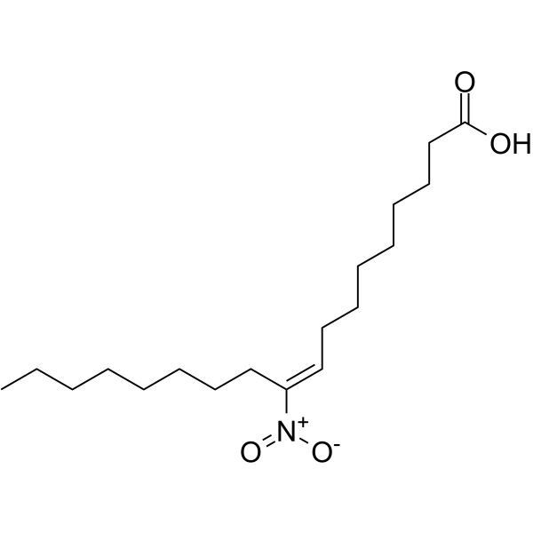
9 (10)-Nitrooleate (9(10)-Nitrated oleic acid) is an endogenous lipid signaling mediator with vasoprotective effects. 9 (10)-Nitrooleate enhances enzymatic activity and improves nitric oxide bioavailability by inducing phosphorylation of Akt and ERK1/2, regulating the multi-site phosphorylation status of eNOS and optimizing its interaction with Hsp90. 9 (10)-Nitrooleate also activates PPARα, PPARδ and PPARγ receptors, thereby regulating adipogenesis, glucose uptake and inflammation-related gene expression, and exhibits immunosuppressive effects by inhibiting neutrophil migration and cytokine secretion. 9 (10)-Nitrooleate is widely used in studies of sepsis and related inflammatory diseases.
For research use only. We do not sell to patients.
- Purity : 99.00%
- CAS No.: 1092676-99-7
- Formula: C18H33NO4
- Molecular Weight:327.46
-
Storage:
Solution, -20°C, 2 years
Biological Activity
Description
IC50 & Target
|
PPARα |
PPARγ |
PPARβ/δ |
eNOS |
In Vitro
9(10)-Nitrooleate (3 μM; 2-16 h) activates Akt and ERK1/2 signaling in human umbilical vein endothelial cells, inducing peak threefold Akt phosphorylation at 2 h and sixfold ERK1/2 phosphorylation at 4 h[1].
9(10)-Nitrooleate (3 μM; 2-16 h) modulates eNOS protein interactions in human umbilical vein endothelial cells, reducing eNOS/Cav-1 association and increasing eNOS/Hsp90 association to enhance eNOS activity[1].
9(10)-Nitrooleate (1 μM; 2 days) significantly increases glucose uptake in differentiated 3T3-L1 adipocytes, with this effect partially dependent on PPARγ activation[2].
9(10)-Nitrooleate (1-10 μM; 30 min) dose-dependently inhibits LPS-induced secretion of TNFα and IL-1β from normal murine peripheral blood PMNs[3].
MedChemExpress (MCE) has not independently confirmed the accuracy of these methods. They are for reference only. Further protocols information, click here.
-
Cell Line:human umbilical vein endothelial cells (HUVEC)
-
Concentration:3 μM
-
Incubation Time:2, 4, 8, 16 h
-
Result:Increased phosphorylation of eNOS-Ser1177 at 2-4 h, gradually increased phosphorylation of eNOS-Ser633 through 16 h, and gradually increased dephosphorylation of eNOS at Ser113 and Thr495, despite decreasing total eNOS protein expression.\n
Induced threefold phosphorylation of Akt-Ser473 at 2 h, followed by sixfold phosphorylation of ERK1/2 (P44-Thr202/P42-Tyr204) at 4 h, with both kinases gradually returning to basal levels after peak induction.
-
Cell Line:3T3-L1 preadipocyte
-
Concentration:1-3 μM
-
Incubation Time:14 days
-
Result:Induced ~60% of 3T3-L1 preadipocyte differentiation at 3 μM, as measured by oil red O staining for intracellular lipid accumulation.
Upregulated expression of adipocyte-specific markers PPARγ2 and aP2.
Chemical Information
-
CAS No. 1092676-99-7
-
Appearance Liquid
-
Molecular Weight 327.46
-
Formula C18H33NO4
-
Color Colorless to light yellow
-
SMILES
OC(CCCCCCC/C([N+]([O-])=O)=C\CCCCCCCC)=O.OC(CCCCCCC/C=C([N+]([O-])=O)\CCCCCCCC)=O
-
Synonyms
9(10)-Nitrated oleic acid
-
Shipping
Room temperature in continental US; may vary elsewhere.
-
Storage
Solution, -20°C, 2 years
Protocols
-
Kinase activity and phosphorylation assays
Kinase activity assays measure the ability of kinases to transfer phosphate groups from ATP to specific substrates, while phosphorylation assays detect the presence and levels of phosphorylated proteins. Common methods include radiolabeled ATP incorporation (e. g. ,), ADP release detection via bioluminescence (e. g. ,[3]), enzyme-linked immunosorbent assays (ELISA) for phospho-specific epitopes (e. g. ,[6]), and microtiter-based formats for high-throughput screening (e. g. ,[8]). The ADP-Glo assay quantifies kinase activity by measuring ADP produced during phosphorylation using a luciferase-based system. Radiometric assays involve autoradiography or scintillation counting after incorporation of 32P-labeled ATP into substrate proteins. ELISA-based approaches rely on phospho-specific antibodies to detect activated kinases in cell lysates or purified samples.
-
RT-PCR
Reverse transcription technology uses RNA as a template to synthesize DNA. RT-PCR is simple, specific and sensitive, and can be used to detect gene expression levels and expression differences in cells; detect RNA virus content; clone cDNA sequences of specific genes.
-
Western Blot
Western blotting (WB) is a commonly used experimental method in molecular biology, biochemistry, and immunogenetics for identifying and quantifying target proteins. It combines gel electrophoresis with immunoassay, enabling researchers to analyze protein expression, post-translational modifications, and molecular weight.
-
3T3-L1 preadipocyte-to-adipocyte differentiation
3T3-L1 preadipocytes are induced to differentiate after growth arrest using adipogenic media containing insulin, dexamethasone, and IBMX; differentiation is assessed by lipid-droplet accumulation, triglyceride increase, Oil Red O staining, and adipocyte-marker induction such as PPARγ and C/EBPα.
-
RNA extraction experimental
By lysing cells, releasing RNA, and removing impurities such as proteins and DNA, high-purity RNA products are finally obtained. The commonly used traditional method is the guanidine isothiocyanate/phenol/chloroform method (Trizol), which is suitable for a variety of animal materials including animal tissues, microorganisms, cultured cells, etc., and most plant materials.
-
LPS-Induced Endotoxemia/Systemic Inflammation
Lipopolysaccharide (LPS)-induced endotoxemia is a widely used in vivo model of acute systemic inflammation in which LPS, a Gram-negative bacterial endotoxin, activates innate immune signaling primarily through TLR4, leading to rapid and transient induction of pro-inflammatory cytokines such as TNF-α, IL-6, and IL-1β in circulation and tissues. This cytokine surge is commonly used as a measurable readout of systemic inflammatory activation and immune dysregulation, and is typically assessed within hours after intraperitoneal LPS administration in mouse models of endotoxemia. The model captures key features of systemic inflammatory response syndrome, including cytokine release, immune cell activation, and downstream tissue responses, and has been used to evaluate anti-inflammatory interventions such as cytokine modulation, lipid mediators, and immune cell-targeting therapies.
-
Cell migration
Cell migration is a method that plays an important role in wound healing, cell differentiation, embryonic development, etc.
-
Protocol for Pharmacokinetic Study
Pharmacokinetic studies quantify how an organism handles a drug over time through absorption, distribution, metabolism, and excretion, and the core experimental readout is the concentration-time profile of parent drug and, when relevant, metabolites in biological matrices such as plasma, whole blood, urine, bile, or tissue. Pharmacokinetic analysis links dose, route, exposure, clearance, half-life, distribution, bioavailability, and systemic exposure to drug efficacy and toxicity hypotheses rather than measuring a signaling pathway directly. The literature links pharmacokinetics to drug-development phenotypes by showing that drug metabolism and pharmacokinetics influence compound progression, exposure-response interpretation, safety margins, dosing strategy, and failure risk during discovery and development. DMPK science contributes to compound optimization by integrating physicochemical properties, in vitro metabolism, transporter behavior, in vivo exposure, and pharmacodynamic contex
-
Protocol for Kinase activity and phosphorylation assays
Kinase activity assays measure transfer of phosphate from ATP to a protein or peptide substrate, generating phosphorylated substrate, ADP, or incorporated radiolabeled phosphate as the readout; phosphorylation assays measure site-specific phosphorylation in cells or tissues as a proxy for kinase-pathway activation, inhibition, or substrate regulation. Phosphorylation can be detected by phospho-specific Western blot, immunoprecipitation kinase assay, phospho-immunofluorescence, phospho-flow cytometry, luminescent ADP detection, radiolabeled ATP incorporation, or reporter-based pathway assays, and these readouts can be applied to cancer cells, primary neurons, mouse tumors, organoids, inflammatory macrophages, ferroptosis studies, and mitophagy studies when the kinase target is biologically relevant.
-
Research Protocol for Inflammation-related Diseases
The NLRP3 inflammasome is a cytosolic innate immune signaling platform that integrates priming signals and danger-signal activation to promote caspase-1 activation, maturation of IL-1β and IL-18, and gasdermin D-mediated pyroptotic cell death. The core experimental logic is to determine whether inflammatory disease phenotypes are driven by increased NLRP3 expression, ASC-containing inflammasome assembly, caspase-1 cleavage, GSDMD cleavage, and extracellular release of IL-1β/IL-18 rather than by nonspecific cell injury alone. The pathway is strongly linked to inflammation-related disease phenotypes because monosodium urate crystals activate NALP3/NLRP3 inflammasome signaling in gout-like crystal inflammation, cholesterol crystals activate NLRP3 inflammasomes in atherogenesis models, and DSS-induced intestinal inflammation has been reported to involve NLRP3 inflammasome activity. However, experimental colitis studies also show context-dependent protective effects of NLRP3 inflammasome co
-
Real Time qPCR (Q-PCR)
Real-time quantitative PCR (qPCR) quantifies an amplifiable nucleic-acid target by monitoring fluorescence during PCR cycling rather than measuring product only after amplification. The increase in fluorescence tracks accumulation of PCR product, and the quantification cycle (Cq; historically also Ct/CP) is related to the initial amount of target: samples containing more starting target generally reach the defined fluorescence threshold in fewer cycles.
-
Lipid Droplets: Oil Red O/Sudan Dye Lipid Staining
Lipid droplets are intracellular organelles with a neutral-lipid core that stores triacylglycerols and sterol esters, and Oil Red O or Sudan dyes detect these hydrophobic lipid deposits by partitioning into retained lipids in fresh or frozen specimens. Oil Red O stains neutral triglycerides and lipids in frozen tissue sections or air-dried cytologic preparations, while Sudan Black B has also been used as a histochemical fat stain for lipid-rich tissue structures.
Purity & Documentation
-
Data Sheet (274 KB)
-
SDS (252 KB)
- English - EN (252 KB)
- Français - FR (252 KB)
- Deutsch - DE (252 KB)
- Norwegian - NO (252 KB)
- Español - ES (252 KB)
- Swedish - SV (252 KB)
- Italian - IT (252 KB)
- Korean - KR (252 KB)
- Portuguese - PT (252 KB)
-
Handling Instructions (2659 KB)
References
[1]. Shin E, et al. Nitrooleate mediates nitric oxide synthase activation in endothelial cells. Lipids. 2014;49(5):457-466. [Content Brief]
[2]. Baker PR, et al. Fatty acid transduction of nitric oxide signaling: multiple nitrated unsaturated fatty acid derivatives exist in human blood and urine and serve as endogenous peroxisome proliferator-activated receptor ligands. J Biol Chem. 2005;280(51):42464-42475. [Content Brief]
[3]. Panati K, et al. The nitrated fatty acid, 10-nitrooleate inhibits the neutrophil chemotaxis via peroxisome proliferator-activated receptor gamma in CLP-induced sepsis in mice. Int Immunopharmacol. 2019;72:159-165. [Content Brief]
Calculators
Concentration (start) × Volume (start) = Concentration (final) × Volume (final)