



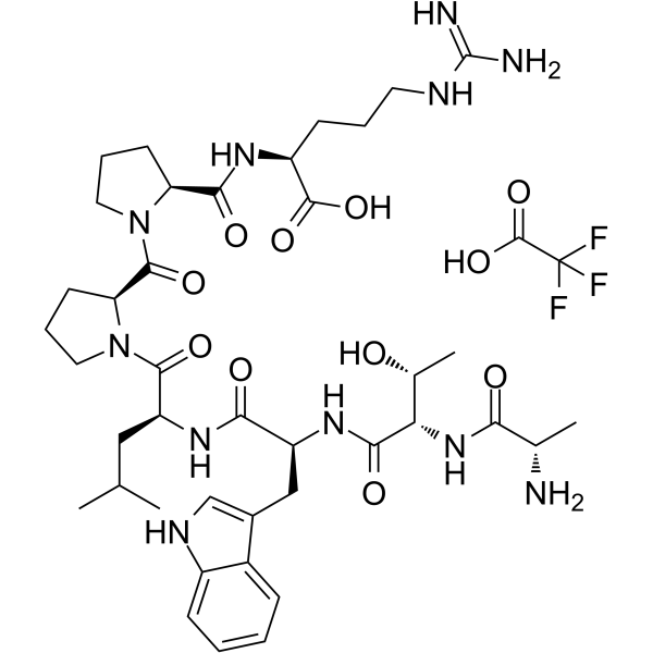
ATWLPPR Peptide TFA
Based on 2 publication(s) in Google Scholar
ATWLPPR Peptide TFA, a heptapeptide, acts as a selective neuropilin-1 inhibitor, inhibits VEGF165 binding to NRP-1, used in the research of angiogenesis. ATWLPPR Peptide TFA has potential in reducing the early retinal damage caused by diabetes.
연구목적의 판매만을 진행합니다. 환자를 대상으로 한 판매는 하지 않습니다.
- Purity : 99.20%
- 화학식: C42H62F3N11O11
- 분자량:954.00
-
보관:
Sealed storage, away from moisture and light.
Powder -80°C, 2 years , -20°C, 1 year* In solvent : -80°C, 6 months; -20°C, 1 month (sealed storage, away from moisture and light)

To place orders, for customer services and technical support, please contact: MedChemExpress USA
Tel: 609-228-6898 E-mail: [email protected] [email protected]


-
 Biological Activity
-
Chemical Information
-
용액&용해도
- Protocol
- 순도&문서
- References
-
 Help & FAQs


Publications Citing Use of MedChemExpress (MCE) ATWLPPR Peptide TFA
More

Biological Activity
제품 설명
IC50 & Target
Neuropilin-1[2]
In Vitro
In Vivo
ATWLPPR prevents the increase of inflammation-associated proteins (GFAP, VEGF and ICAM-1) in the retina[2].
MedChemExpress (MCE) has not independently confirmed the accuracy of these methods. They are for reference only.


Chemical Information
-
Appearance Solid
-
분자량 954.00
-
화학식 C42H62F3N11O11
-
Color White to off-white
-
SMILES
O=C(N[C@@H]([C@H](O)C)C(N[C@@H](CC1=CNC2=CC=CC=C12)C(N[C@@H](CC(C)C)C(N3[C@@H](CCC3)C(N4[C@@H](CCC4)C(N[C@@H](CCCNC(N)=N)C(O)=O)=O)=O)=O)=O)=O)[C@H](C)N.OC(C(F)(F)F)=O
-
선적
Room temperature in continental US; may vary elsewhere.
-
보관
Sealed storage, away from moisture and light
Powder -80°C 2 years -20°C 1 year * In solvent : -80°C, 6 months; -20°C, 1 month (sealed storage, away from moisture and light)


Publications (2)
-
Journal Impact Factor
-
Most Recent
-
-
Sci Adv
Regeneration of the pulmonary vascular endothelium after viral pneumonia requires COUP-TF2. [Abstract]2020 Nov 25;6(48):eabc4493. PMID: 33239293


용액&용해도
In Vitro:
H2O : 100 mg/mL (104.82 mM; Need ultrasonic)
Please refer to the solubility information to select the appropriate solvent. Once prepared, please aliquot and store the solution to prevent product inactivation from repeated freeze-thaw cycles.
Storage method and period of stock solution: -80°C, 6 months; -20°C, 1 month (sealed storage, away from moisture and light). When stored at -80°C, please use it within 6 months. When stored at -20°C, please use it within 1 month.
* Note: If you choose water as the stock solution, please dilute it to the working solution, then filter and sterilize it with a 0.22 μm filter before use.
Please refer to the solubility information to select the appropriate solvent. Once prepared, please aliquot and store the solution to prevent product inactivation from repeated freeze-thaw cycles.
Storage method and period of stock solution: -80°C, 6 months; -20°C, 1 month (sealed storage, away from moisture and light). When stored at -80°C, please use it within 6 months. When stored at -20°C, please use it within 1 month.
* Note: If you choose water as the stock solution, please dilute it to the working solution, then filter and sterilize it with a 0.22 μm filter before use.
Concentration (start) × Volume (start) = Concentration (final) × Volume (final)
In Vivo:
For the following dissolution methods, please prepare the working solution directly:
It is recommended to prepare fresh solutions and use them promptly within a short period of time.
The percentages shown for the solvents indicate their volumetric ratio in the final prepared solution. If precipitation or phase separation occurs during preparation, heat and/or sonication can be used to aid dissolution.
Add each solvent one by one: PBS
Solubility: 100 mg/mL (104.82 mM); Clear solution; Need ultrasonic


순도&문서
-
Data Sheet (283 KB)
-
SDS (252 KB)
- English - EN (252 KB)
- Français - FR (252 KB)
- Deutsch - DE (252 KB)
- Norwegian - NO (252 KB)
- Español - ES (252 KB)
- Swedish - SV (252 KB)
- Italian - IT (252 KB)
- Korean - KR (252 KB)
- Portuguese - PT (252 KB)
-
Handling Instructions (2659 KB)
References
[1]. Starzec A, et al. Structure-function analysis of the antiangiogenic ATWLPPR peptide inhibiting VEGF(165) binding to neuropilin-1 and molecular dynamics simulations of the ATWLPPR/neuropilin-1 complex. Peptides. 2007 Dec;28(12):2397-402. [Content Brief]
[2]. Wang J, et al. The Neuropilin-1 Inhibitor, ATWLPPR Peptide, Prevents Experimental Diabetes-Induced Retinal Injury by Preserving Vascular Integrity and Decreasing Oxidative Stress. PLoS One. 2015 Nov 10;10(11):e0142571. [Content Brief]


Complete Stock Solution Preparation Table
Please refer to the solubility information to select the appropriate solvent. Once prepared, please aliquot and store the solution to prevent product inactivation from repeated freeze-thaw cycles.
Storage method and period of stock solution: -80°C, 6 months; -20°C, 1 month (sealed storage, away from moisture and light). When stored at -80°C, please use it within 6 months. When stored at -20°C, please use it within 1 month.
| Optional Solvent | Concentration Solvent Mass | 1 mg | 5 mg | 10 mg | 25 mg |
|---|---|---|---|---|---|
| H2O | 1 mM | 1.0482 mL | 5.2411 mL | 10.4822 mL | 26.2055 mL |
| 5 mM | 0.2096 mL | 1.0482 mL | 2.0964 mL | 5.2411 mL | |
| 10 mM | 0.1048 mL | 0.5241 mL | 1.0482 mL | 2.6205 mL | |
| 15 mM | 0.0699 mL | 0.3494 mL | 0.6988 mL | 1.7470 mL | |
| 20 mM | 0.0524 mL | 0.2621 mL | 0.5241 mL | 1.3103 mL | |
| 25 mM | 0.0419 mL | 0.2096 mL | 0.4193 mL | 1.0482 mL | |
| 30 mM | 0.0349 mL | 0.1747 mL | 0.3494 mL | 0.8735 mL | |
| 40 mM | 0.0262 mL | 0.1310 mL | 0.2621 mL | 0.6551 mL | |
| 50 mM | 0.0210 mL | 0.1048 mL | 0.2096 mL | 0.5241 mL | |
| 60 mM | 0.0175 mL | 0.0874 mL | 0.1747 mL | 0.4368 mL | |
| 80 mM | 0.0131 mL | 0.0655 mL | 0.1310 mL | 0.3276 mL | |
| 100 mM | 0.0105 mL | 0.0524 mL | 0.1048 mL | 0.2621 mL |
* Note: If you choose water as the stock solution, please dilute it to the working solution, then filter and sterilize it with a 0.22 μm filter before use.


ATWLPPR Peptide TFA Related Classifications
HY-P1663A Related Classifications
 Powered by Bioz