



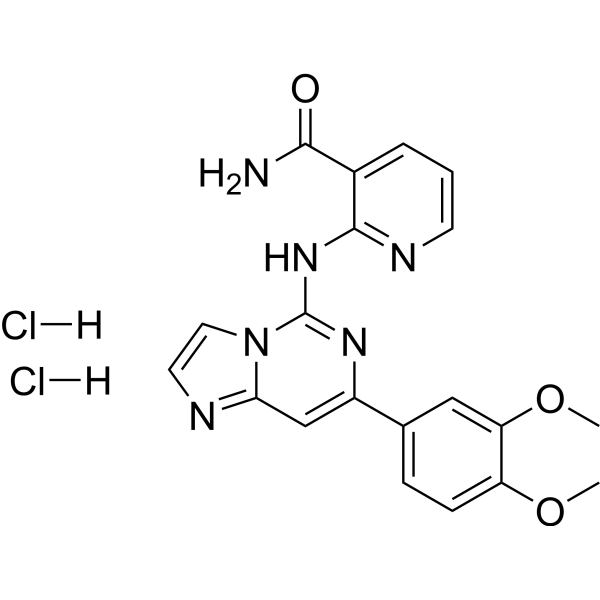
BAY 61-3606 dihydrochloride
Based on 13 publication(s) in Google Scholar
BAY 61-3606 dihydrochloride is an orally available, ATP-competitive, reversible and highly selective Syk inhibitor with a Ki of 7.5 nM an IC50 of 10 nM. BAY 61-3606 dihydrochloride reduces ERK1/2 and Akt phosphorylation in neuroblastoma cell. BAY 61-3606 dihydrochloride induces a large decrease of Syk phosphorylation in K-rn cell lysates. Bay 61-3606 dihydrochloride sensitizes TRAIL-induced apoptosis by downregulating Mcl-1 in breast cancer cells.
For research use only. We do not sell to patients.
- Purity : 99.45%
- CAS No.: 648903-57-5
- Formula: C20H20Cl2N6O3
- Molecular Weight:463.32
-
Storage:
-20°C, sealed storage, away from moisture
* In solvent : -80°C, 6 months; -20°C, 1 month (sealed storage, away from moisture)

To place orders, for customer services and technical support, please contact: MedChemExpress USA
Tel: 609-228-6898 E-mail: [email protected] [email protected]


-
 Biological Activity
-
Chemical Information
-
Solvent & Solubility
- Protocols
- Purity & Documentation
- References
-
 Help & FAQs


-
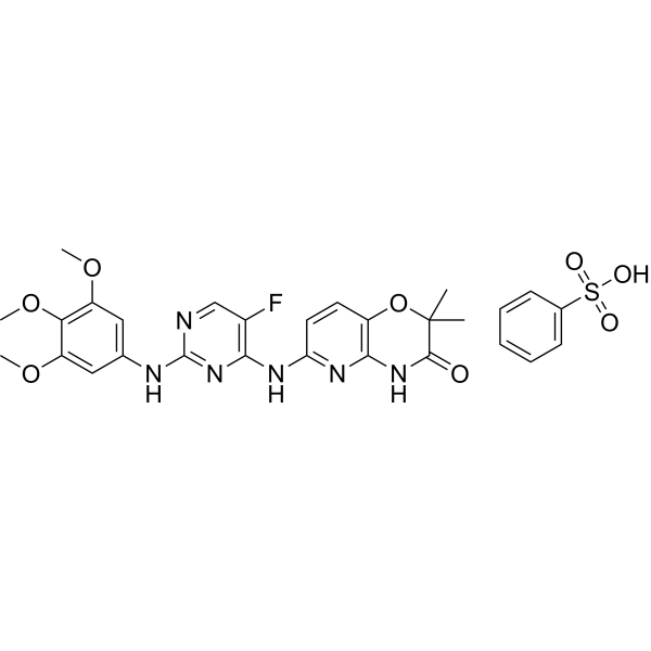
HY-12067
R406Syk/FLT3 Inhibitor -
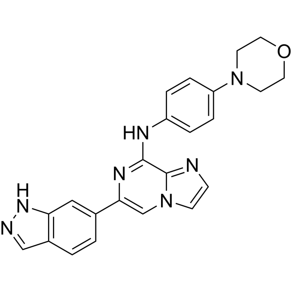
HY-15968
EntospletinibSyk Inhibitor -
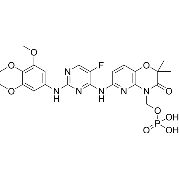
HY-13038A
FostamatinibSyk/FLT3 Inhibitor -
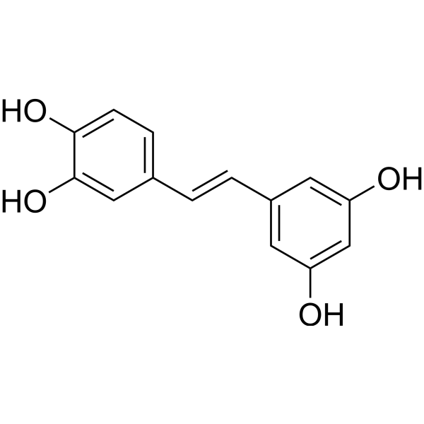
HY-13518
PiceatannolSyk Inhibitor -
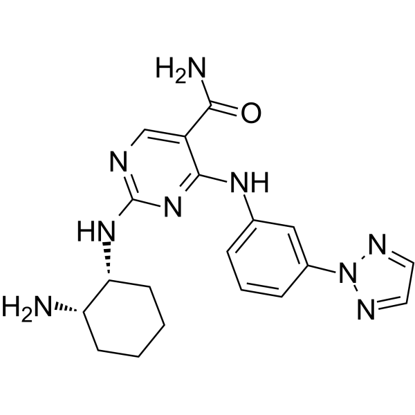
HY-15322
PRT062607Syk Inhibitor -
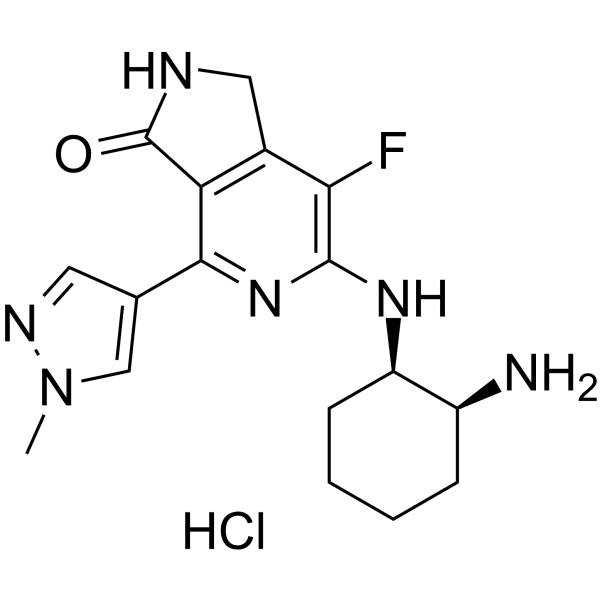
HY-100867A
Mivavotinib monohydrochlorideSYK/FLT3 Inhibitor -
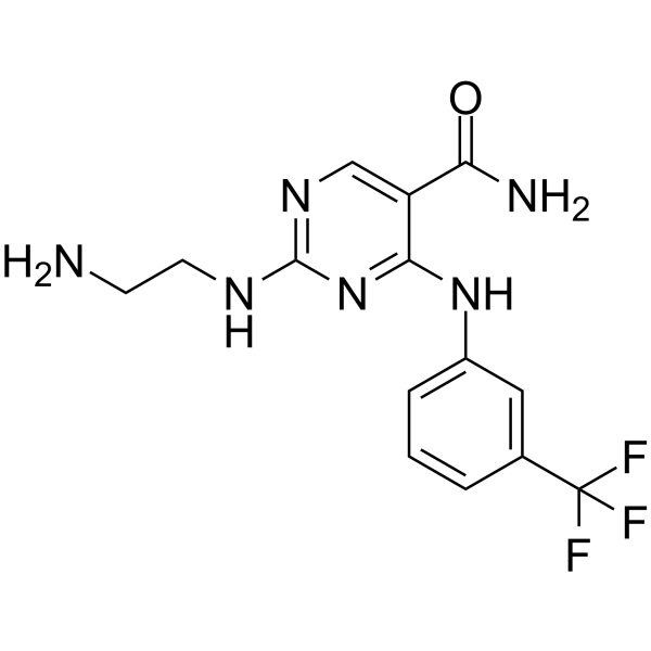
HY-112390A
Syk Inhibitor IISyk Inhibitor -
HY-13038
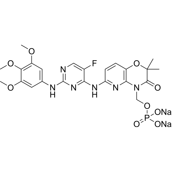
Fostamatinib DisodiumSyk/FLT3 Inhibitor -

HY-13259
MG-132Proteasome Inhibitor -
HY-11108
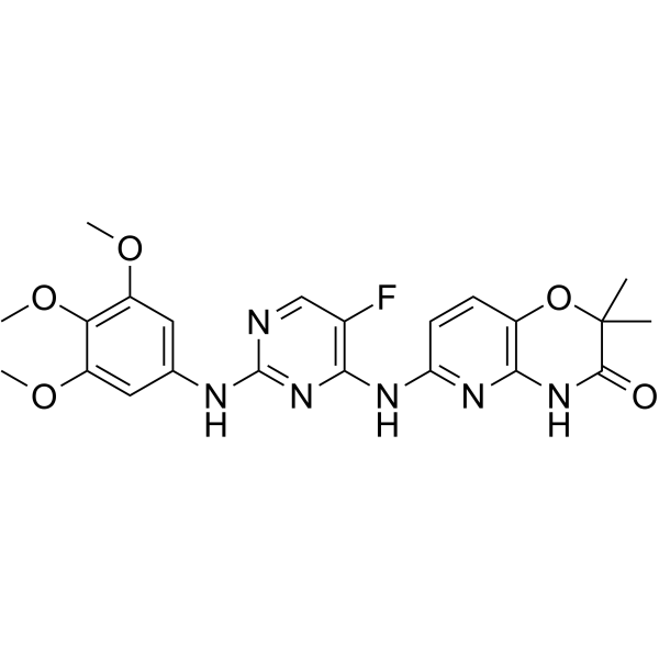
R406 free baseSyk/FLT3 Inhibitor -
HY-80013
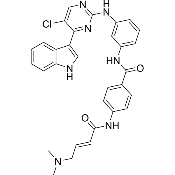
THZ1CDK7 Inhibitor -
HY-15142
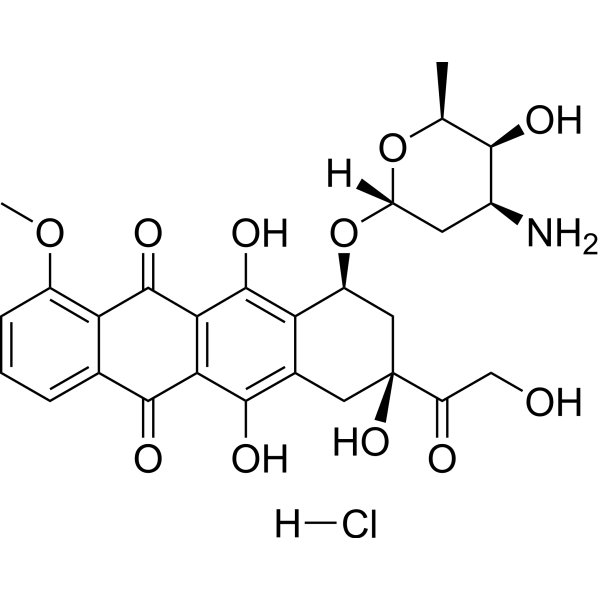
Doxorubicin hydrochlorideTopoisomerase Inhibitor
Publications Citing Use of MedChemExpress (MCE) BAY 61-3606 dihydrochloride
More- Neuro Oncol. 2018 Apr 9;20(5):621-631. [Abstract]
- Mol Ther. 2025 Dec 10:S1525-0016(25)01033-0. [Abstract]
- NPJ Precis Oncol. 2025 Dec 15;9(1):398. [Abstract]
- Proc Natl Acad Sci U S A. 2022 Oct 25;119(43):e2207280119. [Abstract]
- Front Immunol. 2018 Feb 15:9:249. [Abstract]
- Mol Cancer Ther. 2024 Oct 1;23(10):1404-1417. [Abstract]
- JCI Insight. 2024 Jun 13;9(14):e174746. [Abstract]
- Int J Mol Sci. 2021 Mar 24;22(7):3323. [Abstract]
- Pulm Pharmacol Ther. 2026 Jun:93:102421. [Abstract]
- Hematology. 2025 Dec;30(1):2495221. [Abstract]
- University of Pennsylvania. 2026.
- University of Pennsylvania. 2026.
- Research Square Preprint. 2024 Apr 19.


Biological Activity
Description
IC50 & Target
In Vitro
BAY 61-3606 (0.01-10 μM ; 48 hours) significantly reduces the cell viability of SYK-positive SH-SY5Y and SYK-negative SK-N-BE cells in a dose-dependent matter. SH-SY5Y cells expressing high SYK levels are significantly more sensitive to BAY 61-3606 in comparison to SK-N-BEcells expressing very low or no SYK[2].
BAY 61-3606 (0.4 and 0.8 μM; 4 or 24 hours) inhibits SYK activity by reducing ERK1/2 and Akt phosphorylation in neuroblastoma cell SH-SY5Y[2].
BAY 61-3606 (2 μM; 2 hours) induces a large decrease of Syk phosphorylation in K-rn cell lysates[3].
MedChemExpress (MCE) has not independently confirmed the accuracy of these methods. They are for reference only. Further protocols information, click here.
-
Cell Line:SYK-positive SH-SY5Y and SYK-negative SK-N-BE cells
-
Concentration:0.01, 0.1, 1, and 10 μM
-
Incubation Time:48 hours
-
Result:Significantly reduced the cell viability of both cell lines in a dose-dependent matter.
-
Cell Line:SH-SY5Y cells
-
Concentration:0.4 and 0.8 μM
-
Incubation Time:4 or 24 hours
-
Result:Reduced the phosphorylation of ERK1/2 and Akt after a 4 or 24 h treatment.
-
Cell Line:K-rn cell lysates
-
Concentration:2 μM
-
Incubation Time:2 hours
-
Result:Induced a large decrease of Syk phosphorylation.
In Vivo
MedChemExpress (MCE) has not independently confirmed the accuracy of these methods. They are for reference only.
-
Animal Model:Female BALB/c nude mice (5 weeks old) bearing MCF-7 tumor xenograft[4]
-
Dosage:50 mg/kg
-
Administration:Injected intraperitoneally twice a week with Bay 61–3606 (50 mg/kg), TRAIL (10 mg/kg) or a combination of Bay 61-3606 (50 mg/kg) and TRAIL (10 mg/kg); TRAIL was given 2 h after the injection of Bay 61-3606; for two weeks
-
Result:Led to efficacious reductions in tumor growth.


Chemical Information
-
CAS No. 648903-57-5
-
Appearance Solid
-
Molecular Weight 463.32
-
Formula C20H20Cl2N6O3
-
Color Off-white to yellow
-
SMILES
O=C(N)C1=CC=CN=C1NC2=NC(C3=CC=C(C(OC)=C3)OC)=CC4=NC=CN24.[H]Cl.[H]Cl
-
Shipping
Room temperature in continental US; may vary elsewhere.
-
Storage
-20°C, sealed storage, away from moisture
* In solvent : -80°C, 6 months; -20°C, 1 month (sealed storage, away from moisture)


Publications (13)
-
Journal Impact Factor
-
Most Recent
-
Neuro Oncol
SYK inhibition blocks proliferation and migration of glioma cells and modifies the tumor microenvironment. [Abstract]2018 Apr 9;20(5):621-631. PMID: 29401256 -
Mol Ther
FcεRγI promotes canine CD8 chimeric antigen receptor T cell cytotoxicity through a Syk-NF-κB axis. [Abstract]2025 Dec 10:S1525-0016(25)01033-0. PMID: 41376156 -
NPJ Precis Oncol
Siglec-14-LGALS3BP glycoimmune axis shapes tumor-associated macrophage polarization and confers poor outcome in colorectal cancer. [Abstract]2025 Dec 15;9(1):398. PMID: 41398201 -
Proc Natl Acad Sci U S A
Endocytosis triggers V-ATPase-SYK-mediated priming of cGAS activation and innate immune response. [Abstract]2022 Oct 25;119(43):e2207280119. PMID: 36252040 -
Front Immunol
Whole-Genome Expression Profiling in Skin Reveals SYK As a Key Regulator of Inflammation in Experimental Epidermolysis Bullosa Acquisita. [Abstract]2018 Feb 15:9:249. PMID: 29497423 -
Mol Cancer Ther
AKT Inhibition Sensitizes to Polo-Like Kinase 1 Inhibitor Onvansertib in Prostate Cancer. [Abstract]2024 Oct 1;23(10):1404-1417. PMID: 38894678 -
JCI Insight
2024 Jun 13;9(14):e174746. PMID: 38869957 -
Int J Mol Sci
A Combined Activity of Thrombin and P-Selectin Is Essential for Platelet Activation by Pancreatic Cancer Cells. [Abstract]2021 Mar 24;22(7):3323. PMID: 33805059 -
Pulm Pharmacol Ther
Evodiamine protects against lung ischemia-reperfusion injury by attenuating SYK-associated TLR4/NLRP3 inflammasome activation and ferroptosis-related injury signatures. [Abstract]2026 Jun:93:102421. PMID: 41951097 -
Hematology
Shikonin promotes ferroptosis though NSUN2-mediated m5C methylation modification of TFRC in acute myelocytic leukemia. [Abstract]2025 Dec;30(1):2495221. PMID: 40270446 -
-
-


Solvent & Solubility
In Vitro:
H2O : 4.55 mg/mL (9.82 mM; ultrasonic and warming and heat to 60°C)
Please refer to the solubility information to select the appropriate solvent. Once prepared, please aliquot and store the solution to prevent product inactivation from repeated freeze-thaw cycles.
Storage method and period of stock solution: -80°C, 6 months; -20°C, 1 month (sealed storage, away from moisture). When stored at -80°C, please use it within 6 months. When stored at -20°C, please use it within 1 month.
* Note: If you choose water as the stock solution, please dilute it to the working solution, then filter and sterilize it with a 0.22 μm filter before use.
Please refer to the solubility information to select the appropriate solvent. Once prepared, please aliquot and store the solution to prevent product inactivation from repeated freeze-thaw cycles.
Storage method and period of stock solution: -80°C, 6 months; -20°C, 1 month (sealed storage, away from moisture). When stored at -80°C, please use it within 6 months. When stored at -20°C, please use it within 1 month.
* Note: If you choose water as the stock solution, please dilute it to the working solution, then filter and sterilize it with a 0.22 μm filter before use.
Concentration (start) × Volume (start) = Concentration (final) × Volume (final)
In Vivo:
Select the appropriate dissolution method based on your experimental animal and administration route.
- For the following dissolution methods, please ensure to first prepare a clear stock solution using an In Vitro approach and then sequentially add co-solvents:
- To ensure reliable experimental results, the clarified stock solution can be appropriately stored based on storage conditions. As for the working solution for In Vivo experiments, it is recommended to prepare freshly and use it on the same day.
- The percentages shown for the solvents indicate their volumetric ratio in the final prepared solution. If precipitation or phase separation occurs during preparation, heat and/or sonication can be used to aid dissolution.
Add each solvent one by one: 10% DMSO 40% PEG300 5% Tween-80 45% Saline
Solubility: ≥ 0.71 mg/mL (1.53 mM); Clear solution
This protocol yields a clear solution of ≥ 0.71 mg/mL (saturation unknown).
Taking 1 mL working solution as an example, add 100 μL DMSO stock solution (7.1 mg/mL) to 400 μL PEG300, and mix evenly; then add 50 μL Tween-80 and mix evenly; then add 450 μL Saline to adjust the volume to 1 mL.
Preparation of Saline: Dissolve 0.9 g sodium chloride in ddH₂O and dilute to 100 mL to obtain a clear Saline solution.
Add each solvent one by one: 10% DMSO 90% (20% SBE-β-CD in Saline)
Solubility: ≥ 0.71 mg/mL (1.53 mM); Clear solution
This protocol yields a clear solution of ≥ 0.71 mg/mL (saturation unknown).
Taking 1 mL working solution as an example, add 100 μL DMSO stock solution (7.1 mg/mL) to 900 μL 20% SBE-β-CD in Saline, and mix evenly.
Preparation of 20% SBE-β-CD in Saline (4°C, storage for one week): 2 g SBE-β-CD powder is dissolved in 10 mL Saline, completely dissolve until clear.
In Vivo Dissolution Calculator
Please enter the basic information of animal experiments:
-
-
-
-
Recommended: Prepare an additional quantity of animals to account for potential losses during experiments.
Working solution concentration: 0.22 mg/mL

Protocols
-
Kinase activity and phosphorylation assays
Kinase activity assays measure the ability of kinases to transfer phosphate groups from ATP to specific substrates, while phosphorylation assays detect the presence and levels of phosphorylated proteins. Common methods include radiolabeled ATP incorporation (e. g. ,), ADP release detection via bioluminescence (e. g. ,[3]), enzyme-linked immunosorbent assays (ELISA) for phospho-specific epitopes (e. g. ,[6]), and microtiter-based formats for high-throughput screening (e. g. ,[8]). The ADP-Glo assay quantifies kinase activity by measuring ADP produced during phosphorylation using a luciferase-based system. Radiometric assays involve autoradiography or scintillation counting after incorporation of 32P-labeled ATP into substrate proteins. ELISA-based approaches rely on phospho-specific antibodies to detect activated kinases in cell lysates or purified samples.
-
Western Blot
Western blotting (WB) is a commonly used experimental method in molecular biology, biochemistry, and immunogenetics for identifying and quantifying target proteins. It combines gel electrophoresis with immunoassay, enabling researchers to analyze protein expression, post-translational modifications, and molecular weight.
-
Apoptosis
Apoptosis, also called programmed cell death, is generally characterized by distinct morphological characteristics.
-
TUNEL staining for apoptotic DNA fragmentation
TUNEL staining detects DNA strand breaks by using terminal deoxynucleotidyl transferase to add labeled nucleotides to exposed 3′-OH DNA termini, generating either microscopic staining in fixed cells or tissue sections, or fluorescence/cytometric signal in cell suspensions. TUNEL positivity reflects DNA fragmentation but should not be interpreted alone as definitive apoptosis, because TUNEL can also label necrotic, autolytic, mechanically damaged, or DNA-repair-associated DNA breaks.
-
Cell Cytotoxicity Assay
Cytotoxicity assays are usually based on the assessment of cell membrane damage, which can also be indirectly detected by measuring cell viability. Detection methods include MTT assay, CKK-8 assay, LDH assay and ATP assay, etc.
-
Annexin V plus membrane-impermeant dye apoptosis staining
Annexin V-based apoptosis assays rely on the detection of phosphatidylserine (PS) externalization from the inner leaflet of the plasma membrane to the outer leaflet, an early biochemical hallmark of apoptosis. Fluorescently labeled Annexin V binds PS in a calcium-dependent manner, enabling identification of early apoptotic cells by flow cytometry or fluorescence microscopy. When combined with a membrane-impermeant DNA-binding dye (e. g. , propidium iodide), this approach allows discrimination between viable (Annexin V−/dye−), early apoptotic (Annexin V+/dye−), and late apoptotic or necrotic (Annexin V+/dye+) cell populations by assessing membrane integrity and PS exposure.
-
Apoptosis Solutions
Apoptosis is a regulated, generally non-lytic cell-death pathway that removes unwanted, damaged, infected, or abnormal cells through coordinated morphological changes, caspase activation, DNA fragmentation, and membrane remodeling. The intrinsic apoptosis pathway is controlled mainly by mitochondrial outer membrane permeabilization, BCL-2 family proteins, cytochrome c release, apoptosome formation, caspase-9 activation, and downstream executioner caspase-3/7 activation. The extrinsic apoptosis pathway is initiated by death receptors such as Fas, TNFR, and TRAIL receptors, which recruit adaptor proteins and activate caspase-8 before engaging executioner caspases or mitochondrial amplification through BID cleavage. Apoptosis is linked to many phenotypes, including cancer cell killing, tissue homeostasis, immune regulation, neurodegeneration, infection response, and treatment-induced cytotoxicity; unresolved questions include how apoptosis interacts with necroptosis, pyroptosis, ferroptos
-
Protocol for Kinase activity and phosphorylation assays
Kinase activity assays measure transfer of phosphate from ATP to a protein or peptide substrate, generating phosphorylated substrate, ADP, or incorporated radiolabeled phosphate as the readout; phosphorylation assays measure site-specific phosphorylation in cells or tissues as a proxy for kinase-pathway activation, inhibition, or substrate regulation. Phosphorylation can be detected by phospho-specific Western blot, immunoprecipitation kinase assay, phospho-immunofluorescence, phospho-flow cytometry, luminescent ADP detection, radiolabeled ATP incorporation, or reporter-based pathway assays, and these readouts can be applied to cancer cells, primary neurons, mouse tumors, organoids, inflammatory macrophages, ferroptosis studies, and mitophagy studies when the kinase target is biologically relevant.
-
Breast Cancer Modeling
Breast cancer is a heterogeneous cancer, and it has been distinguished into four subtypes: luminal A, luminal B, HER2-positive and basal-like. Molecular mutations, epigenetic alterations, hormone exposure and immune microenvironment are related to the progression of breast cancer.


Purity & Documentation
-
Data Sheet (283 KB)
-
SDS (393 KB)
- English - EN (393 KB)
- Français - FR (393 KB)
- Deutsch - DE (393 KB)
- Norwegian - NO (393 KB)
- Español - ES (393 KB)
- Swedish - SV (393 KB)
- Italian - IT (393 KB)
- Korean - KR (393 KB)
- Portuguese - PT (393 KB)
-
Handling Instructions (2659 KB)
References
[1]. Yamamoto N, et al. The orally available spleen tyrosine kinase inhibitor 2-[7-(3,4-dimethoxyphenyl)-imidazo[1,2-c]pyrimidin-5-ylamino]nicotinamide dihydrochloride (BAY 61-3606) blocks antigen-induced airway inflammation in rodents. J Pharmacol Exp Ther. 2003 Sep;306(3):1174-81. [Content Brief]
[2]. Tümmler C, et al. SYK Inhibition Potentiates the Effect of Chemotherapeutic Drugs on Neuroblastoma Cells in Vitro. Cancers (Basel). 2019 Feb 10;11(2). pii: E202. [Content Brief]
[3]. Gioia R, et al. Quantitative phosphoproteomics revealed interplay between Syk and Lyn in the resistance to AMN107 in chronic myeloid leukemia cells. Blood. 2011 Aug 25;118(8):2211-21. [Content Brief]
[4]. Kim SY, et al. Bay 61-3606 Sensitizes TRAIL-Induced Apoptosis by Downregulating Mcl-1 in Breast Cancer Cells. PLoS One. 2015 Dec 31;10(12):e0146073. [Content Brief]


Complete Stock Solution Preparation Table
Please refer to the solubility information to select the appropriate solvent. Once prepared, please aliquot and store the solution to prevent product inactivation from repeated freeze-thaw cycles.
Storage method and period of stock solution: -80°C, 6 months; -20°C, 1 month (sealed storage, away from moisture). When stored at -80°C, please use it within 6 months. When stored at -20°C, please use it within 1 month.
| Optional Solvent | Concentration Solvent Mass | 1 mg | 5 mg | 10 mg | 25 mg |
|---|---|---|---|---|---|
| H2O | 1 mM | 2.1583 mL | 10.7917 mL | 21.5834 mL | 53.9584 mL |
| 5 mM | 0.4317 mL | 2.1583 mL | 4.3167 mL | 10.7917 mL |
* Note: If you choose water as the stock solution, please dilute it to the working solution, then filter and sterilize it with a 0.22 μm filter before use.


BAY 61-3606 dihydrochloride Related Classifications
HY-14985 Related Classifications
 Powered by Bioz