



Diphenyl Blue
Based on 4 publication(s) in Google Scholar
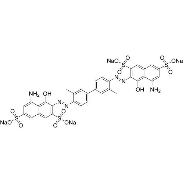
Diphenyl Blue (Trypan Blue) is a cell active dye, the most commonly used dye for the identification of dead cells, of en used to test cell membrane integrity and cell viability. Diphenyl Blue staining is one of the methods for tissue and cell culture. When cells are deactivated or have incomplete cell membranes, Diphenyl Blue can stain them Blue. Normal living cells with intact cell membranes reject Diphenyl blue and do not stain them blue. However, macrophages are capable of phagocytosis of Diphenyl Blue, so it can be used as a living stain for macrophages.
For research use only. We do not sell to patients.
- CAS No.: 72-57-1
- Formula: C34H24N6Na4O14S4
- Molecular Weight:960.81
-
Storage:
4°C, sealed storage, away from moisture and light
* In solvent : -80°C, 6 months; -20°C, 1 month (sealed storage, away from moisture and light)

To place orders, for customer services and technical support, please contact: MedChemExpress USA
Tel: 609-228-6898 E-mail: [email protected] [email protected]


-
 Biological Activity
-
Chemical Information
-
Solvent & Solubility
- Protocols
- Purity & Documentation
- References
-
 Help & FAQs


-
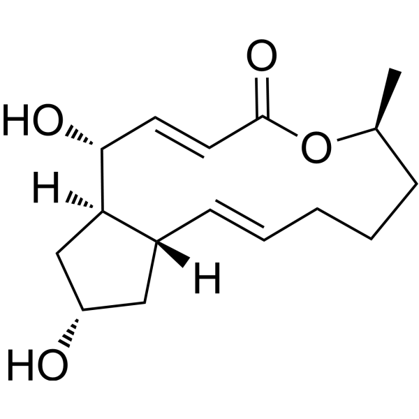
HY-16592
Brefeldin AProtein Transport Inhibitor -

HY-10219
RapamycinmTOR Inhibitor -

HY-Y0320C
Dimethyl sulfoxideAprotic Solvent -

HY-18739
Phorbol 12-myristate 13-acetatePKC Activator -

HY-13259
MG-132Proteasome Inhibitor -
HY-K0010
Protease Inhibitor Cocktail (EDTA-Free, 100× in DMSO) -
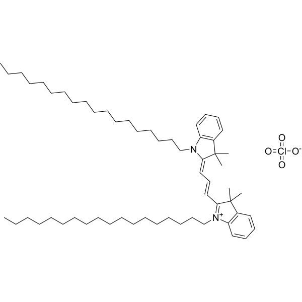
HY-D0083
DiIFluorescent Dye -
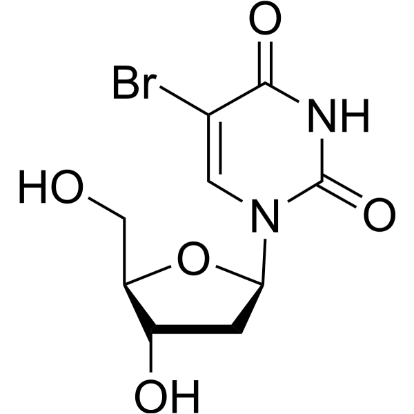
HY-15910
5-BrdUThymidine Analog -
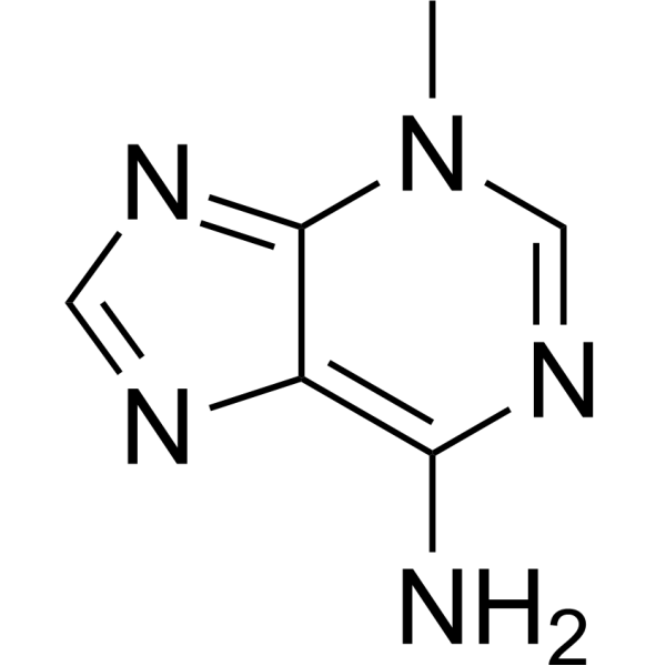
HY-19312
3-MethyladenineAutophagy/PI3K Inhibitor -
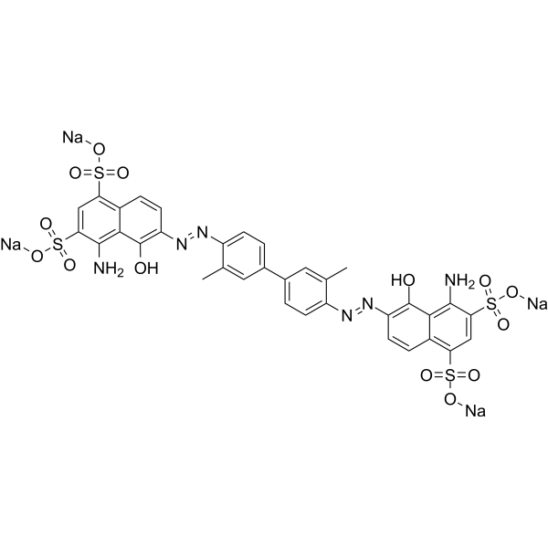
HY-B1102
Evans BlueL-glutamate Uptake Inhibitor -
HY-K3007
0.25% Trypsin-EDTA (1x), phenol red -
HY-N0335
IndigoBlue Dye
Publications Citing Use of MedChemExpress (MCE) Diphenyl Blue
More Customer Validation & Images

-
Cell Proliferation/Viability Assay
-
Histological Imaging/Staining


Biological Activity
Description
In Vitro
Guide (The following is our recommended protocol. This protocol is only a guide and should be modified according to your specific needs).
1. Preparation of Trypan Blue Working Solution
1.1 Prepare 0.4% trypan blue solution with pure water. For example, dissolve 400 mg trypan blue in 100 mL of pure water.
1.2 Dilute the stock solution with preheated serum-free cell culture medium or PBS to prepare 0.04% trypan blue working solution.
Note: Please adjust the concentration of trypan blue working solution according to the actual situation, and prepare it fresh each time you use it.
2. Cell Staining
2.1 Suspension Cells: Collect cells by centrifugation, wash twice with PBS, 5 minutes each time.
Adherent Cells: Discard the culture medium, wash twice with PBS, 5 minutes each time.
2.2 Add trypan blue working solution and incubate at room temperature for 5 minutes.
2.3 Discard the staining solution, wash cells twice with PBS, 5 minutes each time.
2.4 After resuspending the cells in serum-free culture medium or PBS, cell viability can be accurately quantified by direct counting under a microscope or by counting after taking photographs under a microscope.
MedChemExpress (MCE) has not independently confirmed the accuracy of these methods. They are for reference only.
Clinical Trial
| NCT Number | Sponsor | Condition | Start Date |
Phase

|
|---|---|---|---|---|
| NCT01329991 | Plexxikon| | 2011-05 | PHASE1 |


Chemical Information
-
CAS No. 72-57-1
-
Appearance Solid
-
Molecular Weight 960.81
-
Formula C34H24N6Na4O14S4
-
Color Light brown to black
-
SMILES
CC1=CC(C2=CC=C(/N=N/C3=C(S(=O)(O[Na])=O)C=C4C=C(S(=O)(O[Na])=O)C=C(N)C4=C3O)C(C)=C2)=CC=C1/N=N/C5=C(S(=O)(O[Na])=O)C=C6C=C(S(=O)(O[Na])=O)C=C(N)C6=C5O
-
Synonyms
Direct Blue 14; Trypan Blue
-
Shipping
Room temperature in continental US; may vary elsewhere.
-
Storage
4°C, sealed storage, away from moisture and light
* In solvent : -80°C, 6 months; -20°C, 1 month (sealed storage, away from moisture and light)


Publications (4)
-
Journal Impact Factor
-
Most Recent
-
-
Cell Death Dis
Dual targeting of PI3Kδ and PPARα enhances antitumor activity via FoxO1 activation in follicular lymphoma. [Abstract]2026 Mar 23;17(1):341. PMID: 41872189 -
Cell Death Dis
Bcl-2 inhibition combined with PPARα activation synergistically targets leukemic stem cell-like cells in acute myeloid leukemia. [Abstract]2023 Aug 29;14(8):573. PMID: 37644011
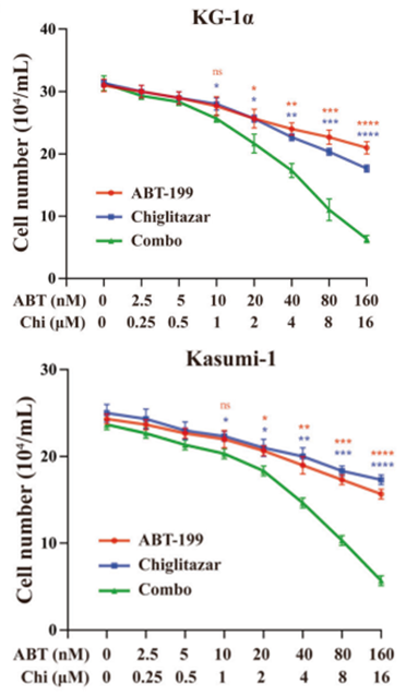
Diphenyl Blue purchased from MedChemExpress. Usage Cited in: Cell Death Dis. 2023 Aug 29;14(8):573. [Abstract]
Living cells of KG-1α and Kasumi-1 were counted with Diphenyl Blue (trypan blue) staining. The results revealed that venetoclax or chiglitazar decreased living cell numbers in a dose-dependent manner and the combination regimen further decreased cell viability.
-
Photochem Photobiol
Photobiomodulation therapy at 632 nm wavelength ameliorates intrauterine adhesion via activation of cAMP/PKA/CREB pathway. [Abstract]2024 Jan-Feb;100(1):214-224. PMID: 37212452
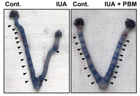
Diphenyl Blue purchased from MedChemExpress. Usage Cited in: Photochem Photobiol. 2024 Jan-Feb;100(1):214-224. [Abstract]
Diphenyl Blue (Trypan Blue; i.p.) dye staining of the uteri at embryonic day 4.5. Arrows indicate the implantation sites.


Solvent & Solubility
In Vitro:
H2O : 25 mg/mL (26.02 mM; ultrasonic and warming and heat to 60°C)
DMSO : 25 mg/mL (26.02 mM; ultrasonic and warming and heat to 60°C; Hygroscopic DMSO has a significant impact on the solubility of product, please use newly opened DMSO)
Please refer to the solubility information to select the appropriate solvent. Once prepared, please aliquot and store the solution to prevent product inactivation from repeated freeze-thaw cycles.
Storage method and period of stock solution: -80°C, 6 months; -20°C, 1 month (sealed storage, away from moisture and light). When stored at -80°C, please use it within 6 months. When stored at -20°C, please use it within 1 month.
* Note: If you choose water as the stock solution, please dilute it to the working solution, then filter and sterilize it with a 0.22 μm filter before use.
Please refer to the solubility information to select the appropriate solvent. Once prepared, please aliquot and store the solution to prevent product inactivation from repeated freeze-thaw cycles.
Storage method and period of stock solution: -80°C, 6 months; -20°C, 1 month (sealed storage, away from moisture and light). When stored at -80°C, please use it within 6 months. When stored at -20°C, please use it within 1 month.
* Note: If you choose water as the stock solution, please dilute it to the working solution, then filter and sterilize it with a 0.22 μm filter before use.
Concentration (start) × Volume (start) = Concentration (final) × Volume (final)


Purity & Documentation
-
Data Sheet (276 KB)
-
SDS (479 KB)
- English - EN (479 KB)
- Français - FR (479 KB)
- Deutsch - DE (479 KB)
- Norwegian - NO (479 KB)
- Español - ES (479 KB)
- Swedish - SV (479 KB)
- Italian - IT (479 KB)
- Korean - KR (479 KB)
- Portuguese - PT (479 KB)
-
Handling Instructions (2659 KB)
References


Complete Stock Solution Preparation Table
Please refer to the solubility information to select the appropriate solvent. Once prepared, please aliquot and store the solution to prevent product inactivation from repeated freeze-thaw cycles.
Storage method and period of stock solution: -80°C, 6 months; -20°C, 1 month (sealed storage, away from moisture and light). When stored at -80°C, please use it within 6 months. When stored at -20°C, please use it within 1 month.
| Optional Solvent | Concentration Solvent Mass | 1 mg | 5 mg | 10 mg | 25 mg |
|---|---|---|---|---|---|
| H2O / DMSO | 1 mM | 1.0408 mL | 5.2039 mL | 10.4079 mL | 26.0197 mL |
| 5 mM | 0.2082 mL | 1.0408 mL | 2.0816 mL | 5.2039 mL | |
| 10 mM | 0.1041 mL | 0.5204 mL | 1.0408 mL | 2.6020 mL | |
| 15 mM | 0.0694 mL | 0.3469 mL | 0.6939 mL | 1.7346 mL | |
| 20 mM | 0.0520 mL | 0.2602 mL | 0.5204 mL | 1.3010 mL | |
| 25 mM | 0.0416 mL | 0.2082 mL | 0.4163 mL | 1.0408 mL |
* Note: If you choose water as the stock solution, please dilute it to the working solution, then filter and sterilize it with a 0.22 μm filter before use.


Diphenyl Blue Related Classifications
HY-D0970 Related Classifications
 Powered by Bioz