



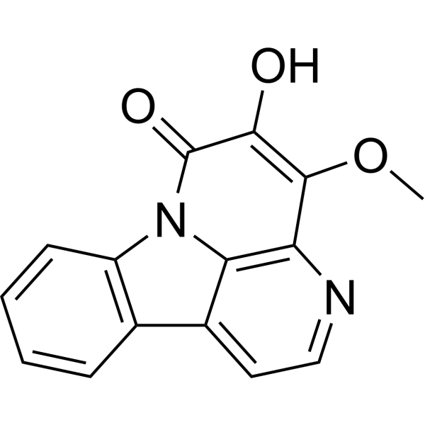
Nigakinone

Based on 1 Customer Validation
Nigakinone is an orally active FXR agonist and NLRP3 inhibitor. Nigakinone suppresses bile acid-induced inflammation and cell damage, regulates the bile acid profile, and alleviates intestinal mucosal barrier injury. Nigakinone synergizes with Irinotecan (HY-16562) to inhibit the proliferation and migration of colorectal cancer cells as well as the growth of subcutaneous xenograft tumors. Nigakinone can be used in research related to ulcerative colitis and colorectal cancer.
For research use only. We do not sell to patients.
- Purity : 99.95%
- CAS No.: 18110-86-6
- Formula: C15H10N2O3
- Molecular Weight:266.25
-
Storage:Powder -20°C, 3 years , 4°C, 2 years ; In solvent -80°C, 6 months , -20°C, 1 month

To place orders, for customer services and technical support, please contact: MedChemExpress USA
Tel: 609-228-6898 E-mail: [email protected] [email protected]


-
 Biological Activity
-
Chemical Information
-
Solvent & Solubility
- Protocols
- Purity & Documentation
- References
-
 Help & FAQs


-

HY-165772
10-Hydroxy-12(Z)-octadecenoic acidUnsaturated Fatty Acid -

HY-125726
Sulfo galactosylceramideGlycolipid -
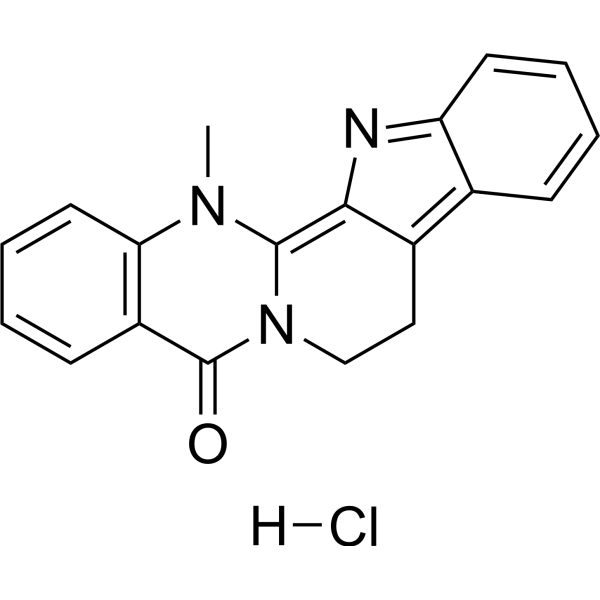
HY-N6029
Dehydroevodiamine hydrochlorideAChE/BACE1 Inhibitor -
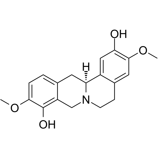
HY-N1255
ScoulerineAntimitotic Agent -
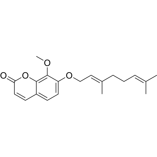
HY-N8388
CollininAnti-tubercular Agent -

HY-133976
Aceclofenac methyl esterAceclofenac Impurity -
HY-N0443
N-MethylcytisineTricyclic Quinolizidine Alkaloid -

HY-171037
Prostaglandin BxProstaglandin Derivative -
HY-125827
13-Methylberberine chlorideInterleukin Related Activator -
HY-N3940
Gelsevirine


Biological Activity
Description
IC50 & Target
[1]|
NLRP3 |
Cellular Effect
|
Cell Line

|
Type | Value | Description | References |
|---|---|---|---|---|
| CNE-2 | IC50 |
7.96 μg/mL
Compound: 39
|
Cytotoxicity against human CNE-2 cells incubated for 48 hrs by MTT assay
Cytotoxicity against human CNE-2 cells incubated for 48 hrs by MTT assay
|
[PMID: 34332400] |
In Vitro
Nigakinone (1 μM; 48 h) acts synergistically with Irinotecan to inhibit the clonogenic activity of HCT116 and Caco-2 colorectal cancer cells with low FXR expression[2].
Nigakinone (0.2-5 μM) significantly upregulates the expression of FXR protein in FXR-low-expressing Caco-2 colorectal cancer cells[2].
Nigakinone (0.2-5 μM) significantly upregulates the expression of FXR protein in HCT116 colon cancer cells with low FXR expression[2].
Nigakinone (10 μM; 24 h) functionally activates hFXR in HEK293T cells and significantly enhances BSEP promoter activity in an hFXR-dependent manner[1].
Nigakinone acts as an FXR agonist in HT-29 colon cancer cells with high FXR expression, and significantly upregulates FGF19 and SHP, the downstream target genes of FXR[2].
MedChemExpress (MCE) has not independently confirmed the accuracy of these methods. They are for reference only.
-
Cell Line:FXR-low HCT116, Caco-2, and HT-29 siFXR (FXR-knockdown) colorectal cancer cells
-
Concentration:0.2, 1, 5 μM nigakinone; 5, 10, 20 μM irinotecan
-
Incubation Time:48 h
-
Result:Exhibited significant synergistic inhibition of cell viability across all tested cell lines.
Showed Q values predominantly exceeding 1.15 and CI values below 1 across different concentration combinations.
Achieved synergy at lower concentrations than silibinin, a comparable dual FXR inducer/agonist.
-
Cell Line:FXR-low HCT116, Caco-2, and HT-29 siFXR colorectal cancer cells
-
Concentration:1 μM nigakinone; 10 μM irinotecan
-
Incubation Time:24 h, 48 h
-
Result:Exhibited significant synergistic inhibition of cell migration at both 24 h and 48 h across all tested cell lines.
Showed Q values ranging from 1.15 to 1.61 and CI values ranging from 0.29 to 0.57.
In Vivo
Nigakinone (60 mg/kg; p.o.; once daily; for 9 consecutive days) alleviates dextran sulfate sodium (DSS)-induced ulcerative colitis in wild-type C57BL/6 mice by inhibiting NLRP3-mediated inflammatory responses, protecting the intestinal mucosal barrier, and regulating the enterohepatic circulation of bile acids via activation of FXR[1].
Nigakinone (60 mg/kg; p.o.; once daily; for 9 consecutive days) fails to alleviate dextran sulfate sodium (DSS)-induced ulcerative colitis in FXR-knockout C57BL/6N mice, confirming that FXR is an essential prerequisite for nigakinone to exert its therapeutic effect on colitis[1].
MedChemExpress (MCE) has not independently confirmed the accuracy of these methods. They are for reference only.
-
Animal Model:Sprague-Dawley (SD) (male, 200-220 g, ulcerative colitis induced by 4% DSS ad libitum for 7 days)[1]
-
Dosage:25 mg/kg; 50 mg/kg; 100 mg/kg
-
Administration:p.o.; daily; 9 days
-
Result:Significantly reduced body weight loss, lowered DAI scores, decreased myeloperoxidase (MPO) and inducible nitric oxide synthase (iNOS) levels, and alleviated colon tissue damage including mucosal ulceration, crypt loss, and inflammatory infiltration.
Significantly reduced colon tissue mRNA expression of NLRP3 and caspase-1, and protein expression of NLRP3, caspase-1, IL-1β, ASC-2, and pro-caspase-1 (100 mg/kg dose).
Significantly increased colon tissue mRNA expression of tight junction proteins ZO-1, Claudin-1, and Occludin, and protected colonic microvilli and tight junctions (100 mg/kg dose).
Significantly reduced total bile acid (TBA) pool size in serum, liver, ileum, and colon; decreased BA proportion in liver and colon while increasing BA proportion in serum and ileum; shifted BA profile to reduce primary BA and increase secondary BA (100 mg/kg dose).
Significantly increased mRNA and protein expression of FXR in liver and ileum; increased liver mRNA expression of BSEP, ileum mRNA expression of FGF15, ASBT, and IBABP; and decreased liver protein expression of CYP7A1 and NTCP (100 mg/kg dose).
Significantly increased colon tissue mRNA and protein expression of FXR.
-
Animal Model:C57BL/6 (WT) (male, 20-22 g, ulcerative colitis induced by 3.5% DSS ad libitum for 7 days)[1]
-
Dosage:60 mg/kg
-
Administration:p.o.; daily; 9 days
-
Result:Significantly reduced body weight loss, lowered DAI scores, reversed colon length shortening, decreased serum levels of MPO, TNF-α, and IL-1β, and reduced colon histological scores and tissue damage.
Significantly increased colon tissue mRNA and protein expression of FXR; reduced mRNA and protein expression of NLRP3, IL-1β, TNF-α, ASC-2, and pro-caspase-1; and increased mRNA and protein expression of tight junction proteins ZO-1 and Claudin-1.
Significantly reduced total BA pool size, decreased BA proportion in liver and colon while increasing BA proportion in serum and ileum, increased liver protein expression of FXR, BSEP, SHP, and OSTβ, decreased liver protein expression of CYP7A1 and NTCP, and increased ileum protein expression of FXR, IBABP, and OSTβ.
-
Animal Model:C57BL/6N (FXR KO) (male, 20-22 g, ulcerative colitis induced by 3.5% DSS ad libitum for 7 days)[1]
-
Dosage:60 mg/kg
-
Administration:p.o.; daily; 9 days
-
Result:Did not significantly reduce body weight loss, lower DAI scores, reverse colon length shortening, decrease serum levels of MPO, TNF-α, and IL-1β, or reduce colon histological scores and tissue damage.
Did not significantly reduce colon tissue mRNA or protein expression of NLRP3, IL-1β, TNF-α, ASC-2, or pro-caspase-1, or increase mRNA or protein expression of tight junction proteins ZO-1 and Claudin-1.
Did not significantly reduce total BA pool size, alter BA proportion distribution in serum, liver, ileum, or colon, or regulate liver or ileum protein expression of bile acid transporters and regulators.
-
Animal Model:BALB/c nude (5-week-old, male, SPF-grade, HCT116 cell-derived subcutaneous xenograft model)[2]
-
Dosage:35 mg/kg (single agent); 35 mg/kg (in combination with irinotecan 40 mg/kg)
-
Administration:i.p.; daily; 21 days (single agent); i.p.; daily (nigakinone) + weekly (irinotecan); 21 days (combination)
-
Result:Reduced tumor weight relative to the control group.
Significantly upregulated FXR protein expression in tumor tissues compared to the control and irinotecan-only groups.
Decreased Ki67 expression compared to the control group.
Increased cleaved caspase 3 expression compared to the control group.
Produced a synergistic reduction in tumor volume relative to irinotecan alone when combined with irinotecan.
Produced a synergistic reduction in tumor weight relative to irinotecan alone when combined with irinotecan.
Further decreased Ki67 expression compared to irinotecan alone when combined with irinotecan.
Further increased cleaved caspase 3 expression compared to irinotecan alone when combined with irinotecan.
Maintained significant FXR upregulation in tumor tissues when combined with irinotecan.
Showed no significant differences in organ indices (heart, liver, lung, spleen, kidneys, brain) or colon length between nigakinone-treated and control groups, indicating good tolerability.
- How to Select the Route of Administration for Mammals
- Cell migration
- DSS-Induced Colitis
- TNBS-Induced Colitis
- Patient-Derived Xenograft (PDX)
- Splenic/Portal-Vein Liver Metastasis Xenograft
- Research Protocol for Inflammation-related Diseases
- Subcutaneous Cell-Line-Derived Xenograft
- Orthotopic Cell-Line Xenograft
- Cell Viability Determination by MTT Colorimetric Assay
- Intraperitoneal/Peritoneal Dissemination Xenograft


Chemical Information
-
CAS No. 18110-86-6
-
Appearance Solid
-
Molecular Weight 266.25
-
Formula C15H10N2O3
-
Color Light yellow to yellow
-
SMILES
O=C1N2C3=C(N=CC=C3C4=C2C=CC=C4)C(OC)=C1O
-
Structure Classification
-
Initial Source
-
Shipping
Room temperature in continental US; may vary elsewhere.
-
Storage
Powder -20°C 3 years 4°C 2 years In solvent -80°C 6 months -20°C 1 month


Solvent & Solubility
In Vitro:
DMSO : 6.25 mg/mL (23.47 mM; ultrasonic and warming and heat to 60°C; Hygroscopic DMSO has a significant impact on the solubility of product, please use newly opened DMSO)
Please refer to the solubility information to select the appropriate solvent. Once prepared, please aliquot and store the solution to prevent product inactivation from repeated freeze-thaw cycles.
Storage method and period of stock solution: -80°C, 6 months; -20°C, 1 month. When stored at -80°C, please use it within 6 months. When stored at -20°C, please use it within 1 month.
Please refer to the solubility information to select the appropriate solvent. Once prepared, please aliquot and store the solution to prevent product inactivation from repeated freeze-thaw cycles.
Storage method and period of stock solution: -80°C, 6 months; -20°C, 1 month. When stored at -80°C, please use it within 6 months. When stored at -20°C, please use it within 1 month.
Concentration (start) × Volume (start) = Concentration (final) × Volume (final)


Purity & Documentation
-
Data Sheet (293 KB)
-
SDS (396 KB)
- English - EN (396 KB)
- Français - FR (396 KB)
- Deutsch - DE (396 KB)
- Norwegian - NO (396 KB)
- Español - ES (396 KB)
- Swedish - SV (396 KB)
- Italian - IT (396 KB)
- Korean - KR (396 KB)
- Portuguese - PT (396 KB)
-
Handling Instructions (2659 KB)
References
[1]. Liu F, et al. Nigakinone alleviates DSS-induced experimental colitis via regulating bile acid profile and FXR/NLRP3 signaling pathways. Phytotherapy research : PTR. 2023 Jan;37(1):15-34. [Content Brief]
[2]. Zhang Y, et al. Nigakinone enhances FXR expression to synergize with irinotecan in suppressing colorectal cancer cells and xenografts. Biochemical pharmacology. 2026 Jul;249:117904. [Content Brief]


Complete Stock Solution Preparation Table
Please refer to the solubility information to select the appropriate solvent. Once prepared, please aliquot and store the solution to prevent product inactivation from repeated freeze-thaw cycles.
Storage method and period of stock solution: -80°C, 6 months; -20°C, 1 month. When stored at -80°C, please use it within 6 months. When stored at -20°C, please use it within 1 month.
| Optional Solvent | Concentration Solvent Mass | 1 mg | 5 mg | 10 mg | 25 mg |
|---|---|---|---|---|---|
| DMSO | 1 mM | 3.7559 mL | 18.7793 mL | 37.5587 mL | 93.8967 mL |
| 5 mM | 0.7512 mL | 3.7559 mL | 7.5117 mL | 18.7793 mL | |
| 10 mM | 0.3756 mL | 1.8779 mL | 3.7559 mL | 9.3897 mL | |
| 15 mM | 0.2504 mL | 1.2520 mL | 2.5039 mL | 6.2598 mL | |
| 20 mM | 0.1878 mL | 0.9390 mL | 1.8779 mL | 4.6948 mL |


Nigakinone Related Classifications
HY-N2128 Related Classifications
 Powered by Bioz