



Ubiquinone-1

Based on 1 Customer Validation
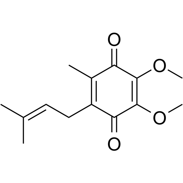
Ubiquinone-1 is an intermediate in the synthesis of Coenzyme Q. Ubiquinone-1 can be reduced by NADPH oxidase, hepatoma cells, Ascorbic acid (HY-B0166).
For research use only. We do not sell to patients.
- Purity : 99.0%
- CAS No.: 727-81-1
- Formula: C14H18O4
- Molecular Weight:250.29
-
Storage:
Solution, -20°C, 2 years

To place orders, for customer services and technical support, please contact: MedChemExpress USA
Tel: 609-228-6898 E-mail: [email protected] [email protected]


-
 Biological Activity
-
Chemical Information
- Protocols
- Purity & Documentation
- References
-
 Help & FAQs


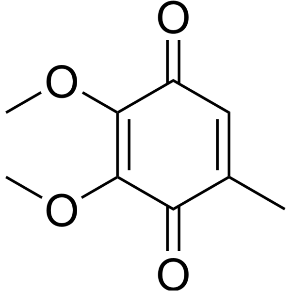
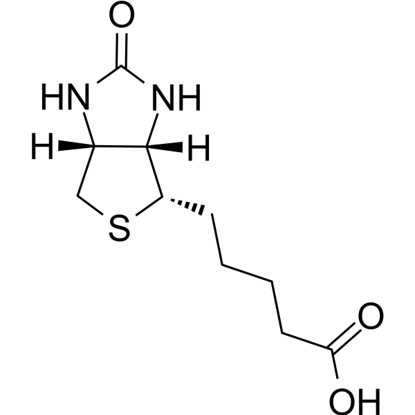
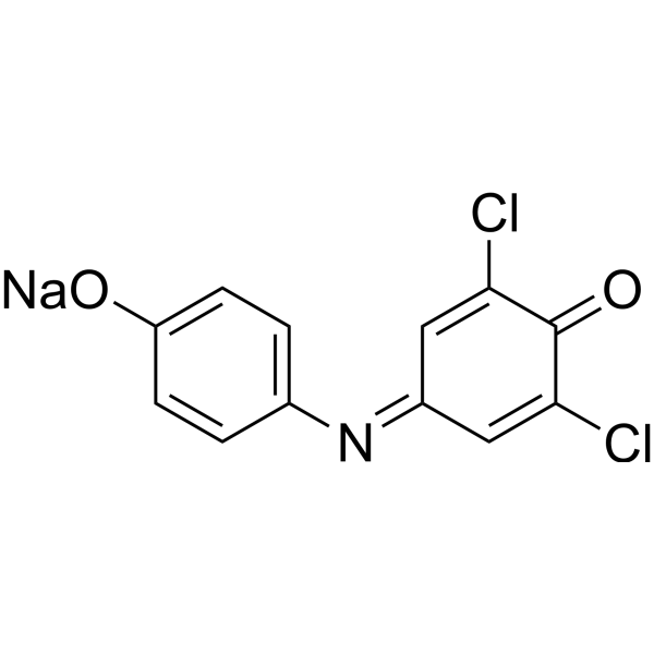

All Endogenous Metabolite Isoforms
More

Biological Activity
Description
IC50 & Target
|
Human Endogenous Metabolite |
In Vitro
Ubiquinone-1 (60 µM) induces external deamino-NADH oxidation in potato tuber mitochondria by altering the specificity of an external Rotenone (HY-B1756)-insensitive NADH dehydrogenase[1].
Ubiquinone-1 (100 µM) is rapidly and efficiently reduced to extracellularly detectable ubiquinol-1 by whole blood, isolated erythrocytes, and human hepatoma HepG2 cells[3].
Ubiquinone-1 (150 µM) is reduced by NADPH oxidase of guinea pig macrophages under anaerobic conditions[4].
Ubiquinone-1 is reduced by Ascorbic acid (HY-B0166) in a molecular oxygen concentration-controlled process[5].
MedChemExpress (MCE) has not independently confirmed the accuracy of these methods. They are for reference only.


Chemical Information
-
CAS No. 727-81-1
-
Appearance Liquid
-
Molecular Weight 250.29
-
Formula C14H18O4
-
Color Orange to red
-
SMILES
O=C1C(OC)=C(OC)C(C(C)=C1C/C=C(C)\C)=O
-
Structure Classification
-
Shipping
Room temperature in continental US; may vary elsewhere.
-
Storage
Solution, -20°C, 2 years


Purity & Documentation
-
Data Sheet (269 KB)
-
SDS (394 KB)
- English - EN (394 KB)
- Français - FR (394 KB)
- Deutsch - DE (394 KB)
- Norwegian - NO (394 KB)
- Español - ES (394 KB)
- Swedish - SV (394 KB)
- Italian - IT (394 KB)
- Korean - KR (394 KB)
- Portuguese - PT (394 KB)
-
Handling Instructions (2659 KB)
References
[1]. Moller IM, et al. Ubiquinone-1 Induces External Deamino-NADH Oxidation in Potato Tuber Mitochondria. Plant Physiol. 1996 Sep;112(1):75-78. [Content Brief]
[2]. R Fato, et al. Steady-state kinetics of the reduction of coenzyme Q analogs by complex I (NADH:ubiquinone oxidoreductase) in bovine heart mitochondria and submitochondrial particles. Biochemistry. 1996 Feb 27;35(8):2705-16. [Content Brief]
[3]. Stocker R, et al. Extracellular reduction of ubiquinone-1 and -10 by human Hep G2 and blood cells. Biochim Biophys Acta. 1993 Aug 20;1158(1):15-22. [Content Brief]
[4]. Murakami M, et al. NADPH oxidase of guinea-pig macrophages catalyses the reduction of ubiquinone-1 under anaerobic conditions. Biochem J. 1986 Jul 15;237(2):541-5. [Content Brief]
[5]. Roginsky VA, et al. Reduction of ubiquinone-1 by ascorbic acid is a catalytic and reversible process controlled by the concentration of molecular oxygen. Redox Rep. 1996 Feb;2(1):55-62. [Content Brief]

Calculators
Concentration (start) × Volume (start) = Concentration (final) × Volume (final)


Ubiquinone-1 Related Classifications
HY-113449 Related Classifications
 Powered by Bioz